Small round cell tumours
Jump to navigation
Jump to search
The printable version is no longer supported and may have rendering errors. Please update your browser bookmarks and please use the default browser print function instead.

Small round cell tumours (SRCT), also small round blue cell tumours (SRBCT), are a group of tumours that have a similar histologic appearance.
General
- Group of tumours that is typically seen in childhood and younger adults.
- Most common small round cell tumour is non-Hodgkin lymphoma.[1]
A short differential diagnosis of small round cell tumours
- Neuroblastoma.
- Wilms tumour (AKA nephroblastoma).
- Alveolar rhabdomyosarcoma.
- Ewing sarcoma / primative neuroectodermal tumour (PNET).
- Lymphoma - non-Hogkin's lymphoma, usu. large cell lymphomas (e.g. diffuse large B cell lymphoma, anaplastic large cell lymphoma), Burkitt's lymphoma.
- Retinoblastoma.
- Hepatoblastoma.
- Desmoplastic small round cell tumour (DSRCT).
- Small cell carcinoma.
Others:
Memory device: 4 -blastomas (hepato-, neuro-, nephro-, retino-), PNET/Ewing sarcoma (medulloblastoma), LADSSS (lymphoma, alveolar rhabdomyosarcoma, DSRCT, small cell carcinoma, small cell osteosarcoma, synovial sarcoma).
A long differential diagnosis of small blue cell tumours
Adapted from Miller with modifications:[4]
Notes:
- This DDx includes small cells that may be spindled.
- The DDx for large (non-pleomorphic) epithelioid cells is found in large epithelioid tumours.
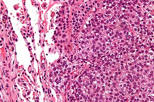
Microscopic
Features:
- Small cells ~ 2X RBC diameter - key feature.
- Scant cytoplasm/high NC ratio.
- Sheets of cells, very cellular.
- Coarse chromatin.
- +/-Nucleolus; usu. not prominent.
- +/-Vascularity.
Tabular comparison
Adapted from Thorner:[9]
| Tumour | Key histologic features | History | Key IHC / special stains |
|---|---|---|---|
| Hepatoblastoma | fetal hepatocytes (~ 1:3 NC ratio, eosinophilic cytoplasm) | liver lesion, usu. < 3 years old | AFP |
| Neuroblastoma | nests, thin fibrovascular septae, lymphocytes, ganglion-like cells (prominent nucleolus), neuropil (eosinophilic, fluffy, finely vacuolated) | adrenal gland, sympathetic chain; usu. < 3 years old | NB-84+, NSE+, S-100- |
| Nephroblastoma (Wilms tumour) | triphasic - (1) blue cells (blastema), (2) spindle cells (stroma), (3) tubular structures (epithelial) | kidney lesion, various syndromes (e.g. Beckwith-Wiedemann syndrome[10]) | WT-1+ |
| Retinoblastoma | Flexner-Wintersteiner rosette (rosette with empty centre (donut hole))[11] | eye lesion; +/-RB1 gene mutations (familial) | |
| Medulloblastoma | Homer-Wright rosettes (rosette with a meshwork of fibers (neuropil) at the centre)[11] | +/-nevoid basal cell carcinoma syndrome; must arise in cerebellum | |
| Ewing sarcoma/PNET | clear cytoplasm (glycogen) | bone lesion, adolescents | PAS+/PASD-, CD99 diffuse membranous |
| Alveolar rhabdomyosarcoma | alveolar-like spaces (small blue cells line spaces supported by fibrous tissue), rhabdomyoblasts (eosinophilic cytoplasm, +/-cross striations (uncommon), eccentric nucleus, +/-elongated/cigar-shaped cells (uncommon)) | adolescents, young adults; often mets at presentation | "DAM" = desmin, actin, myogenin |
| Embryonal rhabdomyosarcoma§ | nests with rounded border, rhabdomyoblasts (+/-elongated/cigar-shaped cells, eosinophilic cytoplasm, +/-cross striations, eccentric nucleus) | <10 years old; usu. localized | "DAM" = desmin, actin, myogenin |
| Desmoplastic small round cell tumour (DSRCT) | nests with "jigsaw" border | peritoneum (abdomen or plevis); male > female; locally invasive | keratin+, EMA+, desmin+ !!! |
| Small cell carcinoma | nuclear moulding, stippled chromatin | adults, smokers | synaptophysin, chromogranin A, CD56, keratins |
| Lymphoma, non-Hogkin's, usu. large cell (e.g. DLBCL) | dyscohesive cells, usu. ~2x normal (resting) lymphocyte, usu. nucleolus | usu. lymphadenopathy | CD45, CD20, TdT, CD3 |
Notes:
- § Uncommonly has the small round cell tumour morphology - included for comparison to alveolar rhabdomyosarcoma.
IHC panel
Gino's panel:[12]
| Lesion | NB84 | Myogenin | Desmin | CD99 | CD45 |
|---|---|---|---|---|---|
| Lymphoma | -ve | -ve | -ve | +ve/-ve | + |
| Ewing sarcoma | +ve/-ve | -ve | -ve | +ve (membranous) | - |
| Neuroblastoma | +ve | -ve | -ve | +ve/-ve | -ve |
| Rhabdomyosarcoma | -ve | +ve | +ve | -ve | -ve |
More general panel:[1]
- CD45, CD20, CD3, CD99, desmin, EMA, pankeratin, synaptophysin, chromogranin, GFAP.
See also
References
- ↑ 1.0 1.1 D'cruze, L.; Dutta, R.; Rao, S.; R, A.; Varadarajan, S.; Kuruvilla, S. (Jul 2013). "The role of immunohistochemistry in the analysis of the spectrum of small round cell tumours at a tertiary care centre.". J Clin Diagn Res 7 (7): 1377-82. doi:10.7860/JCDR/2013/5127.3132. PMID 23998069.
- ↑ URL: http://www.thedoctorsdoctor.com/diseases/small_round_blue_cell_tumor.htm. Accessed on: 2 July 2010.
- ↑ Chen QR, Vansant G, Oades K, et al. (February 2007). "Diagnosis of the small round blue cell tumors using multiplex polymerase chain reaction". J Mol Diagn 9 (1): 80–8. doi:10.2353/jmoldx.2007.060111. PMC 1867426. PMID 17251339. https://www.ncbi.nlm.nih.gov/pmc/articles/PMC1867426/.
- ↑ Miller RT. (Sep 2013). Atlantic Diagnostic Immunohistochemistry Symposium. St. John's, NL, Canada.
- ↑ Mayall, FG.; Gibbs, AR. (Jan 1992). "The histology and immunohistochemistry of small cell mesothelioma.". Histopathology 20 (1): 47-51. PMID 1310669.
- ↑ Kutzner, H.; Mentzel, T.; Kaddu, S.; Soares, LM.; Sangueza, OP.; Requena, L. (Mar 2001). "Cutaneous myoepithelioma: an under-recognized cutaneous neoplasm composed of myoepithelial cells.". Am J Surg Pathol 25 (3): 348-55. PMID 11224605.
- ↑ Reichard, KK.; Burks, EJ.; Foucar, MK.; Wilson, CS.; Viswanatha, DS.; Hozier, JC.; Larson, RS. (Oct 2005). "CD4(+) CD56(+) lineage-negative malignancies are rare tumors of plasmacytoid dendritic cells.". Am J Surg Pathol 29 (10): 1274-83. PMID 16160468.
- ↑ Guillou, L.; Coquet, M.; Chaubert, P.; Coindre, JM. (Aug 1998). "Skeletal muscle regeneration mimicking rhabdomyosarcoma: a potential diagnostic pitfall.". Histopathology 33 (2): 136-44. PMID 9762546.
- ↑ PST. 14 February 2011.
- ↑ URL: http://www.ncbi.nlm.nih.gov/pubmedhealth/PMH0002168/. Accessed on: 4 April 2011.
- ↑ 11.0 11.1 Wippold FJ, Perry A (March 2006). "Neuropathology for the neuroradiologist: rosettes and pseudorosettes". AJNR Am J Neuroradiol 27 (3): 488–92. PMID 16551982.
- ↑ G. Somers. 22 March 2011.