Forensic pathology
Forensic pathology is figuring-out why, when, where and how people died, if the manner of death is not obviously natural.
Death categorization
Deaths are categorized foremost by the manner of death. The manner is the single most important legal categorization for a death. The cause of death is important for understanding what happened. The mechanism of death is the pathophysiologic reason for death and can be inferred from the cause.
Examples:
| Cause of death | Manner of death | Mechanism of death | Scenario |
|---|---|---|---|
| Electrocution | accident | cardiac arrhythmia | man struck by lightning |
| Hyperthermia | accident | arrhythmias, seizures[1] | man lost on hiking trip in desert |
| Epidural hemorrhage due to blunt force trauma to the head | homicide | brain stem compression or cerebral vascular spasm leading to autonomic dysregulation | man hit with a hammer in the head |
| Carbon monoxide toxicity | suicide | cerebral hypoxia (CO binds to hemoglobin impairing oxygen transport) | woman found in car with suicide note, long history of depression, previous suicide attempts |
| Atherosclerotic heart disease | natural | cardiac arrhythmia due to ischemia | man found dead in bed, apartment locked, 95% stenosis of LMCA at autopsy, no other significant autopsy findings |
Manner of death
The manner of death is a legislatively defined classification. It varies slightly between jurisdictions.
| Manner | |||||||||||||||||||||||||||||||||||||||
| Homicide | Suicide | Natural | Accident | Undetermined | |||||||||||||||||||||||||||||||||||
Notes:
- Undetermined - is a waste basket category.
- Homicide - not necessarily murder.
- Can be group into three:
- Intent to kill (homicide, suicide).
- No intent to kill (natural, accidental).
- Undetermined.
Mechanism of death
This is occasionally of interest. It is usually based on physiology.
The mechanism is often asked for asphyxial deaths. The short answer it is: brain stem hypoxia due to ischemia caused by venous obstruction in the neck.[2][3]
Cause of death
- Abbreviated COD.
General
- The cause of death should be what started the sequence of events that lead to death.
Word form for cause of death
Examples:
- C. difficile colitis complicating antibiotic treatment for a dental abscess.[4]
- Complications of laparoscopic cholecystectomy for ascending cholangitis with mesothelioma and atherosclerotic heart disease.[5]
General forms:
- A complicating B for the treatment of C.
- A complicating B for the treatment of C with D and E.
World Health Organization form for cause of death
General form:[6]
- 1a = immediate cause of death.
- 1b = what lead to the immediate cause of death.
- 1c... 1[x] -- where 'x' is the last letter used; 1x = What started the sequence of events. This is known as the underlying cause of death.
- 2 = contributing factors.
Example 1:
- 1a. Ketoacidosis.
- 1b. Diabetes mellitus.
- 2. Alcoholism and acute bronchopneumonia.
Example 2:
- 1a. Hemoperitoneum.
- 1b. Splenic laceration.
- 1c. Blunt force trauma.
- 2. Liver cirrhosis.
Natural deaths
- The cause should be a medical diagnosis, not the mechanism (e.g. cardiac arrest, cachexia, kidney failure).
- The mechanism is irrelevant.
Notes:
- Unnatural causes trump natural ones. If a guy with (nothing more than) a 70% proximal LAD stenosis and an old myocardial infarct is found in the water, they are usually called drowning.
- Cancer is rarely the immediate cause of death - it is usually something else.[7]
- Things (mechanisms) that shouldn't be used: http://www.pallimed.org/2008/03/unacceptable-causes-of-death-other-web.html
Legal frame work
General
- In Ontario, the manner is determined by the coroner.
- Coroners, in Ontario, are MDs -- usually family docs.
- The cause (e.g. "gunshot wound to the head") is determined by the pathologist.
NB - the word coroner is not synoymous with MD. British Columbia has coroners that aren't MDs.
Case classification (Ontario)
Cases are classified as:
- A case = homicide and suspicious for homicide, (all) gunshot wounds.
- B case = adult, non-suspicious.
- C case = child, non-suspicious.
Notes:
- All A cases are done at regional centers by certified forensic pathologists.
Forensic golden triangle
- History.
- Scene.
- Autopsy.
Forensic diagnostic triangle
Most general differential diagnosis:
- Natural:
- Haemorrhage (e.g. cerebral bleed, gastrointestinal bleed, aortic aneurysm).
- Infection (e.g. pneumonia).
- Coronary artery atherosclerosis (cardiac arrhythmias - more common in the forensic context than myocardial infarction (MI); individuals with MIs don't usu. drop dead-- they go to the ER).
- Post myocardial infarction (free wall rupture).
- Ruptured (atherosclerotic) plaque.
- Toxic (memory device: PAIRO):
- Trauma (memory device AGE BS):
- Asphyxial.
- Gunshot wounds (GSWs).
- Environmental (e.g. hypothermia, hyperthermia, drowning, lack of oxygen, electrocution).
- Blunt force trauma.
- Sharp force trauma.
Difficulties arise when more than one point of the triangle is in play, i.e. the forensic pathologist has to earn their pay when an old man with a heart condition is known to be into erotic asphyxia, and dies after doing some drugs and whilst indulging in erotic asyphxiation with a friend...
- If he had an arrhythmia and there was no stressor... natural death.
- If he over did it with the drugs, it is an overdose, ergo accidental.
- If he did the erotic asphyxia a bit too long it is accidental.
- If the friend held the plastic bag over his head just a bit long to asphyxiate him... it is a homicide.
- If he was a lone and depressed... he might have been trying to kill himself, ergo suicide.
Rigor mortis
Definition:
- Muscle rigidity following death (caused by depletion of ATP).
Dependent on:
- Temperature of patient at death.
- Temperature variations in the environment since death.
- Presence of some medical conditions.
- May never develop!
It is the explanation for post-mortem goose bumps.
Summary
- Its onset & presence is highly variable.
- Forensic pathologists do not comment on time of death, as the above times are subject to such a large degree of variability, i.e. the estimates are essentially useless.
Time estimates
A crude guess for time of death based on rigor:[8]
- Warm & flaccid <3 h.
- Warm & stiff 3-8 h.
- Cold & stiff 8-36 h.
- Cold & flaccid > 36 h.
Notes:
- Memory device: 3s: cut points are at 3 hours, 1/3 of a day, 3/2 of a day.
Livor mortis
Definition: pooling of blood in the dependent position, due to blood stasis.
- Onset may preceed death in the context of congestive heart failure.
- If pressure is applied to a dependent area-- no blood can enter there; thus, a pressure area is blanched (i.e. white).
- Can be seen externally, i.e. on the skin, and internally.
- Liver mortis becomes fixed some time after death.
- Liver mortis does NOT tell one the position the decedent was in at the time of death-- only the position the decedent was at the time liver mortis became fixed. If the decedent wasn't moved liver mortis can help determine the position the person was in when they died.
Averages:
- Start: 30 minutes to 2 hours
- Fixed: 8-12 hours.
DDx:
- Blunt force trauma - especially to the inexperienced eye.
- Post-mortem hypostatic bruising.
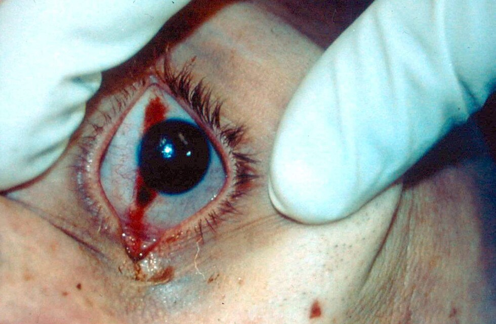
Tache noire
Literally black spot.
Features:[9]
- Brown/black horizontal line of the eye due to drying.
- Arises if the eye remains open after death.
- May mimic a traumatic injury.
Images:
Post-mortem decomposition/preservation
One of three things happens post-mortem:[11]
- Mummification.
- Putrefaction (skeletonisation).
- Green colour due to break down of hemoglobin (biliverdin).[12]
- Adipocere - transformation into wax (due to anaerobic bacterial hydrolysis of fat).
- Useless for toxicology and DNA.
- A mix of the above often occur, i.e. part of the corpse is mummified... part of it decomposed through putrefaction.
Mummification:
- Predominant in dry environments.
- Body becomes dry and leathery.
Putrefaction:
- Body wet/moist after death -- ideal environment for putrefactive bacteria and organisms.
Artefacts
- Prinsloo and Gordon artefact = artefactual post-morten haemorrhage on the posterior surface of the esophagus.[13]
- Minimized by removing cranial contents & thoracic contents before undertaking neck dissection.[14]
- Artefactual fractures (see fractures).
- Dilated anus (in isolation).[15][16]
- Towel clip injury, usu. paired (in organ donors) - may be mistaken for an electroshock weapon (e.g. Taser) wound.[17]
- Subclavian stab for vascular access - may be confused with a gunshot exit wound.
Infants
- Lumpy neck - small superficial nodules on anterior neck ~2-5 mm (???).[18]
- Intussusception of small bowel - often multiple.
Wounds
General
- Wound - definition: defect in skin or mucous membrane[19] - usually due to trauma.
Special types of wounds:
- Gunshot wounds.
- Incised wounds - see sharp force trauma.
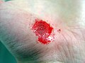
Gross pathologic classification of injuries
Mnemonic CALI:
- Contusion - "bruise", hematoma.
- Abrasion - "scrape", e.g. motorcyclist slide across the roadway... skin scraped-off.
- Can be subclassified as brush abrasions (has skin tags) and crush abrasions (do not have skin tags).
- Skin tags suggest directionality; they are found at the distal point / point of last contact.[21]
- Can be subclassified as brush abrasions (has skin tags) and crush abrasions (do not have skin tags).
- Laceration - "tear", indicates blunt force trauma; contact point may be distant from where skin splits.
- Incised - "cut", e.g. caused by a knife,[22] subdivided as follows:
- "Cut" or "slash" = length > depth.
- "Stab" = depth > length.
- "Chop" = typically have a contusion at the margin of the wound, classically caused by an axe. May be caused by a propeller.[23]
Images
Abrasion. (WC)
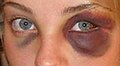
Contusion ("black eye"). (WC)


DDx
How to decide what you're looking at:
- Contusion:
- Can be demonstrated histologically... there are extravascular RBCs.
- If pre-morten there is vital reaction, i.e. WBCs come to clean-up the trauma.
- If the post mortem interval is not known and long-- differentiation from decomposition may be non-trivial/impossible.
- Can be demonstrated histologically... there are extravascular RBCs.
- Abrasion vs. contusion:
- Contusions skin is intact... in abrasion it is not.
- Abrasions and contusions may be co-localized, i.e. in the same place.
- Laceration vs. incision:
- Lacerations have "bridges", incisions do NOT have bridges.
- Bridges are fine strands of tissue that cross the long axis of the skin defect.
- You can think of the wound as partially "sutured" by the bridges of tissue.
- Bridges are fine strands of tissue that cross the long axis of the skin defect.
- Lacerations are usually associated with a contusion and/or crush and have an irregular margin.[24]
- Lacerations are classically on the skull and face. They are rarely on the abdomen.
- Lacerations have "bridges", incisions do NOT have bridges.
Wound dating
- Colour is somewhat useful for contusions (bruises).
- Post-mortem injuries tend to be orange-yellow.[25]
- Wounds age is difficult to determine as wound healing is affected by a large number of variables.
- Old wounds (scars), generally, cannot be dated - one can only say they are old.
Microscopic
Wounds can be grouped into:
- Pre-mortem.
- Post-mortem.
Signs a wound was inflicted during life:
- Blood.
- Hypostasis/decomposition can mess with this, i.e. blood oozing out of vessels post-mortem shouldn't be called an injury.
- Hemosiderin demonstrated by an iron stain - hard sign.
- Inflammation:[26]
- PMNs 6-24 hours after injury.
- PMNs replaced monocytes in 24-48 hours.
Stains
- Iron stain for siderophages (hemosiderin-laden macrophages) -- presence suggests 2-3 days or older.[27]
Bone fractures
Artefactual fractures
- "Undertaker's fracture" - cervical fracture due to rough handling.[28]
- Basal skull fracture due to opening of skull.[29]
- Classically does not cross sella turcica.
- Notably absent features of a real (ante-mortem) fracture: hematoma, brain injury.
- Mechanism to explain trauma not present in history; a fall/tripping not sufficient.
Healing of fractures
Simplified classification
- Primary callus (cartilaginous) - early.
- Secondary callus (bone) - late.
Microscopic
Features:
- Fragmentation of bone.
- +/-Dead bone = lacunae have no osteocytes.[30]
- Takes days for osteocyte loss.
- +/-Inflammatory cells.
- +/-Hemosiderin-laden macrophages.
- +/-Osteoblastic rimming.
DDx:
- Fracture secondary to a tumour:
- Metastatic carcinoma.
- Osteosarcoma - typically does not have osteoblastic rimming.
Notes:
- Radiology is not good at dating fratures;[31] however, it is good at finding 'em.
Pattern and cause
- Paravertebral (bony) nodules = classic location for rib fractures in child abuse.
- Metaphyseal fractures - "classical metaphyseal lesions".[32]
Motor vehicle versus pedestrian
If the pedestrian is standing during the initial impact one classically finds, at bumper level, a lower limb fracture with a Messerer wedge (German: Messerer-Kiel);[33] the wedge points in the direction of the (impact) force.
Location or type
Orbital floor fractures
General
- Classically due to fights, followed by traffic accidents.[35]
- Thought to result from loading on the orbital rim directly or the orbit - both are transmitted to the orbital floor.[34]
Note:
- The orbital floor tends to the be weaker than other components of the orbital cavity wall; thus, it is the most common site of fracture in the orbital cavity wall.
Basal skull fracture
General
Etiology:
- Blunt force trauma - high energy & velocity.
- Seen in motor vehicle collisions, descent from height.
Clinical/external findings:
- Raccoon eyes = periorbital ecchymosis.
- Battle sign = mastoid ecchymosis.
- Associated with orbital roof fractures.[36]
- Cerebrospinal fluid rhinorrhea.
- Hemorrhage from nose and ears.
- Hemotympanum.
Note:
- There is a dictum that states bilateral petrous bone fractures are due to impact to the side of the head - it isn't true.[37]
Hinge fracture of the skull
- A special type of basal skull fracture.
- Complete hinge fractures are considered severe; they are a 4 on the abbreviated injury scale (AIS).[38]
- Classically due to a blow to the chin - resulting in a fracture across the medial fossa and sella turcica.[39]
Pathologic fracture
- A fracture due to an underlying pathology.
Hip fractures
Autopsy
The autopsy article covers procedural things. Heart dissection is covered in the heart article.
Types
Forensic vs. hospital:
- Forensic autopsies are focused on the external exam.
Marking conventions for common findings
There are no universal marking conventions for injuries.
One system in use (the Rose system) is:[40]
- One red line for an incised wound.
- Multiple closely spaced red lines, i.e. red hatching, for abrasions.
- Multiple closely-spaced blue lines, i.e. blue hatching, for contusions.
The above makes sense in that:
- Abrasions and incised wounds typically bleed - are red.
- Contusions (bruises) don't classically bleed and are classically blue.
External exam findings
Colour of the corpse:[41]
- Red (Pink) = carbon monoxide, cyanide, fluoroacetate,[42] hypothermia.
- Purple (intense) = propane.
- Green = hydrogen sulfide.
- Brown = nitrites (methemoglobinemia).
Autopsy terminology
- Gutter butter = adipose tissue in a decomp case; looks like butter topping put on popcorn. A Toronto-ism.
- Gutter blood = blood in the empty thorax - after extraction of the organ block.
- Tardieu spots = postmortem hypostatic hemorrhages;[43] look like petechiae - in dependent areas, i.e. in the zone of livity.
Autopsy on decomposed remains
- AKA "decomp autopsy" or simply "decomp".
General
- Histology usually very limited or useless.
- Often done to exclude trauma.
- Typical scenario: decedent lives alone -- body not discovered for prolonged period of time.
- More likely to be a negative autopsy than non-decomp cases.
Suspicious decomp
Common sense rules for if skin is not intact:
- Blunt dissection (to avoid artefactual injuries to the bones).
- Clean the bones (not with bleach)
- Bones cooked for 1+ hours... with frequent checks to avoid that they become mushy.
Causes of death
Environmental
They include:
- Hypothermia.
- Hyperthermia.
- Drowning - see asphyxial deaths.
- Lack of oxygen - see asphyxial deaths.
- Electrocution.
Gunshot wounds
Gunshot wounds (GSWs) are a relatively uncommon finding in Canada. They are dealt within a separate article.
Asphyxia
- This is a big topic and covered by a separate article.
Classification
- Strangulation - where there are signs of neck compression.
- Includes: ganging, ligature strangulation and manual strangulation.
- Chemical asphyxia - usually no signs of neck compression.
- Includes: carbon monoxide poisoning.
- Suffocation - usually no signs of neck compression.
Blunt force injury
- AKA blunt force trauma.
General
Classification:
- Contusions.
- Laceration.
- Acceleration/deceleration injury, e.g. diffuse axonal injury.
Weapons:
- Fist.
- Foot.
- Baseball bat... pretty much anything.
- Beer bottles are common... and strong enought to fracture a skull.
- Empty bottles have a higher fracture energy than full ones.[44]
Cause of death
Commotio cordis
- Often negative autopsy; no cardiac pathology.
- Etiology: arrhythmia.
- History: trauma to chest.
Note:
- May be spelled Commodio cordis.[47]
- Analogous to commotio medullaris.
Scenarios
Motor vehicle collisions
- Pedestrian vs. motor vehicle: heel to injury measurement, remember to include the thickness of the heel/sole of shoe.[48]
- Dicing injuries: tempered glass used in side window construction fragments into cubes when fractured causing L-shaped wounds.
Descent from height
- Relatively common way to suicide.
- May be an accident, e.g. decedent thought they can fly (due to a psychosis).
- May be a homicide, e.g. decedent was pushed.
Gross
Features:
- Multiple injuries - often including multiple fractures, e.g. basal skull fracture, flail chest.
- +/-Haemothorax - can be proved with a large bore needle.
- Sufficient for cause of death - can be used to do an abbreviated post-mortem.
- +/-Haemoaspiration (due to facial trauma) - presence suggest that decendent was alive shortly after landing/impact and thus likely very alive during the descent.
- Patchy red centrilobular spots on gross examination.
Injury patterns
Seromuscular tear
- AKA seatbeat syndrome.
- Intestinal injury associated with motor vehicle collisions and more specifically seatbelts.
Features:
- Def'n: separation of (inner) muscularis propria from submucosa.[49]
Bite injury
- A special type of blunt force trauma.
- May be seen in the context of a sexual assault.
- A forensic dentist may be able to assist.
In the context of a suspicious case:
- Human vs. animal.
- Bite marks, as evidence, have a limited value for identification purposes.
- In the context of identifying a potential perpetrator, it is essential to swab the bite mark for saliva, which is rich in DNA.[50]
Images
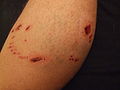
Bite injury. (WC)

Aortic trauma
- Classic location of transection of the aorta is distal the the left subclavian branch point near the insertion of the ligamentum arteriosum (e.g. peri-isthmus).[51]
- Aortic dissection due to trauma is often catastrophic. Several mechanisms have been proposed and there is a body of trauma biomechanics research that explores this.
Trauma with delayed death
- Epidural hemorrhage with a lucid interval.
- Subcapsular splenic hematoma with subsequent rupture.
- Subcapsular hepatic hematoma with subsequent rupture.
- Aortic dissection with subsequent rupture.
Sharp force injury
- AKA sharp force trauma.
General
Injuries caused by:[52]
- Knife.
- Scissors - classic "Z" shape.
- Screwdriver.
- Glass.
Gross
Features:[52]
- Incised wound (see: Classification of wounds).
- "Clean" edge (no contusion, no abrasion).
- Well-demarcated edges.
- +/-Hilt mark.
- Due to contact of hilt.
Subclassified into - see classification of wounds:
- Cut/slash.
- Stab.
- Chop - a mixed injury, sharp force and blunt force.
Images
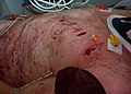
Sharp force trauma - thorax. (WC)

Head injuries
Accidental vs. intentional
Features of non-accidental injuries:[53]
- Lacerations:
- More than three.
- Length >= 7 cm or more.
- Location:
- Above hat brim line (HBL).
- Ear.
- Left-sided.
- Fractures:
- Comminuted or depressed calvarial fractures.
- Location:
- Fractures located above the HBL.
- Left-sided fractures.
- Facial fractures.
- Contusions:
- Greater than four facial contusions.
- Other:
- "Postcranial osseous" [sic] (non-rib, non-skull) and/or visceral trauma.
Note: The paper doesn't give odds ratios for the the different features -- like in the rational clinical exam series... it is a shame.
Diffuse axonal injury
- Abbreviated DAI.
General
Clinical:
- Vegetative state.
- Imaging findings: no anatomical cause apparent (in some cases).
Etiology:
- Hypothesized to arise from high shear loading of white mater tracts.[54]
Gross
Macroscopic findings:[54]
- Tears - corpus callosum.
- Haemorrhage.
Other (chronic) changes:[55][citation needed]
- Thalamus - shrinkage.
- Enlargement of third ventricle.
DDx (medical imaging):[56]
Microscopic
Microscopic findings:[54]
- Axonal retraction balls.
- "Microglial stars".
- Degeneration of fibre tracts.
Grading:[57]
- Grade 1: only microscopic findings.
- Grade 2: macroscopic corpus callosum injury + microscopic findings of DAI.
- Grade 3: macroscopic corpus callosum and midbrain injuries + microscopic findings of DAI.
Stains
- Bielschowsky stain to highlight axonal swellings - appear 12-18 hours after injury.[58]
IHC
Intracranial hemorrhage
Intracranial hemorrhage may be a consequence of blunt force trauma.
Classification:
Cerebral contusion
General
- Due to blunt force trauma.
Gross
Features:
- Focal superficial hemorrhage.
- Location, usually, frontal lobe and temporal lobe.[61]
Notes:
- Classically, come in pairs:[61]
- Coup contusion - at the site of the (primary) impact
- Contrecoup contusion - secondary internal impact.
- Example - fall on back of head:
- Occipital lobe contusion = coup contusion.
- Frontal lobe contusion = contrecoup contusion.
- May be associated with contusions of the:[62]
- Deep brain structures, known as an "intermediary coup".
- Dorsal surface of the cerebral hemispheres, known as "gliding contusions".
- Resolve as a yellow lesion (like at other sites), known as a plaque jaune in the brain.
- Classically, inferior aspect of the frontal lobe.
DDx:
- Hemorrhagic stroke - usually temporal lobe and/or parietal lobe.
Traumatic brain injury in infants
- Shaken-impact syndrome, AKA shaken baby syndrome.
Commotio medullaris
Features:[63]
- Sudden death after head trauma that is insufficient to explain death.
- Etiology: unknown - thought to be related to apnea.
Note:
- Analogous to commotio cordis.
Excited delirium
General
- Diagnosis is considered somewhat controversial outside of the forensic pathology community.[65]
- The diagnosis has garnered attention in the context of electroshock weapon use, as Taser International (a manufacturer of electroshock weapons) has often ascribed the deaths involving its weapons to it - when it is alleged that their electroshock weapon caused the death.
- There is no "official" definition for excited delirium.
- Most agree it includes fever.
One paper defines it in relation neuroleptic malignant syndrome:[64]
- Fever.
- Disorientation and confusion.
- Increased energy/superhuman strength.
Excited delirium - hypothesis:
- Thought to arise in the context of severe chronic mental disorders (e.g. schizophrenia) and protracted cocaine binges.[66]
- Thought to result from alteration of dopamine receptor density. The D2 receptor in particular, which is thought to be important in temperature regulation, is decreased in psychotic cocaine abusers.[64]
Toxicology & biochemistry
General
Things usually collected at autopsy:
- Blood in EDTA tube (genetic testing).
- Urine toxicology:
- Useful to evaluate myoglobin.
- Vitreous:
- Biochemistry.
- Ketones.
- Urea (???).
- Bile:[67]
- Acetaminophen overdoses.
- Opiate overdoses.
Myoglobin DDx:
- Neuroleptic malignant syndrome.
- Malignant hyperthermia.
- Serotonin syndrome.
Biochemistry
- Diabetes mellitus:[68]
- Plasma:
- Hemoglobin A1c - increased.
- Acetone - increased.
- Beta-hydroxybutyrate - increased.
- Also increased in alcoholic ketoacidosis (though ketones low).
- Urine:
- Aceto-acetate - increased.
- Plasma:
Death by insulin overdose:[69]
- C-peptide - low.
- Insulin - high.
Serum
- Potassium - rises quickly and rapidly after death; completely useless.
- Sodium - tends to decrease after death; usually useless.
- Glucose - drops quickly; useless unless sky high.
- Urea, creatinine and urate - stable for ~48 hours post-mortem.[70]
Vitreous
- Creatinine and urea - approximate those at time of death.[70]
- Glucose - used to assess for hyperglycemia (due to diabetic coma) in life.[71]
Toxicology
- Should be submitted with anatomical findings and history.
Common submissions:
- Alcohol only.
- Suspected toxicologic death - need details on drugs.
Mandated by case
In Ontario, the following are mandated by the case:[72]
- Sudden death of child under five years old.
- Workplace death.
- Fatal motor vehicle collision - esp. driver.
- Aviation death - esp. pilot & co-pilot.
- Fire-related death (carboxyhemoglobin).
Toxins
Ethanol toxicity
- Usually measured (in Canada) as: mass of EtOH (mg)/volume of blood (mL).
- Limit (Ontario): 80 milligrams of alcohol in 100 millilitres of blood (0.08 gm/100 mL).[73]
- Enough to be fatal ~ 350 mg/dL ~= 76 mmol/L.
Ethanol intoxication as a table[74]
| Concentration | Concentration | Concentration | Concentration | |
| Legal limit - Ontario[75] | 80 mg/dL | ~17 mmol/L | 0.8 g/L | 0.08 g/dL |
| Mild | < 180 mg/dL | < 39 mmol/L | < 1.8 g/L | < 0.18 g/dL |
| Moderate | 180-350 mg/dL | 39-76 mmol/L | 1.8-3.5 g/L | 0.18-0.35 g/dL |
| Severe | 350-450 mg/dL | 76-98 mmol/L | 3.5-4.5 g/L | 0.35-0.45 g/dL |
Notes:
- 1 mg/dL = 1/4.607 mmol/L.
- Ethanol's molar mass = 46.07 g/mol.
Methanol toxicity
- Minimum lethal dose: 40 mg/dl.[76]
- Typically an accidental death; person consumes methanol as an ethanol substitute.
- Blindness.
- Putamen necrosis (bilateral).[77]
- +/-Pancreatic injury.[78]
Cocaine toxicity
- No agreed upon toxic dose[79] - due to tolerance.
- Usual mechanism cardiac failure.
Features - heart:
- Usually anatomically normal heart.
- +/-Advanced coronary artery atherosclerosis for age.
- +/-Myocardial infarction.
- +/-Contraction band necrosis.
- +/-Cardiac hypertrophy.
Other:
- +/-Nasal septum perforation.
- +/-Track marks (other drug use).
- +/-Finger burns (during preparation of crack).
- +/-Drug paraphernalia, e.g. crack pipe.
Ethylene glycol toxicity
- For a more general discussion see urine crystals
- Not done in routine toxicology screening.
- Birefringent calcium oxalate crystals found in kidney (with polarized light).[80]
Anaphylaxis
- Allergic reaction, e.g. peanut allergy.
Diagnosis - serology:[81]
- IgE.
- Tryptase.
Natural death
There is a lot that can kill ya... but only a few of those are quickly, i.e. within a hour or so.
Generally, these things are:
- Cardiovascular:
- Arrhythmia.
- Myocardial infarction.
- Haemorrhage.
- Ruptured aneurysm.
- Hypertensive heart disease.
- Respiratory:
- Pulmonary embolism (PE).
- Asthma.
- GI:
- Haemorrhage.
- Esophageal varices.
- Gastric varices.
- Haemorrhage.
- Neurologic:
- Intracranial haemorrhage.
- Ruptured aneurysm.
- Spontaneous subdural hemorrhage.
- Stroke:
- Haemorrhagic.
- Thrombotic (more common than hemorrhagic).
- Intracranial haemorrhage.
Forensic entomology
- Study of the bugs that eat corpses.
- Bugs may hide a wound... it is important to know where they like to be.
Forensic anthropology
Forensic anthropology is looking at skeletal remains. It may be useful of identification and, rarely, the cause of death. Important in skeletonized remains and decomp cases.
Forensic taphonomy
- The study of post-mortem decay to assist in a medicolegal investigation.
- Taphonomy = postmortem fate of biological remains; derived from the Greek word taphos (grave).[82]
See also
References
- ↑ URL: http://www.forensicmed.co.uk/pathology/mechanisms-of-death/. Accessed on: 19 April 2012.
- ↑ URL: http://www.forensicmed.co.uk/pathology/mechanisms-of-death/. Accessed on: 1 May 2012.
- ↑ URL: http://www.forensicmed.co.uk/pathology/pressure-to-the-neck/. Accessed on: 1 May 2012.
- ↑ MSP. 8 September 2010.
- ↑ TR. 3 September 2010.
- ↑ Pollanen MS (June 2005). "Deciding the cause of death after autopsy--revisited". J Clin Forensic Med 12 (3): 113–21. doi:10.1016/j.jcfm.2005.02.004. PMID 15914304.
- ↑ Shannon, P. 2009.
- ↑ Saukko, Pekka; Knight, Bernard (2004). Knight's Forensic Pathology (3rd ed.). A Hodder Arnold Publication. pp. 61. ISBN 978-0340760444.
- ↑ 9.0 9.1 URL: http://emedicine.medscape.com/article/1680032-overview. Accessed on: 6 March 2012.
- ↑ URL: http://www.demussen.net/carbon-monoxide/chemical-changes-in-body-fluids.html. Accessed on: 6 March 2012.
- ↑ Burton, Julian L.; Rutty, Guy N. (2010). The Hospital Autopsy A Manual of Fundamental Autopsy Practice (3rd ed.). Oxford University Press. pp. 102. ISBN 978-0340965146.
- ↑ NOIR BA, GARAY ER, ROYER M (May 1965). [linkinghub.elsevier.com/retrieve/pii/0304416565900097 "SEPARATION AND PROPERTIES OF CONJUGATED BILIVERDIN"]. Biochim. Biophys. Acta 100: 403–10. PMID 14347937. linkinghub.elsevier.com/retrieve/pii/0304416565900097.
- ↑ Piette MH, De Letter EA (November 2006). "Drowning: still a difficult autopsy diagnosis". Forensic Sci. Int. 163 (1-2): 1–9. doi:10.1016/j.forsciint.2004.10.027. PMID 16378701.
- ↑ Burton, Julian L.; Rutty, Guy N. (2010). The Hospital Autopsy A Manual of Fundamental Autopsy Practice (3rd ed.). Oxford University Press. pp. 118. ISBN 978-0340965146.
- ↑ URL: http://www.kingstonwhigstandard.com/ArticleDisplay.aspx?archive=true&e=736464. Accessed on: 6 October 2010.
- ↑ Elder, DE. (Nov 2007). "Interpretation of anogenital findings in the living child: Implications for the paediatric forensic autopsy.". J Forensic Leg Med 14 (8): 482-8. doi:10.1016/j.jflm.2007.03.005. PMID 17961873.
- ↑ MSP. 12 October 2010.
- ↑ MSP. 6 October 2010.
- ↑ URL: http://dictionary.reference.com/browse/wound. Accessed on: 20 April 2012.
- ↑ Burton, Julian L.; Rutty, Guy N. (2010). The Hospital Autopsy A Manual of Fundamental Autopsy Practice (3rd ed.). Oxford University Press. pp. 108. ISBN 978-0340965146.
- ↑ 21.0 21.1 Burton, Julian L.; Rutty, Guy N. (2010). The Hospital Autopsy A Manual of Fundamental Autopsy Practice (3rd ed.). Oxford University Press. pp. 105. ISBN 978-0340965146.
- ↑ DiMaio, Vincent J.M.; Dana, Suzanna E. (2006). Handbook of Forensic Pathology (2nd ed.). CRC Press. pp. 154. ISBN 978-0849392870.
- ↑ Ihama, Y.; Ninomiya, K.; Noguchi, M.; Fuke, C.; Miyazaki, T. (Oct 2009). "Fatal propeller injuries: three autopsy case reports.". J Forensic Leg Med 16 (7): 420-3. doi:10.1016/j.jflm.2009.04.006. PMID 19733336.
- ↑ Burton, Julian L.; Rutty, Guy N. (2010). The Hospital Autopsy A Manual of Fundamental Autopsy Practice (3rd ed.). Oxford University Press. pp. 109. ISBN 978-0340965146.
- ↑ Campobasso, CP.; Marchetti, D.; Introna, F.; Colonna, MF. (Mar 2009). "Postmortem artifacts made by ants and the effect of ant activity on decompositional rates.". Am J Forensic Med Pathol 30 (1): 84-7. doi:10.1097/PAF.0b013e318187371f. PMID 19237864.
- ↑ Mitchell, Richard; Kumar, Vinay; Fausto, Nelson; Abbas, Abul K.; Aster, Jon (2011). Pocket Companion to Robbins & Cotran Pathologic Basis of Disease (8th ed.). Elsevier Saunders. pp. 26. ISBN 978-1416054542.
- ↑ Betz, P. (1994). "Histological and enzyme histochemical parameters for the age estimation of human skin wounds.". Int J Legal Med 107 (2): 60-8. PMID 7529545.
- ↑ URL: http://www.the-crankshaft.info/2010/07/postmortem-changes_29.html. Accessed on: 29 September 2010.
- ↑ MSP. 29 September 2010.
- ↑ Fondi, C.; Franchi, A. (Jan 2007). "Definition of bone necrosis by the pathologist.". Clin Cases Miner Bone Metab 4 (1): 21-6. PMID 22460748.
- ↑ Prosser, I.; Maguire, S.; Harrison, SK.; Mann, M.; Sibert, JR.; Kemp, AM. (Apr 2005). "How old is this fracture? Radiologic dating of fractures in children: a systematic review.". AJR Am J Roentgenol 184 (4): 1282-6. PMID 15788611. http://www.ajronline.org/cgi/content/full/184/4/1282.
- ↑ Kleinman, PK.; Marks, SC. (May 1996). "A regional approach to classic metaphyseal lesions in abused infants: the distal tibia.". AJR Am J Roentgenol 166 (5): 1207-12. PMID 8615271.
- ↑ Karger, B.; Teige, K.; Fuchs, M.; Brinkmann, B. (Jun 2001). "Was the pedestrian hit in an erect position before being run over?". Forensic Sci Int 119 (2): 217-20. PMID 11376986.
- ↑ 34.0 34.1 Punke, C.; Fritsche, A.; Martin, H.; Schmitz, KP.; Pau, HW.; Kramp, B. (Dec 2007). "[Investigation of the mechanisms involved in isolated orbital floor fracture. Simulation using a finite element model of the human skull].". HNO 55 (12): 938-44. doi:10.1007/s00106-007-1545-5. PMID 17333039.
- ↑ Gosau, M.; Schöneich, M.; Draenert, FG.; Ettl, T.; Driemel, O.; Reichert, TE. (Jun 2011). "Retrospective analysis of orbital floor fractures--complications, outcome, and review of literature.". Clin Oral Investig 15 (3): 305-13. doi:10.1007/s00784-010-0385-y. PMID 20165966.
- ↑ URL: http://emedicine.medscape.com/article/1680107-overview#showall. Accessed on: 28 March 2012.
- ↑ Harvey, FH.; Jones, AM. (Apr 1980). "Typical basal skull fracture of both petrous bones: an unreliable indicator of head impact site.". J Forensic Sci 25 (2): 280-6. PMID 7391790.
- ↑ Adams, VI.; Carrubba, C. (Sep 1998). "The Abbreviated Injury Scale: application to autopsy data.". Am J Forensic Med Pathol 19 (3): 246-51. PMID 9760090.
- ↑ URL: http://wiki.answers.com/Q/Hinge_fracture_of_skull_is_seen_in_accidents_involving. Accessed on: 28 March 2012.
- ↑ TR. 1 September 2010.
- ↑ Shkrum, Michael J.; Ramsay, David A. (2006). Forensic Pathology of Trauma: Common Problems for the Pathologist (1st ed.). Humana Press. pp. 33. ISBN 978-1588294586.
- ↑ Proudfoot AT, Bradberry SM, Vale JA (2006). "Sodium fluoroacetate poisoning". Toxicol Rev 25 (4): 213–9. PMID 17288493.
- ↑ Pollanen MS, Perera SD, Clutterbuck DJ (December 2009). "Hemorrhagic lividity of the neck: controlled induction of postmortem hypostatic hemorrhages". Am J Forensic Med Pathol 30 (4): 322–6. doi:10.1097/PAF.0b013e3181c17ec2. PMID 19901802.
- ↑ Bolliger SA, Ross S, Oesterhelweg L, Thali MJ, Kneubuehl BP (April 2009). "Are full or empty beer bottles sturdier and does their fracture-threshold suffice to break the human skull?". J Forensic Leg Med 16 (3): 138–42. doi:10.1016/j.jflm.2008.07.013. PMID 19239964.
- ↑ Kohl P, Nesbitt AD, Cooper PJ, Lei M (May 2001). "Sudden cardiac death by Commotio cordis: role of mechano-electric feedback". Cardiovasc. Res. 50 (2): 280–9. PMID 11334832.
- ↑ Maron BJ, Estes NA (March 2010). "Commotio cordis". N. Engl. J. Med. 362 (10): 917–27. doi:10.1056/NEJMra0910111. PMID 20220186. http://www.nejm.org/doi/full/10.1056/NEJMra0910111.
- ↑ Perron AD, Brady WJ, Erling BF (September 2001). "Commodio cordis: an underappreciated cause of sudden cardiac death in young patients: assessment and management in the ED". Am J Emerg Med 19 (5): 406–9. doi:10.1053/ajem.2001.24455. PMID 11555799.
- ↑ Ontario Forensic Pathology Service (2009). Ontario Forensic Pathology Service: Practice Manual for Pathologists (2nd ed.). Queen's Printer for Ontario. pp. 18.
- ↑ Slavin, RE.; Borzotta, AP. (Sep 2002). "The seromuscular tear and other intestinal lesions in the seatbelt syndrome: a clinical and pathologic study of 29 cases.". Am J Forensic Med Pathol 23 (3): 214-22. doi:10.1097/01.PAF.0000023001.32202.2D. PMID 12198344.
- ↑ Pretty, IA.. "Forensic dentistry: 2. Bitemarks and bite injuries.". Dent Update 35 (1): 48-50, 53-4, 57-8 passim. PMID 18277695.
- ↑ Kodali S, Jamieson WR, Leia-Stephens M, Miyagishima RT, Janusz MT, Tyers GF (November 1991). "Traumatic rupture of the thoracic aorta. A 20-year review: 1969-1989". Circulation 84 (5 Suppl): III40–6. PMID 1934437.
- ↑ 52.0 52.1 Burton, Julian L.; Rutty, Guy N. (2010). The Hospital Autopsy A Manual of Fundamental Autopsy Practice (3rd ed.). Oxford University Press. pp. 111-2. ISBN 978-0340965146.
- ↑ Guyomarc'h P, Campagna-Vaillancourt M, Kremer C, Sauvageau A (March 2010). "Discrimination of falls and blows in blunt head trauma: a multi-criteria approach". J. Forensic Sci. 55 (2): 423–7. doi:10.1111/j.1556-4029.2009.01310.x. PMID 20141554.
- ↑ 54.0 54.1 54.2 Blumbergs PC, Jones NR, North JB (July 1989). "Diffuse axonal injury in head trauma". J. Neurol. Neurosurg. Psychiatr. 52 (7): 838–41. PMC 1031929. PMID 2769276. https://www.ncbi.nlm.nih.gov/pmc/articles/PMC1031929/.
- ↑ Rose, Alan G. (2008). Atlas of Gross Pathology with Histologic Correlation (1st ed.). Cambridge University Press. pp. 639. ISBN 978-0521868792.
- ↑ Kumar, S.; Gupta, V.; Aggarwal, S.; Singh, P.; Khandelwal, N.. "Fat embolism syndrome mimicker of diffuse axonal injury on magnetic resonance imaging.". Neurol India 60 (1): 100-2. doi:10.4103/0028-3886.93597. PMID 22406792.
- ↑ URL: http://wiki.cns.org/wiki/index.php/Diffuse_axonal_injury_%28DAI%29. Accessed on: 13 February 2012.
- ↑ Shkrum, Michael J.; Ramsay, David A. (2006). Forensic Pathology of Trauma: Common Problems for the Pathologist (1st ed.). Humana Press. pp. 562. ISBN 978-1588294586.
- ↑ Gleckman AM, Bell MD, Evans RJ, Smith TW (February 1999). "Diffuse axonal injury in infants with nonaccidental craniocerebral trauma: enhanced detection by beta-amyloid precursor protein immunohistochemical staining". Arch. Pathol. Lab. Med. 123 (2): 146–51. PMID 10050789.
- ↑ 60.0 60.1 Mac Donald, CL.; Dikranian, K.; Song, SK.; Bayly, PV.; Holtzman, DM.; Brody, DL. (May 2007). "Detection of traumatic axonal injury with diffusion tensor imaging in a mouse model of traumatic brain injury.". Exp Neurol 205 (1): 116-31. doi:10.1016/j.expneurol.2007.01.035. PMC 1995439. PMID 17368446. https://www.ncbi.nlm.nih.gov/pmc/articles/PMC1995439/.
- ↑ 61.0 61.1 DiMaio, Vincent J.M.; Dana, Suzanna E. (2006). Handbook of Forensic Pathology (2nd ed.). CRC Press. pp. 102. ISBN 978-0849392870.
- ↑ DiMaio, Vincent J.M.; Dana, Suzanna E. (2006). Handbook of Forensic Pathology (2nd ed.). CRC Press. pp. 103. ISBN 978-0849392870.
- ↑ Shkrum, Michael J.; Ramsay, David A. (2006). Forensic Pathology of Trauma: Common Problems for the Pathologist (1st ed.). Humana Press. pp. 613. ISBN 978-1588294586.
- ↑ 64.0 64.1 64.2 Wetli CV, Mash D, Karch SB (July 1996). "Cocaine-associated agitated delirium and the neuroleptic malignant syndrome". Am J Emerg Med 14 (4): 425–8. PMID 8768172.
- ↑ Stanbrook, MB.; Hébert, PC.; Kale, R.; Sibbald, B.; Flegel, K.; MacDonald, N. (May 2008). "Tasers in medicine: an irreverent call for proposals.". CMAJ 178 (11): 1401-2, 1403-4. doi:10.1503/cmaj.080592. PMC 2374865. PMID 18450833. https://www.ncbi.nlm.nih.gov/pmc/articles/PMC2374865/.
- ↑ Pollanen, MS.; Chiasson, DA.; Cairns, JT.; Young, JG. (Jun 1998). "Unexpected death related to restraint for excited delirium: a retrospective study of deaths in police custody and in the community.". CMAJ 158 (12): 1603-7. PMC 1229410. PMID 9645173. http://www.pubmedcentral.nih.gov/articlerender.fcgi?artid=1229410.
- ↑ Burton, Julian L.; Rutty, Guy N. (2010). The Hospital Autopsy A Manual of Fundamental Autopsy Practice (3rd ed.). Oxford University Press. pp. 220. ISBN 978-0340965146.
- ↑ Burton, Julian L.; Rutty, Guy N. (2010). The Hospital Autopsy A Manual of Fundamental Autopsy Practice (3rd ed.). Oxford University Press. pp. 221. ISBN 978-0340965146.
- ↑ Burton, Julian L.; Rutty, Guy N. (2010). The Hospital Autopsy A Manual of Fundamental Autopsy Practice (3rd ed.). Oxford University Press. pp. 224. ISBN 978-0340965146.
- ↑ 70.0 70.1 Burton, Julian L.; Rutty, Guy N. (2010). The Hospital Autopsy A Manual of Fundamental Autopsy Practice (3rd ed.). Oxford University Press. pp. 222. ISBN 978-0340965146.
- ↑ Zilg, B.; Alkass, K.; Berg, S.; Druid, H. (Mar 2009). "Postmortem identification of hyperglycemia.". Forensic Sci Int 185 (1-3): 89-95. doi:10.1016/j.forsciint.2008.12.017. PMID 19167848.
- ↑ Ontario Forensic Pathology Service (2009). Ontario Forensic Pathology Service: Practice Manual for Pathologists (2nd ed.). Queen's Printer for Ontario. pp. 14.
- ↑ URL: http://www.mto.gov.on.ca/english/safety/impaired/fact-sheet.shtml. Accessed on: 28 September 2010.
- ↑ URL: http://www.southend.nhs.uk/pathologyhandbook/clinical_chemistry/Guidelines/ethanol_poisoning.htm. Accessed on: 19 October 2010.
- ↑ URL: http://www.mto.gov.on.ca/english/safety/impaired/fact-sheet.shtml. Accessed on: 28 September 2010.
- ↑ URL: [http://path.upmc.edu/cases/case242/dx.html http://path.upmc.edu/cases/case242/dx.html. Accessed on: 13 January 2012.
- ↑ Blanco, M.; Casado, R.; Vázquez, F.; Pumar, JM. (Feb 2006). "CT and MR imaging findings in methanol intoxication.". AJNR Am J Neuroradiol 27 (2): 452-4. PMID 16484428.
- ↑ Hantson, P.; Mahieu, P. (2000). "Pancreatic injury following acute methanol poisoning.". J Toxicol Clin Toxicol 38 (3): 297-303. PMID 10866330.
- ↑ Stephens BG, Jentzen JM, Karch S, Wetli CV, Mash DC (March 2004). "National Association of Medical Examiners position paper on the certification of cocaine-related deaths". Am J Forensic Med Pathol 25 (1): 11–3. PMID 15075681.
- ↑ Saukko, Pekka; Knight, Bernard (2004). Knight's Forensic Pathology (3rd ed.). A Hodder Arnold Publication. pp. 589. ISBN 978-0340760444.
- ↑ Simons FE (February 2010). "Anaphylaxis". J. Allergy Clin. Immunol. 125 (2 Suppl 2): S161–81. doi:10.1016/j.jaci.2009.12.981. PMID 20176258.
- ↑ Milroy CM (August 1999). "Forensic taphonomy: the postmortem fate of human remains". BMJ 319 (7207): 458. PMC 1127062. PMID 10445946. https://www.ncbi.nlm.nih.gov/pmc/articles/PMC1127062/.
External links
- Forensic pathology (cap-acp.org).
- Fractures (drexelmed.edu).
- Mechanisms of death (forensicmed.co.uk).