Necrosis

Necrosis is a type of cell death that is characterized by inflammation.
It is always pathologic. The differential diagnosis of necrosis is very broad; it is important to not be focused only on cancer and infections.
Classic types
As per Robbins:[1]
- Coagulative.
- Liquefactive.
- Gangreneous.
- Caseous.
- Fatty necrosis.
- Fibrinoid.
Coagulative
Features:[2]
- General: most common; proteinlysis > autolysis/heterolysis.
- Where/when: all tissue except the brain.
- Microscopic: cell outlines present.
Liquefactive
Features:[2]
- General: proteinlysis < autolysis/heterolysis.
- Where/when: infections, brain, abscess.
- Microscopic: nothing left; pink on H&E.
Other classic types
- Gangrenous: coagulative necrosis in ischemic limb.
- Caseous: cheesy material; tuberculosis.
- Fat: adipose tissue, e.g. pancreatitis.
- Fibrinoid: immune complex mediated; bright-pink in vessel walls.
Other types
- Cystic medial necrosis.
- Acute tubular necrosis.
- Contraction band necrosis.
- Dirty necrosis.
- Radiation necrosis.
Gross
Coagulative necrosis:
- Soft.
- Grey or white.
Liquefactive necrosis:
- Mushy, grey.
- Porridge-like consistency.
Image:
Microscopic
Features:[3]
- Dead cells - (too much pink on H&E) - one of the following:
- Anucleate cells ("Ghost cells") - outlines of cells only.
- Usually subtle.
- Fluffy appearance.
- Karyolysis - nucleus disintegrating.
- Karyorrhexis - nucleus fragmenting.
- Pyknosis - nuclear strinkage.
- Weak sign.
- Anucleate cells ("Ghost cells") - outlines of cells only.
- Inflammation - typically neutrophils (very common).
DDx of necrosis:
- Fibrin.
Notes:
- Inflammation is a reactive phenomenon; it requires blood flow.
- Post-mortem it is not found.
Images
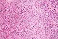
Necrosis in cat scratch disease. (WC/Nephron)

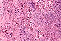
Necrosis in histiocytic necrotizing lymphadenitis. (WC/Nephron)
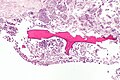
Necrosis in SLE lymphadenopathy. (WC/Nephron)
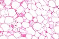
Fat necrosis - high mag. (WC/Nephron)
Necrotic bone. (WC/Nephron)
Radiation necrosis in the CNS. (WC/Tdvorak)


www:
- Necrosis at the centre of a granuloma (sun.ac.za).
- Necrosis (biomedical-engineering-online.com).
- Necrosis (nature.com).
- Necrosis (ouhsc.edu).[4]
Stains
- Martius scarlet blue stain - fibroid necrosis = red.
Sign out
LESION, ANTERIOR RECTUS WALL, SURGICAL BIOPSY:
- EXTENSIVE FAT NECROSIS.
- NO EVIDENCE OF MALIGNANCY.
Micro
The sections show fibroadipose tissue with abundant foamy histiocytes and necrotic adipocytes. Scattered chronic inflammatory cells, including plasma cells eosinophils and lymphocytes, are present. Focally hemosiderin-laden macrophages are identified. Multi-nucleated giant cells are seen. No definite epithelium is identified. Some reactive fibroblasts are present. No significant nuclear atypia is identified.
See also
References
- ↑ Cotran, Ramzi S.; Kumar, Vinay; Fausto, Nelson; Nelso Fausto; Robbins, Stanley L.; Abbas, Abul K. (2005). Robbins and Cotran pathologic basis of disease (7th ed.). St. Louis, Mo: Elsevier Saunders. pp. 21-22. ISBN 0-7216-0187-1.
- ↑ 2.0 2.1 Mitchell, Richard; Kumar, Vinay; Fausto, Nelson; Abbas, Abul K.; Aster, Jon (2011). Pocket Companion to Robbins & Cotran Pathologic Basis of Disease (8th ed.). Elsevier Saunders. pp. 6. ISBN 978-1416054542.
- ↑ Mitchell, Richard; Kumar, Vinay; Fausto, Nelson; Abbas, Abul K.; Aster, Jon (2011). Pocket Companion to Robbins & Cotran Pathologic Basis of Disease (8th ed.). Elsevier Saunders. pp. 4. ISBN 978-1416054542.
- ↑ URL: http://moon.ouhsc.edu/kfung/jty1/Com08/Com801-1-Diss.htm. Accessed on: 3 November 2010.