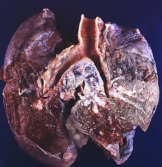
Lung tumours

Lung tumours comes to pathology to get diagnosed.
This article deals with the surgical pathology (core biopsies, lung resections). Pulmonary cytopathology is dealt with in the pulmonary cytopathology article.
An introduction to lung pathology is found in the pulmonary pathology article.
Lung tumours overview
Schematic overview of lung cancer (clinical)
| Lung cancer | |||||||||||||||||||||||||||||||||||||||||
| Primary | Metastatic | ||||||||||||||||||||||||||||||||||||||||
| NSCLC | SCLC | ||||||||||||||||||||||||||||||||||||||||
- NSCLC = non-small cell lung cancer.
- SCLS = small cell lung cancer.
Basic pathologic approach to lung cancer
| Lung cancer | |||||||||||||||||||||||||||||||
| Adenocarcinoma | Squamous cell carcinoma | SCLC | LCLC | ||||||||||||||||||||||||||||
- LCLC = large cell lung cancer.
- SCLS = small cell lung cancer.
Notes:
- Most lung cancer fits into one of the above categories.
- All types may be metastatic. Pathologists usually don't have to sort this out, as the clinican often knows whether a given lesion is metastatic (when correlated with radiology).
- Lung cancers may have a mixed morphology, e.g. SCLS may have squamous component.[1]
- Categorization as non-small cell lung cancer (NSCLC) should be avoided, as treatment is now somewhat dependent on subcategorization.[2]
Major types (primary)
Mnemonic ASSL:
- Adenocarcinoma.
- Squamous cell carcinoma.
- Small cell carcinoma (SCLC).
- Large cell carcinoma (LCLC).
Epidemiology
- Adenocarcinoma is the most common (primary lung cancer).[3]
- Adenocarcinoma is the non-smoker tumour - SCLC and squamous are more strongly associated with smoking.
Children:
- Most common lung tumour in children: metastasis (80-85% of lung tumours in children[4]
- Most common primary tumours in children: inflammatory myofibroblastic tumour, pleuropulmonary blastoma, lung carcinoid.[5]
Distribution
- Distribution - think about the location of letters in mnemonic ASSL.
- Adenocarcinoma is usually periperal, i.e. smaller airways.
- Squamous cell carcinoma and small cell carcinoma are typically central.
Margins in lung
Margin in pneumonectomy specimens include:
- Vessels (artery, vein).
- Airway (bronchus).
- Soft tissue (lymphatics, fibrous tissue and lymph nodes).[6]
Notes:
- The traditional teaching is there are only hollow structure margins (artery, vein, airway) - yet the bronchial margin has been divided into mucosal and extramucosal.[7]
- Peribronchovascular soft tissue involvement is a poor prognosticator but not an independent predictor if considered within the TNM staging.[6]
Management of primary lung cancer
Management in the past was determined by categorization into:
- Small cell cancer.
- Non-small cell cancer (includes adenocarcinoma, squamous cell carcinoma, large cell carcinoma).
Microscopic features overview
Adenocarcinoma
- Glands or cytoplasm with mucin.
Squamous cell carcinoma
- Distinct cell borders with intercellular bridges.
- Eosinophilic cytoplasm.
Small cell carcinoma
- Very cellular.
- Large NC ratio - very small amount of cytoplasm.
- Cells fragile - they tend to look "smudged" (Azzopardi phenomenon).
IHC
There is a great review paper by Jagirdar.[8]
Small cell carcinoma
Note:
- CD56 - cytoplasmic.[10]
Adenocarcinoma
Squamous cell carcinoma
Primary vs. secondary
- TTF-1 is considered useful.[8]
- 75% +ve adenocarcinoma
- 11% +ve SSC
- 50% +ve large cell carcinoma
- 0% +ve mesothelioma
- significant rates of +ve in some metastatic tumours -- see article by Jagirdar.
Note:
- TTF-1 - should be nuclear staining; cytoplasmic staining is non-specific.[13]
Neuroendocrine tumours
Overview
- This is a group of tumours that has benign (e.g. carcinoid tumour of the lung) to malignant (e.g. small cell lung carcinoma) behaviour.[14]
- They are thought to arise from pulmonary neuroendocrine cells.[15]
Classification
The grouping can be divided into four types:[16]
- Small cell carcinoma.
- Large cell neuroendocrine carcinoma.
- Typical carcinoid.
- Atypical carcinoid.
Notes:
- Typical carcinoid-like lesions <5 mm are called carcinoid tumourlets.
Cytologic features
Cytologic features useful for differentiation:
- Small cell carcinoma: necrosis, scant cytoplasm, mitoses.
- Typical carcinoid: often more cytoplasm, no necrosis, low mitotic rate (MIB1: scant staining).
- Atypical carcinoid: higher mitotic rate/MIB1 than typical carcinoid,[17] no necrosis.
Notes:[16]
- Large cell and small cell tumours behave in a similar fashion; large cell can be considered a morphological variant of small cell.
- 9/10 of carcinoids are typical and usually have a good prognosis, i.e. do not metastasize.
- Central location (vis-a-vis peripheral location) tends favours typical carcinoid over atypical carcinoid.
Malignant tumours
Adenocarcinoma of the lung
- AKA lung adenocarcinoma.
Bronchioloalveolar carcinoma
- Abbreviated BAC.
The term is no longer used in the new classification;[18] it is now "adenocarcinoma in situ" - see lung adenocarcinoma.
Squamous cell carcinoma of the lung
Small cell carcinoma of the lung
Malignant mesothelioma
- Should not be confused with benign multicystic mesothelioma and benign papillary mesothelioma.
Non-small cell lung carcinoma
- AKA poorly differentiated carcinoma of the lung.
Adenosquamous carcinoma of the lung
Lung metastasis
- AKA pulmonary metastasis.
Malignant potential
Atypical alveolar hyperplasia
Atypical carcinoid lung tumour
- AKA atypical carcinoid tumour of the lung.
Solitary fibrous tumour of the pleura
Benign tumours
Pulmonary apical cap
A lesion that can mimic a lung neoplasm.
Pulmonary carcinoid tumourlet
- AKA carcinoid tumourlet.
Typical carcinoid lung tumour
Clear cell sugar tumour of the lung
- AKA clear cell sugar tumour.
- Abbreviated CCST.
See also
- Lung - introduction to the lung, includes a basic approach.
- Medical lung disease.
- Thyroid.
- Basics.
- Heart.
- Missed endobronchial biopsy.
References
- ↑ Righi L, Volante M, Rapa I, Scagliotti GV, Papotti M (August 2007). "Neuro-endocrine tumours of the lung. A review of relevant pathological and molecular data". Virchows Arch. 451 Suppl 1: S51–9. doi:10.1007/s00428-007-0445-0. PMID 17684766.
- ↑ URL: http://www.nature.com/modpathol/journal/v21/n2s/full/3801018a.html. Accessed on: 8 September 2010.
- ↑ Lutschg JH (January 2009). "Lung cancer". N. Engl. J. Med. 360 (1): 87-8; author reply 88. doi:10.1056/NEJMc082208. PMID 19118313.
- ↑ Dishop, MK.; Kuruvilla, S. (Jul 2008). "Primary and metastatic lung tumors in the pediatric population: a review and 25-year experience at a large children's hospital.". Arch Pathol Lab Med 132 (7): 1079-103. doi:10.1043/1543-2165(2008)132[1079:PAMLTI]2.0.CO;2. PMID 18605764.
- ↑ Giuseppucci, C.; Reusmann, A.; Giubergia, V.; Barrias, C.; Krüger, A.; Siminovich, M.; Botto, H.; Cadario, M. et al. (May 2016). "Primary lung tumors in children: 24 years of experience at a referral center.". Pediatr Surg Int 32 (5): 451-7. doi:10.1007/s00383-016-3884-3. PMID 26971789.
- ↑ 6.0 6.1 Sakai, Y.; Ohbayashi, C.; Kanomata, N.; Kajimoto, K.; Sakuma, T.; Maniwa, Y.; Nishio, W.; Tauchi, S. et al. (Jul 2011). "Significance of microscopic invasion into hilar peribronchovascular soft tissue in resection specimens of primary non-small cell lung cancer.". Lung Cancer 73 (1): 89-95. doi:10.1016/j.lungcan.2010.11.002. PMID 21129810.
- ↑ Kaiser, LR.; Fleshner, P.; Keller, S.; Martini, N. (Feb 1989). "Significance of extramucosal residual tumor at the bronchial resection margin.". Ann Thorac Surg 47 (2): 265-9. PMID 2537610.
- ↑ 8.0 8.1 Jagirdar J (March 2008). "Application of immunohistochemistry to the diagnosis of primary and metastatic carcinoma to the lung". Arch. Pathol. Lab. Med. 132 (3): 384-96. PMID 18318581. http://journals.allenpress.com/jrnlserv/?request=get-abstract&issn=0003-9985&volume=132&page=384.
- ↑ Hiroshima K, Iyoda A, Shida T, et al (October 2006). "Distinction of pulmonary large cell neuroendocrine carcinoma from small cell lung carcinoma: a morphological, immunohistochemical, and molecular analysis". Mod. Pathol. 19 (10): 1358-68. doi:10.1038/modpathol.3800659. PMID 16862075.
- ↑ URL: http://jcp.bmjjournals.com/content/58/9/978.full. Accessed: 11 February 2010.
- ↑ Turner BM, Cagle PT,Fukuoka J, et al (February 2012). "Napsin A, a New Marker for Lung Adenocarcinoma, Is Complementary and More Sensitive and Specific Than Thyroid Transcription Factor 1 in the Differential Diagnosis of Primary Pulmonary Carcinoma: Evaluation of 1674 Cases by Tissue Microarray". Arch Pathol Lab Med. 136 (10): 163-71. PMID 22288963.
- ↑ Al-Zahrani IH (July 2008). "The value of immunohistochemical expression of TTF-1, CK7 and CK20 in the diagnosis of primary and secondary lung carcinomas". Saudi Med J 29 (7): 957-61. PMID 18626520.
- ↑ Compérat E, Zhang F, Perrotin C, et al. (October 2005). "Variable sensitivity and specificity of TTF-1 antibodies in lung metastatic adenocarcinoma of colorectal origin". Mod. Pathol. 18 (10): 1371–6. doi:10.1038/modpathol.3800422. PMID 15861215. http://www.nature.com/modpathol/journal/v18/n10/full/3800422a.html.
- ↑ URL: http://emedicine.medscape.com/article/426400-overview. Accessed on: 20 January 2010.
- ↑ Chong S, Lee KS, Chung MJ, Han J, Kwon OJ, Kim TS (2006). "Neuroendocrine tumors of the lung: clinical, pathologic, and imaging findings". Radiographics 26 (1): 41–57; discussion 57–8. doi:10.1148/rg.261055057. PMID 16418242.
- ↑ 16.0 16.1 URL: http://www.cancer.org/docroot/CRI/content/CRI_2_4_1X_What_is_lung_carcinoid_tumor_56.asp. Accessed on: 16 February 2011.
- ↑ Geddie, W. February 2010.
- ↑ Travis, WD.; Brambilla, E.; Noguchi, M.; Nicholson, AG.; Geisinger, K.; Yatabe, Y.; Powell, CA.; Beer, D. et al. (Sep 2011). "International association for the study of lung cancer/American Thoracic Society/European Respiratory Society: international multidisciplinary classification of lung adenocarcinoma: executive summary.". Proc Am Thorac Soc 8 (5): 381-5. doi:10.1513/pats.201107-042ST. PMID 21926387.
- ↑ Travis, WD. (Oct 2010). "Advances in neuroendocrine lung tumors.". Ann Oncol 21 Suppl 7: vii65-71. doi:10.1093/annonc/mdq380. PMID 20943645.
- ↑ Mori, M.; Rao, SK.; Popper, HH.; Cagle, PT.; Fraire, AE. (Feb 2001). "Atypical adenomatous hyperplasia of the lung: a probable forerunner in the development of adenocarcinoma of the lung.". Mod Pathol 14 (2): 72-84. doi:10.1038/modpathol.3880259. PMID 11235908.