Difference between revisions of "Stomach"
| (73 intermediate revisions by 2 users not shown) | |||
| Line 1: | Line 1: | ||
'''Stomach''' is an important organ for pathologists. It is often inflamed and may be a site that cancer arises from. Gastroenterologists often biopsy the organ. | [[Image:Gray1051.png|thumb|300px|A drawing of the stomach.]] | ||
'''Stomach''' is an important organ for pathologists. It is often inflamed and may be a site that cancer arises from. Gastroenterologists often biopsy the organ. Surgeons take-out the organ. It connects the [[esophagus]] to the [[duodenum]]. An introduction to gastrointestinal pathology is in the ''[[gastrointestinal pathology]]'' article. | |||
=Normal stomach= | =Normal stomach= | ||
| Line 53: | Line 54: | ||
Notes: | Notes: | ||
*Intraepithelial lymphocytes in the gastric mucosa have a clear halo around 'em.<ref>Sternberg H4P 2nd Ed., P.484</ref> | *Intraepithelial lymphocytes in the gastric mucosa have a clear halo around 'em.<ref>Sternberg H4P 2nd Ed., P.484</ref> | ||
*Memory device: '''F''' | *Memory device: '''F'''oveolar cells have '''f'''riends, i.e. they are close to other foveolar cells. | ||
===Gastric antrum versus gastric body=== | ===Gastric antrum versus gastric body=== | ||
| Line 67: | Line 68: | ||
| few or none | | few or none | ||
| parietal cells: intensely<br> eosinophilic cytoplasm | | parietal cells: intensely<br> eosinophilic cytoplasm | ||
| [ | | [[Image:Normal_gastric_mucosa_intermed_mag.jpg|thumb|center|60px|Parietal cells. (WC)]] | ||
|- | |- | ||
| '''Chief cell''' | | '''Chief cell''' | ||
| Line 73: | Line 74: | ||
| absent | | absent | ||
| chief cells: basophilic cytoplasm, <br>[[IHC]]: +ve for ''pepsinogen I'' | | chief cells: basophilic cytoplasm, <br>[[IHC]]: +ve for ''pepsinogen I'' | ||
| [ | | [[Image:Chief_cells.JPG|thumb|center|100px|Chief cells. (WC)]] | ||
|- | |- | ||
| '''G cell''' | | '''G cell''' | ||
| Line 79: | Line 80: | ||
| present | | present | ||
| fried egg appearance (clear cytoplasm,<br> round nucleus); look at high power - <br>usu. middle 1/3 of gland,<ref>URL: [http://www.lab.anhb.uwa.edu.au/mb140/CorePages/GIT/git.htm http://www.lab.anhb.uwa.edu.au/mb140/CorePages/GIT/git.htm]. Accessed on: 3 December 2010.</ref><br> IHC: +ve for ''gastrin''. | | fried egg appearance (clear cytoplasm,<br> round nucleus); look at high power - <br>usu. middle 1/3 of gland,<ref>URL: [http://www.lab.anhb.uwa.edu.au/mb140/CorePages/GIT/git.htm http://www.lab.anhb.uwa.edu.au/mb140/CorePages/GIT/git.htm]. Accessed on: 3 December 2010.</ref><br> IHC: +ve for ''gastrin''. | ||
| [ | | [[Image:G_cell_hyperplasia_-_very_high_mag.jpg|thumb|center|60px|G cell hyperplasia. (WC)]] | ||
|- | |- | ||
| '''Surface''' | | '''Surface''' | ||
| Line 85: | Line 86: | ||
| blunted villi | | blunted villi | ||
| antrum is somewhat <br>duodenum-like | | antrum is somewhat <br>duodenum-like | ||
| [ | | [[Image:Normal_gastric_mucosa_intermed_mag.jpg |thumb|center|60px|Body - flat. (WC)]] | ||
|- | |- | ||
| '''Gastric glands <br>/ mucosa''' | | '''Gastric glands <br>/ mucosa''' | ||
| Line 99: | Line 100: | ||
===Sign out=== | ===Sign out=== | ||
====Short version==== | ====Short version==== | ||
<pre> | |||
Stomach, Biopsy: | |||
- Antral-type gastric mucosa within normal limits. | |||
</pre> | |||
<pre> | |||
Stomach, Biopsy: | |||
- Body and antral-type gastric mucosa within normal limits. | |||
</pre> | |||
<pre> | |||
Stomach, Biopsy: | |||
- Antral-type gastric mucosa within normal limits. | |||
- NEGATIVE for Helicobacter-like organisms. | |||
</pre> | |||
=====Block letters===== | |||
<pre> | <pre> | ||
STOMACH, BIOPSY: | STOMACH, BIOPSY: | ||
| Line 128: | Line 146: | ||
====Sleeve gastrectomy==== | ====Sleeve gastrectomy==== | ||
{{Main|Sleeve gastrectomy}} | |||
=Introduction= | =Introduction= | ||
| Line 199: | Line 213: | ||
Note: | Note: | ||
*Heaped edges - suggestive of cancer. | *Heaped edges - suggestive of [[stomach cancer|cancer]]. | ||
Image | ====Endoscopic image==== | ||
<gallery> | |||
Image:Deep_gastric_ulcer.png | Gastric ulcer. (WC) | |||
</gallery> | |||
===Microscopic=== | ===Microscopic=== | ||
| Line 211: | Line 227: | ||
==Gastritis== | ==Gastritis== | ||
{{Main|Gastritis}} | |||
{{Main|Chronic gastritis}} | |||
{{Main|Acute gastritis}} | |||
==Helicobacter gastritis== | ==Helicobacter gastritis== | ||
| Line 362: | Line 237: | ||
{{Main|Intestinal metaplasia of the stomach}} | {{Main|Intestinal metaplasia of the stomach}} | ||
==Inflammatory bowel disease | ==Inflammatory bowel disease and the stomach== | ||
:See ''[[inflammatory bowel disease]]''. | :See ''[[inflammatory bowel disease]]''. | ||
*Histopathologic findings are usually non-specific. | *Histopathologic findings are usually non-specific. | ||
*Conventional thinking ''was'' upper GI involvement = Crohn's disease; this is changing.<ref name=pmid20962621>{{cite journal |author=Lin J, McKenna BJ, Appelman HD |title=Morphologic findings in upper gastrointestinal biopsies of patients with ulcerative colitis: a controlled study |journal=Am. J. Surg. Pathol. |volume=34 |issue=11 |pages=1672–7 |year=2010 |month=November |pmid=20962621 |doi=10.1097/PAS.0b013e3181f3de93 |url=}}</ref> | *Conventional thinking ''was'' upper GI involvement = [[Crohn's disease]]; this is changing.<ref name=pmid20962621>{{cite journal |author=Lin J, McKenna BJ, Appelman HD |title=Morphologic findings in upper gastrointestinal biopsies of patients with ulcerative colitis: a controlled study |journal=Am. J. Surg. Pathol. |volume=34 |issue=11 |pages=1672–7 |year=2010 |month=November |pmid=20962621 |doi=10.1097/PAS.0b013e3181f3de93 |url=}}</ref> | ||
===Endoscopic/gross=== | |||
Features - Crohn's:<ref name=Ref_GLP80>{{Ref GLP|80}}</ref> | |||
*+/-Linear fissures, erosions, ulcers, cobblestoning. | |||
*May mimic ''[[linitis plastica]]''. | |||
===Microscopic=== | ===Microscopic=== | ||
Features:<ref> | Features:<ref>Kirsch R. 13 December 2010.</ref> | ||
*Focal inflammation. | *Focal inflammation. | ||
**Common finding - non-specific. | **Common finding - non-specific. | ||
*+/-[[Granulomas]]. | *+/-[[Granulomas]]. | ||
Note: | |||
*Granulomas in Crohn's gastritis present 7-34% of the time.<ref name=Ref_GLP80>{{Ref GLP|80}}</ref> | |||
* | |||
====Images==== | ====Images==== | ||
<gallery> | <gallery> | ||
Image: | Image: Crohn's gastritis -- low mag.jpg | CG - low mag. (WC) | ||
Image: | Image: Crohn's gastritis -- intermed mag.jpg | CG - intermed. mag. (WC) | ||
Image: Crohn's gastritis -- high mag.jpg | CG - high mag. (WC) | |||
Image: Crohn's gastritis -- very high mag.jpg | CG - very high mag. (WC) | |||
</gallery> | </gallery> | ||
=== | =Miscellaneous= | ||
This is a grab bag of stuff seen in the stomach. Some of it is quite rare. | |||
==Gastric antral vascular ectasia== | |||
{{Main|Gastric antral vascular ectasia}} | |||
==Reactive gastropathy== | |||
{{Main|Reactive gastropathy}} | |||
==Autoimmune metaplastic atrophic gastritis== | ==Autoimmune metaplastic atrophic gastritis== | ||
*[[AKA]] ''autoimmune gastritis''. | |||
*[[AKA]] ''autoimmune gastritis''. | {{Main|Autoimmune metaplastic atrophic gastritis}} | ||
==Collagenous gastritis== | ==Collagenous gastritis== | ||
{{Main|Collagenous gastritis}} | |||
==Gastritis cystitis profunda== | ==Gastritis cystitis profunda== | ||
| Line 622: | Line 291: | ||
==Ménétrier's disease== | ==Ménétrier's disease== | ||
{{Main|Ménétrier's disease}} | |||
==Gastric xanthoma== | ==Gastric xanthoma== | ||
| Line 665: | Line 297: | ||
*[[AKA]] ''xanthelasma''. | *[[AKA]] ''xanthelasma''. | ||
*[[AKA]] ''stomach lipidosis''. | *[[AKA]] ''stomach lipidosis''. | ||
{{Main|Gastric xanthoma}} | |||
==Gastric ischemia== | ==Gastric ischemia== | ||
| Line 708: | Line 317: | ||
==Portal hypertensive gastropathy== | ==Portal hypertensive gastropathy== | ||
*Abbreviated ''PHG''. | *Abbreviated ''PHG''. | ||
{{Main|Portal hypertensive gastropathy}} | |||
==Amyloidosis of the stomach== | |||
*[[AKA]] ''gastric amyloidosis''. | |||
{{Main|Amyloidosis}} | |||
===General=== | ===General=== | ||
* | *Very rare. | ||
* | *Etiologies: various - see [[amyloidosis]]. | ||
===Gross=== | ===Gross/endoscopy=== | ||
*Red/swollen gastric folds.<ref name=pmid22863214>{{Cite journal | last1 = Kamata | first1 = T. | last2 = Suzuki | first2 = H. | last3 = Yoshinaga | first3 = S. | last4 = Nonaka | first4 = S. | last5 = Fukagawa | first5 = T. | last6 = Katai | first6 = H. | last7 = Taniguchi | first7 = H. | last8 = Kushima | first8 = R. | last9 = Oda | first9 = I. | title = Localized gastric amyloidosis differentiated histologically from scirrhous gastric cancer using endoscopic mucosal resection: a case report. | journal = J Med Case Rep | volume = 6 | issue = 1 | pages = 231 | month = | year = 2012 | doi = 10.1186/1752-1947-6-231 | PMID = 22863214 | PMC = 3438062 | URL = http://www.jmedicalcasereports.com/content/6/1/231 }} </ref> | |||
Endoscopic DDx: | |||
* | *[[Stomach cancer]].<ref name=pmid14606114>{{Cite journal | last1 = Wu | first1 = D. | last2 = Lou | first2 = JY. | last3 = Chen | first3 = J. | last4 = Fei | first4 = L. | last5 = Liu | first5 = GJ. | last6 = Shi | first6 = XY. | last7 = Lin | first7 = HT. | title = A case report of localized gastric amyloidosis. | journal = World J Gastroenterol | volume = 9 | issue = 11 | pages = 2632-4 | month = Nov | year = 2003 | doi = | PMID = 14606114 }}</ref><ref name=pmid22814919>{{Cite journal | last1 = Sawada | first1 = T. | last2 = Adachi | first2 = Y. | last3 = Akino | first3 = K. | last4 = Arimura | first4 = Y. | last5 = Ishida | first5 = T. | last6 = Ishii | first6 = Y. | last7 = Endo | first7 = T. | title = Endoscopic features of primary amyloidosis of the stomach. | journal = Endoscopy | volume = 44 Suppl 2 UCTN | issue = | pages = E275-6 | month = | year = 2012 | doi = 10.1055/s-0032-1309750 | PMID = 22814919 | URL = https://www.thieme-connect.com/DOI/DOI?10.1055/s-0032-1309750 }}</ref> | ||
==== | |||
</ | |||
===Microscopic=== | ===Microscopic=== | ||
Features: | Features: | ||
* | *Lamina propria expanded by amorphous paucicellular material. | ||
Image: | |||
* | *[http://www.jmedicalcasereports.com/content/6/1/231/figure/F5 Stomach amyloidosis (jmedicalcasereports.com)].<ref name=pmid22863214/> | ||
===Stains=== | |||
*[[ | *[[Congo red stain]] +ve. | ||
=== | ==Eosinophilic gastritis== | ||
{{Main|Eosinophilic gastritis}} | |||
==Proton pump inhibitor effect== | |||
*Abbreviated ''PPI effect''. | |||
{{Main|Proton pump inhibitor effect}} | |||
=Gastric polyps= | =Gastric polyps= | ||
| Line 769: | Line 363: | ||
==Hyperplastic polyp of the stomach== | ==Hyperplastic polyp of the stomach== | ||
{{Main|Hyperplastic polyp | {{Main|Hyperplastic polyp of the stomach}} | ||
==Fundic gland polyp== | ==Fundic gland polyp== | ||
{{Main|Fundic gland polyp}} | |||
=Neoplastic= | =Neoplastic= | ||
| Line 851: | Line 377: | ||
==Gastric dysplasia== | ==Gastric dysplasia== | ||
{{Main|Stomach adenoma}} | |||
==Gastric neuroendocrine tumour== | ==Gastric neuroendocrine tumour== | ||
*[[AKA]] ''neuroendocrine tumour of the stomach''. | *[[AKA]] ''neuroendocrine tumour of the stomach'' and ''gastric NET''. | ||
===General=== | ===General=== | ||
*Behaviour dependent on the subtype. | *Behaviour dependent on the subtype. | ||
| Line 1,070: | Line 461: | ||
Features: | Features: | ||
*Sheets of lymphoid cells. | *Sheets of lymphoid cells. | ||
*"Lymphoepithelial lesion" - gastric crypts invaded by a monomorphous population of lymphocytes.<ref>Bailey, D. 6 August 2010.</ref> | *"[[Lymphoepithelial lesion]]" - gastric crypts invaded by a monomorphous population of lymphocytes.<ref>Bailey, D. 6 August 2010.</ref> | ||
**Features: | **Features: | ||
**# Cluster of lymphocytes - three cells or more - '''key feature'''. | **# Cluster of lymphocytes - three cells or more - '''key feature'''. | ||
| Line 1,086: | Line 477: | ||
Others: | Others: | ||
*CD3 (T cells) - scatter positivity. | *CD3 (T cells) - scatter positivity. | ||
*CD20 (B cells) +ve. | *[[CD20]] (B cells) +ve. | ||
*CD138 (plasma cells). | *CD138 (plasma cells). | ||
*kappa, lambda -- often one is predominant, suggesting clonality. | *kappa, lambda -- often one is predominant, suggesting clonality. | ||
| Line 1,105: | Line 496: | ||
! Other | ! Other | ||
|- | |- | ||
| Hereditary diffuse gastric cancer (HDGC) syndrome | | [[Hereditary diffuse gastric cancer syndrome|Hereditary diffuse gastric cancer (HDGC) syndrome]] | ||
| CDH1 (E-cadherin)<ref>{{OMIM|192090}}</ref> | | CDH1 (E-cadherin)<ref>{{OMIM|192090}}</ref> | ||
| diffuse - more specifically [[signet ring cell carcinoma]] | | diffuse - more specifically [[signet ring cell carcinoma]] | ||
| Line 1,131: | Line 522: | ||
|- | |- | ||
| Familial breast and ovarian cancer 2<ref name=omim600185>{{OMIM|600185}}</ref> | | Familial breast and ovarian cancer 2<ref name=omim600185>{{OMIM|600185}}</ref> | ||
| BRCA2 | | [[BRCA2]] | ||
| ? | | ? | ||
| ? | | ? | ||
|} | |} | ||
==Gastric | ==Gastric carcinoma== | ||
:Includes ''gastric adenocarcinoma''. | |||
{{Main|Gastric carcinoma}} | |||
=See also= | =See also= | ||
Latest revision as of 15:51, 26 January 2022

Stomach is an important organ for pathologists. It is often inflamed and may be a site that cancer arises from. Gastroenterologists often biopsy the organ. Surgeons take-out the organ. It connects the esophagus to the duodenum. An introduction to gastrointestinal pathology is in the gastrointestinal pathology article.
Normal stomach
Gross anatomy
- Cardia - first part of the stomach; joins with esophagus.
- Fundus - superior portion - not attached directly to the esophagus.
- Body - contains parietal cells.
- Pylorus - distal (think pyloric stenosis); it joins with the duodenum.
- AKA antrum.
Image
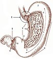
Stomach anatomy (WC)

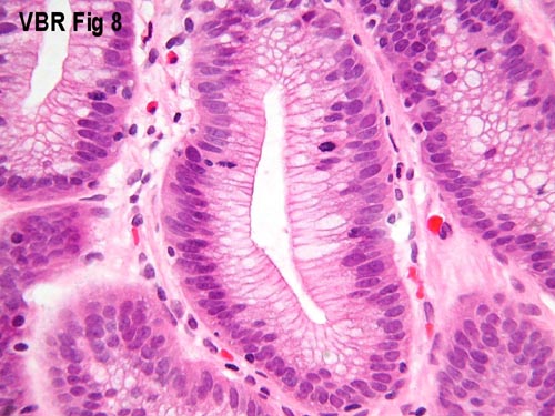
Microscopic
Foveolar cells versus intestinal goblet cells
- Intestinal goblet cells - clear mucin.
- Foveolar cells - eosinophilic contents.
Stomach versus intestine
A tabular comparison:[1]
| Feature | Intestine | Stomach |
|---|---|---|
| Spacing | Goblets cell - spaced | Foveolar cells - beside one another |
| Morphology of epithelial cells | columnar | tall columnar (Champagne flute) |
| Vesicle at luminal surface | touching/small opening | wide open |
| PAS-D | -ve (???) | +ve[2] |
| Villin stain[3][4] | +ve | -ve |
| Images | Tubular adenoma - goblet cells on right of image (WC) |
Gastric biopsy (microscopy-uk.org.uk), Stomach with cancer - PAS (WC), Stomach (WC) |
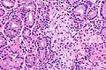
Notes:
- Intraepithelial lymphocytes in the gastric mucosa have a clear halo around 'em.[5]
- Memory device: Foveolar cells have friends, i.e. they are close to other foveolar cells.
Gastric antrum versus gastric body
| Cell | Body | Antrum | Histology | Image |
|---|---|---|---|---|
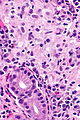
| Parietal cell | abundant | few or none | parietal cells: intensely eosinophilic cytoplasm |
|
| Chief cell | present | absent | chief cells: basophilic cytoplasm, IHC: +ve for pepsinogen I |
|
| G cell | absent | present | fried egg appearance (clear cytoplasm, round nucleus); look at high power - usu. middle 1/3 of gland,[6] IHC: +ve for gastrin. |
|
| Surface | flat | blunted villi | antrum is somewhat duodenum-like |
|
| Gastric glands / mucosa |
thick | thin | not so useful for discrimination |
body - thick, body & antrum |


Notes:
- G cells may superficially resemble intraepithelial lymphocytes.
- G cell nucleus is usu. perfectly round and slightly larger (diameter of 12 micrometers?) than a lymphocyte nucleus (diameter ~ 9-10 micrometers?).
Sign out
Short version
Stomach, Biopsy:
- Antral-type gastric mucosa within normal limits.
Stomach, Biopsy:
- Body and antral-type gastric mucosa within normal limits.
Stomach, Biopsy:
- Antral-type gastric mucosa within normal limits.
- NEGATIVE for Helicobacter-like organisms.
Block letters
STOMACH, BIOPSY:
- BODY AND ANTRAL-TYPE GASTRIC MUCOSA WITHIN NORMAL LIMITS.
STOMACH, BIOPSY:
- BODY AND ANTRAL-TYPE GASTRIC MUCOSA WITHIN NORMAL LIMITS.
- NEGATIVE FOR HELICOBACTER-LIKE ORGANISMS.
STOMACH, BIOPSY:
- ANTRAL-TYPE GASTRIC MUCOSA WITHIN NORMAL LIMITS.
- NEGATIVE FOR HELICOBACTER-LIKE ORGANISMS.
Long version
STOMACH, BIOPSY:
- BODY/ANTRAL-TYPE GASTRIC MUCOSA.
- INFLAMMATION: ABSENT.
- ATROPHY: ABSENT.
- INTESTINAL METAPLASIA: ABSENT.
- HELICOBACTER-LIKE ORGANISMS: NOT IDENTIFIED WITH ROUTINE STAINS.
- NEGATIVE FOR DYSPLASIA AND NEGATIVE FOR MALIGNANCY.
Sleeve gastrectomy
Introduction
Useful stains for stomach
- Cresyl violet stain[7] - used to find H. pylori.[8]
- Alcian blue stain - used to find mucin[9] which is present in intestinal metaplasia
Things to look for...
- Parietal cells (indicate you're in the body of the stomach) - pink (eosinophilic) cytoplasm.
- Lack of parietal cells -- DDx: Bx of antrum (pylorus), Bx of cardia, pernicious anemia.
- Goblet cells = intestinal metaplasia.
- Architectural distortion of gastric glands - suspect cancer.
- Signet ring cells = (usually) gastric carcinoma.
- Can be very easy to miss in some biopsies.
- Inflammation + small bacteria = suspect H. pylori gastritis.
Some patterns
Gastric atrophy
General
- Has a wide differential diagnosis.
Microscopic
Can take three general forms:
- Intestinal metaplasia - see intestinal metaplasia section.
- Pseudopyloric metaplasia; gastric body looks like gastric antrum.
- Characterized by foveolar hyperplasia.
- Cell loss without replacement.
- Clue is deep inflammation in the body.
Plasma cells in the stomach
DDx of plasmacytosis:
- Plasma cell neoplasm.
- Syphilis.
- Chronic gastritis.
Granulomatous gastritis
- Usual DDx of granulomatous disease (see Basics article):
- DNF AAII:
- Drugs, Neoplasms, Foreign body, Autoimmune, Allergic, Infectious, Idiopathic.
- DNF AAII:
Important ones:
- Autoimmune - Crohn's disease.
- Infectious - Tuberculosis.
- Idiopathic - Sarcoidosis.
Non-neoplastic disease
Peptic ulcer disease
- Abbreviated PUD.
- For duodenal manifestations see Peptic duodenitis.
General
- Benign.
Complications:
- Hemorrhage.
- Obstruction.
- Perforation - can be fatal.
Etiology - typically:[11]
Gross
Features:
- Typically in the duodenum; duodenum:stomach = ~4:1.
- Epithelial defect with punched-out edges (suggestive of a benign process).
Note:
- Heaped edges - suggestive of cancer.
Endoscopic image
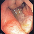
Gastric ulcer. (WC)

Microscopic
Features:
- Loss of epithelium.
- Inflammation.
- +/-Helicobacter organisms - see Helicobacter gastritis.
Gastritis
Helicobacter gastritis
Intestinal metaplasia of the stomach
Inflammatory bowel disease and the stomach
- Histopathologic findings are usually non-specific.
- Conventional thinking was upper GI involvement = Crohn's disease; this is changing.[12]
Endoscopic/gross
Features - Crohn's:[13]
- +/-Linear fissures, erosions, ulcers, cobblestoning.
- May mimic linitis plastica.
Microscopic
Features:[14]
- Focal inflammation.
- Common finding - non-specific.
- +/-Granulomas.
Note:
- Granulomas in Crohn's gastritis present 7-34% of the time.[13]
Images
CG - low mag. (WC)
CG - intermed. mag. (WC)
CG - high mag. (WC)
CG - very high mag. (WC)


Miscellaneous
This is a grab bag of stuff seen in the stomach. Some of it is quite rare.
Gastric antral vascular ectasia
Reactive gastropathy
Autoimmune metaplastic atrophic gastritis
- AKA autoimmune gastritis.
Collagenous gastritis
Gastritis cystitis profunda
- AKA Gastritic cystica profunda.[citation needed]
General
- May be associated with glandular proliferation as well.[15] (???)
- Super rare.
- Similar to cystitis cystica.
Microscopic
Features:
- Cystic spaces lined by foveolar epithelium.
Ménétrier's disease
Gastric xanthoma
Gastric ischemia
- Gastric necrosis redirects here.
General
Microscopic
Features:
Image:
Portal hypertensive gastropathy
- Abbreviated PHG.
Amyloidosis of the stomach
- AKA gastric amyloidosis.
General
- Very rare.
- Etiologies: various - see amyloidosis.
Gross/endoscopy
- Red/swollen gastric folds.[19]
Endoscopic DDx:
Microscopic
Features:
- Lamina propria expanded by amorphous paucicellular material.
Image:
Stains
- Congo red stain +ve.
Eosinophilic gastritis
Proton pump inhibitor effect
- Abbreviated PPI effect.
Gastric polyps
Similar to colonic polyps - see intestinal polyps.
DDx polyp (similar to colon & rectum):
- Hyperplastic - most common, characterised by abundant elongated foveola + glands.
- Hamartomatous - weriod stuff.
- Inflammatory fibroid polyp - inflammation, myxoid stroma.
- Fundic gland polyp - cystic dilation, flat epithelium.
- Gastric adenoma - polypoid gastric dysplasia.
Inflammatory fibroid polyp
Hyperplastic polyp of the stomach
Fundic gland polyp
Neoplastic
The spectrum from benign to malignant is divided into five:[22]
- Benign.
- Indefinite for gastric epithelial dysplasia.
- Low-grade gastric epithelial dysplasia.
- High-grade gastric epithelial dysplasia.
- Gastric carcinoma.
Gastric dysplasia
Gastric neuroendocrine tumour
- AKA neuroendocrine tumour of the stomach and gastric NET.
General
- Behaviour dependent on the subtype.
- Uncommon.
Overview of subtypes
Divided into four types:[23]
| Tumour type | Relative prevalence | Multifocality | Tumour size | Typical location | Clinical | Other | Histology |
|---|---|---|---|---|---|---|---|
| Type 1 | ~75% | yes | small (5-10 mm) | body | benign typically, female:male ~ 4:1, 50-60 years | chronic atrophic gastritis - usu. autoimmune | WDNET, WDNEC |
| Type 2 | rare | yes | small ~15 mm | body | aggressive, ~50 years old | assoc. MEN I, hyperchlorhydia | WDNEC, WDNET |
| Type 3 | 10-15% | no | small and large | variable location | aggressive if >2.0 cm, males > females | normal gastrin levels | WDNET |
| Type 4 | extremely rare | no | large | variable location | aggressive (mets usu. at time of Dx), males > females | elevated gastrin d/t parietal cell dysfunction | PDNEC |
Notes:
- WDNET = well-differentiated neuroendocrine tumour.
- WDNEC = well-differentiated neuroendocrine carcinoma.
- PDNEC = poorly-differentiated neuroendocrine carinoma.
Microscopic
Neoplastic rare
Gastric calcifying fibrous tumour
Gastric cancer
Gastric lymphoma
General
- Associated with helicobacter infection.[24]
- Usually MALT lymphoma (mucosa-associated lymphoid tissue lymphoma).
Microscopic
Features:
- Sheets of lymphoid cells.
- "Lymphoepithelial lesion" - gastric crypts invaded by a monomorphous population of lymphocytes.[25]
- Features:
- Cluster of lymphocytes - three cells or more - key feature.
- Single lymphocytes don't count.
- Clearing around the lymphocyte cluster.
- Cluster of lymphocytes - three cells or more - key feature.
- Associated with MALT lymphoma;[26] however, not specific.
- Features:
DDx:
IHC
- Panker -- most useful.
Others:
- CD3 (T cells) - scatter positivity.
- CD20 (B cells) +ve.
- CD138 (plasma cells).
- kappa, lambda -- often one is predominant, suggesting clonality.
- BCL2 +ve.
Treatment
- Triple therapy (two antibiotics, proton pump inhibitor (PPI)).[29]
- Surgery - if triple therapy fails.
Review paper: PMID 16950858.
Hereditary gastric cancer
Several syndromes are associated with gastric cancer:[30]
| Disease | Gene | Histology | Other |
|---|---|---|---|
| Hereditary diffuse gastric cancer (HDGC) syndrome | CDH1 (E-cadherin)[31] | diffuse - more specifically signet ring cell carcinoma | most important; assoc. invasive lobular carcinoma[32] |
| Lynch syndrome | MSH2, MLH1, others | ? | colorectal carcinoma, endometrial carcinoma |
| Familial adenomatous polyposis | APC | ? | adenomatous polyps |
| Peutz-Jeghers syndrome | STK11 | ? | stomach hamartomas - not precursor |
| Li-Fraumeni syndrome | TP53 (p53) | ? | AKA SBLA syndrome = sarcomas, breast, brain, leukemia, laryngeal, lung, adrenocortical carcinoma |
| Familial breast and ovarian cancer 2[33] | BRCA2 | ? | ? |
Gastric carcinoma
- Includes gastric adenocarcinoma.
See also
References
- ↑ ALS. 4 Feb 2009.
- ↑ Rubio, CA. (Jun 2007). "Gastric duodenal metaplasia in duodenal adenomas.". J Clin Pathol 60 (6): 661-3. doi:10.1136/jcp.2006.039388. PMC 1955048. PMID 16837629. http://www.ncbi.nlm.nih.gov/pmc/articles/PMC1955048/.
- ↑ Osborn M, Mazzoleni G, Santini D, Marrano D, Martinelli G, Weber K (1988). "Villin, intestinal brush border hydrolases and keratin polypeptides in intestinal metaplasia and gastric cancer; an immunohistologic study emphasizing the different degrees of intestinal and gastric differentiation in signet ring cell carcinomas". Virchows Arch A Pathol Anat Histopathol 413 (4): 303–12. PMID 2459839.
- ↑ Braunstein, EM.; Qiao, XT.; Madison, B.; Pinson, K.; Dunbar, L.; Gumucio, DL. (May 2002). "Villin: A marker for development of the epithelial pyloric border.". Dev Dyn 224 (1): 90-102. doi:10.1002/dvdy.10091. PMID 11984877.
- ↑ Sternberg H4P 2nd Ed., P.484
- ↑ URL: http://www.lab.anhb.uwa.edu.au/mb140/CorePages/GIT/git.htm. Accessed on: 3 December 2010.
- ↑ http://www.histology-world.com/stains/stains.htm
- ↑ Goggin N, Rowland M, Imrie C, Walsh D, Clyne M, Drumm B (December 1998). "Effect of Helicobacter pylori eradication on the natural history of duodenal ulcer disease". Arch. Dis. Child. 79 (6): 502-5. PMC 1717771. PMID 10210995. http://adc.bmj.com/cgi/pmidlookup?view=long&pmid=10210995.
- ↑ http://www.histology-world.com/stains/stains.htm
- ↑ http://www.histology-world.com/stains/stains.htm
- ↑ Malfertheiner, P.; Chan, FK.; McColl, KE. (Oct 2009). "Peptic ulcer disease.". Lancet 374 (9699): 1449-61. doi:10.1016/S0140-6736(09)60938-7. PMID 19683340.
- ↑ Lin J, McKenna BJ, Appelman HD (November 2010). "Morphologic findings in upper gastrointestinal biopsies of patients with ulcerative colitis: a controlled study". Am. J. Surg. Pathol. 34 (11): 1672–7. doi:10.1097/PAS.0b013e3181f3de93. PMID 20962621.
- ↑ 13.0 13.1 Iacobuzio-Donahue, Christine A.; Montgomery, Elizabeth A. (2005). Gastrointestinal and Liver Pathology: A Volume in the Foundations in Diagnostic Pathology Series (1st ed.). Churchill Livingstone. pp. 80. ISBN 978-0443066573.
- ↑ Kirsch R. 13 December 2010.
- ↑ URL: http://www.springerlink.com/content/u2v2525241754557/ Accessed on: 19 November 2010.
- ↑ Steen, S.; Lamont, J.; Petrey, L. (Jan 2008). "Acute gastric dilation and ischemia secondary to small bowel obstruction.". Proc (Bayl Univ Med Cent) 21 (1): 15-7. PMC 2190544. PMID 18209748. https://www.ncbi.nlm.nih.gov/pmc/articles/PMC2190544/.
- ↑ 17.0 17.1 Papanikolaou, IS.; Foukas, PG.; Sioulas, A.; Beintaris, I.; Panagopoulos, P.; Karamanolis, G.; Panayiotides, IG.; Dimitriadis, G. et al. (2011). "A case of gastric ischemic necrosis.". Endoscopy 43 Suppl 2 UCTN: E342. doi:10.1055/s-0030-1256795. PMID 22020717.
- ↑ Herman, J.; Chavalitdhamrong, D.; Jensen, DM.; Cortina, G.; Manuyakorn, A.; Jutabha, R. (Apr 2011). "The significance of gastric and duodenal histological ischemia reported on endoscopic biopsy.". Endoscopy 43 (4): 365-8. doi:10.1055/s-0030-1256040. PMID 21360426.
- ↑ 19.0 19.1 Kamata, T.; Suzuki, H.; Yoshinaga, S.; Nonaka, S.; Fukagawa, T.; Katai, H.; Taniguchi, H.; Kushima, R. et al. (2012). "Localized gastric amyloidosis differentiated histologically from scirrhous gastric cancer using endoscopic mucosal resection: a case report.". J Med Case Rep 6 (1): 231. doi:10.1186/1752-1947-6-231. PMC 3438062. PMID 22863214. https://www.ncbi.nlm.nih.gov/pmc/articles/PMC3438062/.
- ↑ Wu, D.; Lou, JY.; Chen, J.; Fei, L.; Liu, GJ.; Shi, XY.; Lin, HT. (Nov 2003). "A case report of localized gastric amyloidosis.". World J Gastroenterol 9 (11): 2632-4. PMID 14606114.
- ↑ Sawada, T.; Adachi, Y.; Akino, K.; Arimura, Y.; Ishida, T.; Ishii, Y.; Endo, T. (2012). "Endoscopic features of primary amyloidosis of the stomach.". Endoscopy 44 Suppl 2 UCTN: E275-6. doi:10.1055/s-0032-1309750. PMID 22814919.
- ↑ Rugge, M.; Correa, P.; Dixon, MF.; Hattori, T.; Leandro, G.; Lewin, K.; Riddell, RH.; Sipponen, P. et al. (Feb 2000). "Gastric dysplasia: the Padova international classification.". Am J Surg Pathol 24 (2): 167-76. PMID 10680883.
- ↑ URL: http://www.cap.org/apps/docs/committees/cancer/cancer_protocols/2011/StomachNET_11protocol.pdf. Accessed on: 29 March 2012.
- ↑ Mbulaiteye, SM.; Hisada, M.; El-Omar, EM. (2009). "Helicobacter Pylori associated global gastric cancer burden.". Front Biosci 14: 1490-504. PMID 19273142.
- ↑ Bailey, D. 6 August 2010.
- ↑ Papadaki, L.; Wotherspoon, AC.; Isaacson, PG. (Nov 1992). "The lymphoepithelial lesion of gastric low-grade B-cell lymphoma of mucosa-associated lymphoid tissue (MALT): an ultrastructural study.". Histopathology 21 (5): 415-21. PMID 1452124.
- ↑ Kim, K.; Kim, EJ.; Kim, MJ.; Song, HJ.; Lee, YS.; Jung, KW.; Yu, E. (Dec 2009). "Clinicopathological features of syphilitic gastritis in Korean patients.". Pathol Int 59 (12): 884-9. doi:10.1111/j.1440-1827.2009.02462.x. PMID 20021615.
- ↑ Long, BW.; Johnston, JH.; Wetzel, W.; Flowers, RH.; Haick, A. (Sep 1995). "Gastric syphilis: endoscopic and histological features mimicking lymphoma.". Am J Gastroenterol 90 (9): 1504-7. PMID 7661178.
- ↑ Zullo, A.; Hassan, C.; Andriani, A.; Cristofari, F.; De Francesco, V.; Ierardi, E.; Tomao, S.; Morini, S. et al. (Aug 2009). "Eradication therapy for Helicobacter pylori in patients with gastric MALT lymphoma: a pooled data analysis.". Am J Gastroenterol 104 (8): 1932-7; quiz 1938. doi:10.1038/ajg.2009.314. PMID 19532131.
- ↑ Sereno, M.; Aguayo, C.; Guillén Ponce, C.; Gómez-Raposo, C.; Zambrana, F.; Gómez-López, M.; Casado, E. (Sep 2011). "Gastric tumours in hereditary cancer syndromes: clinical features, molecular biology and strategies for prevention.". Clin Transl Oncol 13 (9): 599-610. PMID 21865131.
- ↑ Online 'Mendelian Inheritance in Man' (OMIM) 192090
- ↑ Guilford, P.; Hopkins, J.; Harraway, J.; McLeod, M.; McLeod, N.; Harawira, P.; Taite, H.; Scoular, R. et al. (Mar 1998). "E-cadherin germline mutations in familial gastric cancer.". Nature 392 (6674): 402-5. doi:10.1038/32918. PMID 9537325.
- ↑ Online 'Mendelian Inheritance in Man' (OMIM) 600185