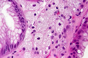
Gastric xanthoma
Jump to navigation
Jump to search
| Gastric xanthoma | |
|---|---|
| Diagnosis in short | |
 Gastric xanthoma. H&E stain. | |
|
| |
| LM | gastric lamina propria with lipid-laden macrophages |
| LM DDx | signet ring cell carcinoma, Whipple disease, MAI infection |
| IHC | CD68 +ve, AE1/AE3 -ve |
| Gross | yellowish nodule or plaque - usu. lesser curvature |
| Site | stomach |
|
| |
| Prevalence | uncommon |
| Prognosis | benign |
Gastric xanthoma, abbreviated GX, is an uncommon benign lesion of the stomach.
It is also known as xanthelasma and stomach lipidosis.
General
- Uncommon.
- Benign.
Gross/endoscopic
Microscopic
Features:[1]
- Collections of lipid-laden macrophages within the gastric lamina propria.
DDx:
Images
www:
IHC
- CD68 +ve.
- Panker (AE1/AE3) -ve.
- To exclude carcinoma.
Sign out
Stomach, Cardia Lesion, Biopsy:
     - Polypoid gastric xanthoma.
     - NEGATIVE for Helicobacter-like organisms.
     - NEGATIVE for intestinal metaplasia.
     - NEGATIVE for dysplasia and NEGATIVE for malignancy.

Comment:
The histiocytes in were demonstrated with a CD68 immunostain.
An AE1/AE3 immunostain and a Ki-67 immunostain have a benign pattern.
See also
References
- ↑ 1.0 1.1 Iacobuzio-Donahue, Christine A.; Montgomery, Elizabeth A. (2005). Gastrointestinal and Liver Pathology: A Volume in the Foundations in Diagnostic Pathology Series (1st ed.). Churchill Livingstone. pp. 111. ISBN 978-0443066573.
- ↑ 2.0 2.1 Drude, RB.; Balart, LA.; Herrington, JP.; Beckman, EN.; Burns, TW. (Jun 1982). "Gastric xanthoma: histologic similarity to signet ring cell carcinoma.". J Clin Gastroenterol 4 (3): 217-21. PMID 6284833.


