|
|
| (120 intermediate revisions by 2 users not shown) |
| Line 1: |
Line 1: |
| '''Stomach''' is an important organ for pathologists. It is often inflamed and may be a site that cancer arises from. Gastroenterologists often biopsy the organ. Surgeon take-out the organ. It connects the [[esophagus]] to the [[duodenum]]. An introduction to gastrointestinal pathology is in the ''[[gastrointestinal pathology]]'' article. | | [[Image:Gray1051.png|thumb|300px|A drawing of the stomach.]] |
| | '''Stomach''' is an important organ for pathologists. It is often inflamed and may be a site that cancer arises from. Gastroenterologists often biopsy the organ. Surgeons take-out the organ. It connects the [[esophagus]] to the [[duodenum]]. An introduction to gastrointestinal pathology is in the ''[[gastrointestinal pathology]]'' article. |
|
| |
|
| =Normal stomach= | | =Normal stomach= |
| Line 9: |
Line 10: |
| **[[AKA]] antrum. | | **[[AKA]] antrum. |
|
| |
|
| Image: [http://en.wikipedia.org/wiki/File:Illu_stomach.jpg Stomach anatomy (WP)]. | | ===Image=== |
| | <gallery> |
| | Image:Illu_stomach.jpg | Stomach anatomy (WC) |
| | </gallery> |
|
| |
|
| ==Microscopic== | | ==Microscopic== |
| Line 50: |
Line 54: |
| Notes: | | Notes: |
| *Intraepithelial lymphocytes in the gastric mucosa have a clear halo around 'em.<ref>Sternberg H4P 2nd Ed., P.484</ref> | | *Intraepithelial lymphocytes in the gastric mucosa have a clear halo around 'em.<ref>Sternberg H4P 2nd Ed., P.484</ref> |
| *Memory device: '''F'''olveolar cells have '''f'''riends, i.e. they are close to other foveolar cells. | | *Memory device: '''F'''oveolar cells have '''f'''riends, i.e. they are close to other foveolar cells. |
|
| |
|
| ===Gastric antrum versus gastric body=== | | ===Gastric antrum versus gastric body=== |
| Line 64: |
Line 68: |
| | few or none | | | few or none |
| | parietal cells: intensely<br> eosinophilic cytoplasm | | | parietal cells: intensely<br> eosinophilic cytoplasm |
| | [http://commons.wikimedia.org/wiki/File:Parietal_cells.jpg], [http://commons.wikimedia.org/wiki/File:Normal_gastric_mucosa_intermed_mag.jpg] | | | [[Image:Normal_gastric_mucosa_intermed_mag.jpg|thumb|center|60px|Parietal cells. (WC)]] |
| |- | | |- |
| | '''Chief cell''' | | | '''Chief cell''' |
| Line 70: |
Line 74: |
| | absent | | | absent |
| | chief cells: basophilic cytoplasm, <br>[[IHC]]: +ve for ''pepsinogen I'' | | | chief cells: basophilic cytoplasm, <br>[[IHC]]: +ve for ''pepsinogen I'' |
| | [http://commons.wikimedia.org/wiki/File:Chief_cells.JPG] | | | [[Image:Chief_cells.JPG|thumb|center|100px|Chief cells. (WC)]] |
| |- | | |- |
| | '''G cell''' | | | '''G cell''' |
| Line 76: |
Line 80: |
| | present | | | present |
| | fried egg appearance (clear cytoplasm,<br> round nucleus); look at high power - <br>usu. middle 1/3 of gland,<ref>URL: [http://www.lab.anhb.uwa.edu.au/mb140/CorePages/GIT/git.htm http://www.lab.anhb.uwa.edu.au/mb140/CorePages/GIT/git.htm]. Accessed on: 3 December 2010.</ref><br> IHC: +ve for ''gastrin''. | | | fried egg appearance (clear cytoplasm,<br> round nucleus); look at high power - <br>usu. middle 1/3 of gland,<ref>URL: [http://www.lab.anhb.uwa.edu.au/mb140/CorePages/GIT/git.htm http://www.lab.anhb.uwa.edu.au/mb140/CorePages/GIT/git.htm]. Accessed on: 3 December 2010.</ref><br> IHC: +ve for ''gastrin''. |
| | [http://commons.wikimedia.org/wiki/File:G_cell_hyperplasia_-_very_high_mag.jpg] | | | [[Image:G_cell_hyperplasia_-_very_high_mag.jpg|thumb|center|60px|G cell hyperplasia. (WC)]] |
| |- | | |- |
| | '''Surface''' | | | '''Surface''' |
| Line 82: |
Line 86: |
| | blunted villi | | | blunted villi |
| | antrum is somewhat <br>duodenum-like | | | antrum is somewhat <br>duodenum-like |
| | [http://commons.wikimedia.org/wiki/File:Normal_gastric_mucosa_intermed_mag.jpg body - flat] | | | [[Image:Normal_gastric_mucosa_intermed_mag.jpg |thumb|center|60px|Body - flat. (WC)]] |
| |- | | |- |
| | '''Gastric glands <br>/ mucosa''' | | | '''Gastric glands <br>/ mucosa''' |
| Line 96: |
Line 100: |
| ===Sign out=== | | ===Sign out=== |
| ====Short version==== | | ====Short version==== |
| | <pre> |
| | Stomach, Biopsy: |
| | - Antral-type gastric mucosa within normal limits. |
| | </pre> |
| | |
| | <pre> |
| | Stomach, Biopsy: |
| | - Body and antral-type gastric mucosa within normal limits. |
| | </pre> |
| | |
| | <pre> |
| | Stomach, Biopsy: |
| | - Antral-type gastric mucosa within normal limits. |
| | - NEGATIVE for Helicobacter-like organisms. |
| | </pre> |
| | |
| | =====Block letters===== |
| | <pre> |
| | STOMACH, BIOPSY: |
| | - BODY AND ANTRAL-TYPE GASTRIC MUCOSA WITHIN NORMAL LIMITS. |
| | </pre> |
| | |
| <pre> | | <pre> |
| STOMACH, BIOPSY: | | STOMACH, BIOPSY: |
| - BODY AND ANTRAL-TYPE GASTRIC MUCOSA WITHIN NORMAL LIMITS. | | - BODY AND ANTRAL-TYPE GASTRIC MUCOSA WITHIN NORMAL LIMITS. |
| | - NEGATIVE FOR HELICOBACTER-LIKE ORGANISMS. |
| </pre> | | </pre> |
|
| |
|
| Line 119: |
Line 146: |
|
| |
|
| ====Sleeve gastrectomy==== | | ====Sleeve gastrectomy==== |
| *Indication: morbid [[obesity]].
| | {{Main|Sleeve gastrectomy}} |
| <pre>
| |
| STOMACH, GREATER CURVE, SLEEVE GASTRECTOMY:
| |
| - STOMACH WALL WITHIN NORMAL LIMITS.
| |
| </pre>
| |
|
| |
|
| =Introduction= | | =Introduction= |
| Line 190: |
Line 213: |
|
| |
|
| Note: | | Note: |
| *Heaped edges - suggestive of cancer. | | *Heaped edges - suggestive of [[stomach cancer|cancer]]. |
|
| |
|
| Image: | | ====Endoscopic image==== |
| *[http://commons.wikimedia.org/wiki/File:Deep_gastric_ulcer.png Gastric ulcer (WC)].
| | <gallery> |
| | Image:Deep_gastric_ulcer.png | Gastric ulcer. (WC) |
| | </gallery> |
|
| |
|
| ===Microscopic=== | | ===Microscopic=== |
| Line 202: |
Line 227: |
|
| |
|
| ==Gastritis== | | ==Gastritis== |
| ===Etiology===
| | {{Main|Gastritis}} |
| A specific cause is uncommonly identified histologically.
| | {{Main|Chronic gastritis}} |
| | | {{Main|Acute gastritis}} |
| Gastritis causes:<ref name=Ref_PBoD812-3>{{Ref PBoD|812-3}}</ref>
| |
| *Infectious:
| |
| **H. pylori infection.
| |
| **[[Tuberculosis]].
| |
| **Salmonellosis.
| |
| **[[CMV]].
| |
| *Endocrine-related:
| |
| **[[Pernicious anemia]].
| |
| **[[Diabetes mellitus]] - gastric atony.
| |
| *Trauma, e.g. NG tube.
| |
| *Vascular, ischemia.
| |
| *Autoimmune:
| |
| **[[Crohn's disease]].
| |
| *Toxins:
| |
| **[[Alcohol]].
| |
| **Medications ([[NSAID]]s).
| |
| **Medications.
| |
| **Uremia.
| |
| **[[Smoking]] (heavy).
| |
| *Radiation.
| |
| | |
| ===Endoscopic appearance===
| |
| *Erythematous.
| |
| | |
| ===Microscopic===
| |
| *Inflammatory cells - see below.
| |
| | |
| ====Acute gastritis====
| |
| *[[AKA]] ''active gastritis''.
| |
| | |
| Features:
| |
| *Neutrophils - especially when intraepithelial.
| |
| | |
| =====Focal active gastritis=====
| |
| DDx:
| |
| #Drugs,<ref>{{Cite journal | last1 = Parfitt | first1 = JR. | last2 = Driman | first2 = DK. | title = Pathological effects of drugs on the gastrointestinal tract: a review. | journal = Hum Pathol | volume = 38 | issue = 4 | pages = 527-36 | month = Apr | year = 2007 | doi = 10.1016/j.humpath.2007.01.014 | PMID = 17367604 }}
| |
| </ref> esp. [[NSAIDs]].
| |
| #Infectious.
| |
| #Inflammatory bowel disease.
| |
| | |
| ====Chronic gastritis====
| |
| Features:
| |
| *[[Plasma cells]] (in lamina propria).
| |
| **Various criteria:
| |
| **#Two plasma cells kissing, i.e. two plasma cells touching/overlapping.
| |
| **#Three is a crowd, i.e. three plasma cells in close proximity.
| |
| | |
| Note:
| |
| *Approximately 20% of cases with an inflamed cardia will have [[intestinal metaplasia]].<ref name=pmid10566710>{{Cite journal | last1 = Voutilainen | first1 = M. | last2 = Färkkilä | first2 = M. | last3 = Mecklin | first3 = JP. | last4 = Juhola | first4 = M. | last5 = Sipponen | first5 = P. | title = Chronic inflammation at the gastroesophageal junction (carditis) appears to be a specific finding related to Helicobacter pylori infection and gastroesophageal reflux disease. The Central Finland Endoscopy Study Group. | journal = Am J Gastroenterol | volume = 94 | issue = 11 | pages = 3175-80 | month = Nov | year = 1999 | doi = 10.1111/j.1572-0241.1999.01513.x | PMID = 10566710 }}</ref>
| |
| | |
| =====Lymphocytic gastritis=====
| |
| ======General======
| |
| The DDx is limited:
| |
| #[[Helicobacter gastritis]].
| |
| #[[Celiac disease]].
| |
| #[[NSAID]]s.{{fact}}
| |
| #Idiopathic.
| |
| #HIV/AIDS.
| |
| | |
| ======Microscopic======
| |
| Features:<ref>El-Zimaity. 18 October 2010.</ref>
| |
| *25 lymphocytes / 100 epithelial cells.
| |
| | |
| ====Sydney criteria for gastritis====
| |
| A bunch of pathologists in Sydney came-up with criteria... and these were revised in Houston.<ref name=pmid8827022>{{cite journal |author=Dixon MF, Genta RM, Yardley JH, Correa P |title=Classification and grading of gastritis. The updated Sydney System. International Workshop on the Histopathology of Gastritis, Houston 1994 |journal=Am. J. Surg. Pathol. |volume=20 |issue=10 |pages=1161-81 |year=1996 |month=October |pmid=8827022 |doi= |url=http://meta.wkhealth.com/pt/pt-core/template-journal/lwwgateway/media/landingpage.htm?issn=0147-5185&volume=20&issue=10&spage=1161}}</ref>
| |
| | |
| =====Classification=====
| |
| Updated Sydney classification:<ref name=pmid8827022/>
| |
| {| class="wikitable"
| |
| | '''Feature''' || '''Non-atrophic Helicobacter''' || '''Atrophic Helicobacter''' || '''Autoimmune'''
| |
| |-
| |
| | Inflammation pattern || antral or diffuse || antrum & corpus, mild inflammation || corpus only
| |
| |-
| |
| | Atrophy & metaplasia || nil || atrophy present, metaplasia at incisura || corpus only
| |
| |-
| |
| |}
| |
| Notes:
| |
| *''Corpus'' = gastric body.
| |
| *''Incisura'' = angular incisure, incisura angularis (Latin) - notched transition point on lesser curvature of the stomach between pylorus and body.<ref>[http://en.wikipedia.org/wiki/Angular_incisure http://en.wikipedia.org/wiki/Angular_incisure]</ref>
| |
| | |
| =====Severity=====
| |
| The Sydney group suggests grading severity with the following language:<ref name=pmid8827022/>
| |
| *Mild.
| |
| *Moderate.
| |
| *Marked.
| |
| | |
| These terms are applied to the parameters described in a biopsy. The Sydney criteria lists ''H. pylori'', ''neutrophils'', ''mononuclear cells'', ''antrum (atrophy)'', ''corpus (atrophy)'' and ''intestinal metaplasia''. The paper that discusses this also give a visual analogue scale.
| |
| | |
| Parameters & Severity (adapted from Dixon et al.<ref name=pmid8827022/>):
| |
| {| class="wikitable sortable"
| |
| ! Feature
| |
| ! Mild
| |
| ! Moderate
| |
| ! Marked
| |
| |-
| |
| | H. pylori
| |
| | few touching
| |
| | many touching
| |
| | piles
| |
| |-
| |
| | Neutrophils
| |
| | few
| |
| | bunches
| |
| | crowded
| |
| |-
| |
| | Mononuclear cells
| |
| | not touching
| |
| | kissing
| |
| | partying
| |
| |-
| |
| |}
| |
| | |
| ===Sign out===
| |
| ====Mild chronic====
| |
| <pre>
| |
| STOMACH, BIOPSY:
| |
| - BODY AND ANTRAL-TYPE GASTRIC MUCOSA WITH MILD CHRONIC INFLAMMATION.
| |
| - NEGATIVE FOR INTESTINAL METAPLASIA.
| |
| - NEGATIVE FOR HELICOBACTOR-LIKE ORGANISMS.
| |
| - NEGATIVE FOR DYSPLASIA AND NEGATIVE FOR MALIGNANCY.
| |
| </pre>
| |
| | |
| ====Moderate chronic active====
| |
| <pre>
| |
| STOMACH, BIOPSY:
| |
| - BODY AND ANTRAL-TYPE GASTRIC MUCOSA WITH MODERATE CHRONIC ACTIVE INFLAMMATION.
| |
| - NEGATIVE FOR INTESTINAL METAPLASIA.
| |
| - NEGATIVE FOR HELICOBACTOR-LIKE ORGANISMS.
| |
| - NEGATIVE FOR DYSPLASIA AND NEGATIVE FOR MALIGNANCY.
| |
| </pre>
| |
| | |
| ====Micro====
| |
| The sections show gastric body type mucosa with small clusters of plasma cells. There are no intraepithelial neutrophils. Goblet cells are not identified. The epithelium matures normally to the surface. No Helicobacter organisms are seen.
| |
|
| |
|
| ==Helicobacter gastritis== | | ==Helicobacter gastritis== |
| *Often abbreviated ''[[HP]]''.
| | {{Main|Helicobacter gastritis}} |
| ===General===
| |
| *Several Helicobacter species can cause gastritis:
| |
| **''[[Helicobacter pylori]]'' - most common.
| |
| **''Helicobacter heilmannii''.
| |
| | |
| Epidemiologic associations - ''Helicobacter'' infections are associated with:<ref>{{Ref PBoD|814}}</ref>
| |
| *Gastritis.
| |
| *Peptic ulcers.
| |
| *Cancer.
| |
| **Carcinoma.
| |
| **[[MALT lymphoma]].
| |
| | |
| ===Gross===
| |
| *Thickened gastric folds.
| |
| *Erythema.
| |
| | |
| ===Microscopic===
| |
| Features:
| |
| *''Helicobacter pylori'':
| |
| **Usually have v-shape (seagull-like shape).
| |
| ***May have a curved shape (comma-like shape) or U-shape.<ref name=pmid21290743>{{Cite journal | last1 = Mobley | first1 = HLT. | last2 = Mendz | first2 = GL. | last3 = Hazell | first3 = SL. | last4 = Andersen | first4 = LP. | last5 = Wadström | first5 = T. | title = Basic Bacteriology and Culture | journal = | volume = | issue = | pages = | month = | year = | doi = | PMID = 21290743 | url = http://www.ncbi.nlm.nih.gov/books/NBK2444/}} </ref>
| |
| *''Helicobacter heilmannii'':<ref name=pmid16224223 >{{Cite journal | last1 = Singhal | first1 = AV. | last2 = Sepulveda | first2 = AR. | title = Helicobacter heilmannii gastritis: a case study with review of literature. | journal = Am J Surg Pathol | volume = 29 | issue = 11 | pages = 1537-9 | month = Nov | year = 2005 | doi = | PMID = 16224223 }}</ref>
| |
| **Corkscrew appearance.
| |
| | |
| Tips:
| |
| *One needs to look at 400x magnification. Even at 400x they are possible to miss.
| |
| **Helicobacter are damn small. They are smaller than the nucleus of the gastric foveollar cell.
| |
| *Look for mucus - they preferentially reside there.
| |
| **This is usually close to the opening of the gastric pits.
| |
| *Helicobacter are found in groups. When you see several that are the same size and shape you can be sure they are real.
| |
| | |
| Notes:
| |
| *Helicobacter can be in antrum and/or body.<ref>{{cite journal |author=Maaroos HI, Kekki M, Villako K, Sipponen P, Tamm A, Sadeniemi L |title=The occurrence and extent of Helicobacter pylori colonization and antral and body gastritis profiles in an Estonian population sample |journal=Scand. J. Gastroenterol. |volume=25 |issue=10 |pages=1010-7 |year=1990 |month=October |pmid=2263873 |doi= |url=}}</ref>
| |
| *Helicobacter don't like the intestinal mucosa ''or'' mucosa that has undergone [[intestinal metaplasia]]; you're unlikely to find 'em there.
| |
| | |
| DDx:
| |
| *Dirt - variable size.
| |
| | |
| Images:
| |
| *[http://commons.wikimedia.org/wiki/File:Immunohistochemical_detection_of_Helicobacter_%281%29_histopatholgy.jpg H. pylori - IHC (WC)].
| |
| *Helicobacter gastritis:
| |
| **[http://commons.wikimedia.org/wiki/File:Gastritis_helicobacter_-_high_mag.jpg Gastritis due to HP (WC)].
| |
| **[http://commons.wikimedia.org/wiki/File:Gastritis_helicobacter_-_very_high_mag_cropped.jpg HP visible (WC)].
| |
| *[http://commons.wikimedia.org/wiki/Category:Helicobacter_gastritis Set of images - HP gastritis (WC)].
| |
| *[http://gut.bmj.com/content/58/12/1669/F2.large.jpg Helicobacter heilmannii (bmj.com)].<ref>URL: [http://gut.bmj.com/content/58/12/1669.extract http://gut.bmj.com/content/58/12/1669.extract]. Accessed on: 2 March 2012.</ref>
| |
| | |
| ===Stains===
| |
| *[[Cresyl violet stain]] - background and organisms blue.
| |
| *[[Warthin-Starry stain]] - background yellow, organisms black.
| |
| | |
| ===IHC===
| |
| *Helicobacter pylori IHC stain +ve.
| |
| | |
| Note:
| |
| *Reportly also stains ''Helicobacter heilmannii''.<ref name=pmid16224223 >{{Cite journal | last1 = Singhal | first1 = AV. | last2 = Sepulveda | first2 = AR. | title = Helicobacter heilmannii gastritis: a case study with review of literature. | journal = Am J Surg Pathol | volume = 29 | issue = 11 | pages = 1537-9 | month = Nov | year = 2005 | doi = | PMID = 16224223 }}</ref>
| |
| | |
| ===Sign out===
| |
| ====Body====
| |
| <pre>
| |
| STOMACH, BIOPSY:
| |
| - BODY-TYPE MUCOSA WITH MODERATE CHRONIC ACTIVE GASTRITIS.
| |
| - ABUNDANT HELICOBACTER-LIKE ORGANISMS PRESENT.
| |
| - NEGATIVE FOR INTESTINAL METAPLASIA.
| |
| - NEGATIVE FOR DYSPLASIA AND NEGATIVE FOR MALIGNANCY.
| |
| </pre>
| |
| | |
| ====Body====
| |
| <pre>
| |
| STOMACH, BIOPSY:
| |
| - ANTRAL-TYPE MUCOSA WITH MODERATE CHRONIC ACTIVE GASTRITIS.
| |
| - ABUNDANT HELICOBACTER-LIKE ORGANISMS PRESENT.
| |
| - NEGATIVE FOR INTESTINAL METAPLASIA.
| |
| - NEGATIVE FOR DYSPLASIA AND NEGATIVE FOR MALIGNANCY.
| |
| </pre>
| |
| | |
| ====Micro====
| |
| The sections show antral-type gastric mucosa with abundant lamina propria plasma cells and
| |
| focal intraepithelial neutrophils. Cocci and bacilli are present. Some of the bacilli
| |
| are Helicobactor-like. The epithelium matures normally to the surface. No goblet cells
| |
| are identified.
| |
|
| |
|
| ==Intestinal metaplasia of the stomach== | | ==Intestinal metaplasia of the stomach== |
| *[[AKA]] ''gastric [[intestinal metaplasia]]''.
| | {{Main|Intestinal metaplasia of the stomach}} |
| *Abbreviated ''IM''.
| |
| ===General===
| |
| *Often part of surgical pathology report, e.g. "negative for intestinal metaplasia" or "intestinal metaplasia present".
| |
| *May be associated with Helicobacter spp. infection -- though Helicobacter don't like intestinal type mucosa, i.e. H. pylori are not typically found in regions with intestinal metaplasia.
| |
| *May be reversible - some epidemiological evidence.<ref name=pmid12477745>{{Cite journal | last1 = Walker | first1 = MM. | title = Is intestinal metaplasia of the stomach reversible? | journal = Gut | volume = 52 | issue = 1 | pages = 1-4 | month = Jan | year = 2003 | doi = | PMID = 12477745 | PMC = 1773527
| |
| }}</ref>
| |
| | |
| Significance:
| |
| *Moderate risk increase for carcinoma; risk less than for Barrett's esophagus.<ref name=pmid20203636>{{cite journal |author=Correa P, Piazuelo MB, Wilson KT |title=Pathology of gastric intestinal metaplasia: clinical implications |journal=Am. J. Gastroenterol. |volume=105 |issue=3 |pages=493–8 |year=2010 |month=March |pmid=20203636 |pmc=2895407 |doi=10.1038/ajg.2009.728 |url=http://www.ncbi.nlm.nih.gov/pmc/articles/PMC2895407/?tool=pubmed}}</ref>
| |
| **Odds ratio for corpus (~5.8x) higher than antrum (2.3x) when compared to individuals without IM.<ref name=pmid21575058>{{Cite journal | last1 = Sakitani | first1 = K. | last2 = Hirata | first2 = Y. | last3 = Watabe | first3 = H. | last4 = Yamada | first4 = A. | last5 = Sugimoto | first5 = T. | last6 = Yamaji | first6 = Y. | last7 = Yoshida | first7 = H. | last8 = Maeda | first8 = S. | last9 = Omata | first9 = M. | title = Gastric cancer risk according to the distribution of intestinal metaplasia and neutrophil infiltration. | journal = J Gastroenterol Hepatol | volume = 26 | issue = 10 | pages = 1570-5 | month = Oct | year = 2011 | doi = 10.1111/j.1440-1746.2011.06767.x | PMID = 21575058 }}</ref>
| |
| | |
| ===Microscopic===
| |
| Features:
| |
| *Goblet cells are present in the stomach - '''key feature'''.<ref>URL: [http://esynopsis.uchc.edu/eAtlas/GI/1280.htm http://esynopsis.uchc.edu/eAtlas/GI/1280.htm]. Accessed on: 16 August 2010.</ref>
| |
| **In [[H&E stain|H&E]] sections the vacuole often stains light grey.
| |
| **Foveolar epithelium should be present in the same fragment.
| |
| *+/-Paneth cells - deep in the glands.<ref name=pmid19918317/>
| |
| **Very rarely present.
| |
| **Very uncommon in isolation.
| |
| | |
| Note:
| |
| *Intestinal metaplasia (IM) is occasionally subdivided:<ref name=pmid10680883>{{Cite journal | last1 = Rugge | first1 = M. | last2 = Correa | first2 = P. | last3 = Dixon | first3 = MF. | last4 = Hattori | first4 = T. | last5 = Leandro | first5 = G. | last6 = Lewin | first6 = K. | last7 = Riddell | first7 = RH. | last8 = Sipponen | first8 = P. | last9 = Watanabe | first9 = H. | title = Gastric dysplasia: the Padova international classification. | journal = Am J Surg Pathol | volume = 24 | issue = 2 | pages = 167-76 | month = Feb | year = 2000 | doi = | PMID = 10680883 }}</ref>
| |
| **''Complete IM'' = goblet cells and (intestinal) brush border.
| |
| **''Incomplete IM'' = mucus vacuoles of various sizes, no (intestinal) brush border.
| |
| | |
| DDx:
| |
| *[[Normal duodenum|Normal small bowel mucosa]] - no foveolar epithelium present.
| |
| *[[Gastric dysplasia]], intestinal type.
| |
| | |
| Image:
| |
| *[http://commons.wikimedia.org/wiki/File:Gastric_adenocarcinoma.jpg Intestinal metaplasia in the stomach - crappy quality (WC)].
| |
| | |
| ===Stains===
| |
| *Alcian blue (pH 2.5)/PAS +ve.<ref name=pmid14736279>{{Cite journal | last1 = Rivera-Hueto | first1 = F. | last2 = Lag-Asturiano | first2 = E. | last3 = Utrilla-Alcolea | first3 = JC. | last4 = Herrerías-Gutiérrez | first4 = JM. | title = Advanced gastric carcinoma with a complete intestinal metaplasia phenotype associated with early intestinal-type carcinoma. | journal = Arch Pathol Lab Med | volume = 128 | issue = 2 | pages = 218-21 | month = Feb | year = 2004 | doi = 10.1043/1543-2165(2004)128218:AGCWAC2.0.CO;2 | PMID = 14736279 }}</ref>
| |
| **May be used to divide into ''complete'' (type I) and ''incomplete'' (type II).<ref name=pmid7139576>{{Cite journal | last1 = Iida | first1 = F. | last2 = Kusama | first2 = J. | title = Gastric carcinoma and intestinal metaplasia. Significance of types of intestinal metaplasia upon development of gastric carcinoma. | journal = Cancer | volume = 50 | issue = 12 | pages = 2854-8 | month = Dec | year = 1982 | doi = | PMID = 7139576 }}</ref><ref>{{Ref Odze|276}}</ref>
| |
| *Alican blue stain +ve.{{fact}}
| |
|
| |
|
| Image:
| | ==Inflammatory bowel disease and the stomach== |
| *[http://commons.wikimedia.org/wiki/File:Barrett%27s_mucosa,_Alcian_blue_stain.jpg Barrett's mucosa - Alcian blue stain (WC)].
| |
| | |
| ===IHC===
| |
| *CDX2 +ve (-ve in normal stomach).<ref name=pmid12477745>{{Cite journal | last1 = Walker | first1 = MM. | title = Is intestinal metaplasia of the stomach reversible? | journal = Gut | volume = 52 | issue = 1 | pages = 1-4 | month = Jan | year = 2003 | doi = | PMID = 12477745 | PMC = 1773527
| |
| }}</ref>
| |
| **Strong assoc. with ''[[Helicobacter gastritis]]'' as well as IM.<ref name=pmid12047325>{{Cite journal | last1 = Satoh | first1 = K. | last2 = Mutoh | first2 = H. | last3 = Eda | first3 = A. | last4 = Yanaka | first4 = I. | last5 = Osawa | first5 = H. | last6 = Honda | first6 = S. | last7 = Kawata | first7 = H. | last8 = Kihira | first8 = K. | last9 = Sugano | first9 = K. | title = Aberrant expression of CDX2 in the gastric mucosa with and without intestinal metaplasia: effect of eradication of Helicobacter pylori. | journal = Helicobacter | volume = 7 | issue = 3 | pages = 192-8 | month = Jun | year = 2002 | doi = | PMID = 12047325 }}</ref>
| |
| | |
| Others:
| |
| *Lysozyme +ve - marks paneth cells.<ref name=pmid19918317>{{Cite journal | last1 = Rubio | first1 = CA. | last2 = Befrits | first2 = R. | title = Increased lysozyme expression in gastric biopsies with intestinal metaplasia and pseudopyloric metaplasia. | journal = Int J Clin Exp Med | volume = 2 | issue = 3 | pages = 248-53 | month = | year = 2009 | doi = | PMID = 19918317 }}</ref>
| |
| | |
| ===Sign out===
| |
| <pre>
| |
| STOMACH, BIOPSY:
| |
| - BODY-TYPE MUCOSA WITH INTESTINAL METAPLASIA, FOCAL.
| |
| - MINIMAL CHRONIC GASTRITIS (BODY OF STOMACH).
| |
| - NEGATIVE FOR HELICOBACTER-LIKE ORGANISMS.
| |
| - NEGATIVE FOR DYSPLASIA AND NEGATIVE FOR MALIGNANCY.
| |
| </pre>
| |
| | |
| <pre>
| |
| STOMACH, BIOPSY:
| |
| - ANTRAL-TYPE MUCOSA WITH INTESTINAL METAPLASIA, EXTENSIVE.
| |
| - MILD CHRONIC (ANTRAL) GASTRITIS.
| |
| - NEGATIVE FOR HELICOBACTER-LIKE ORGANISMS.
| |
| - NEGATIVE FOR DYSPLASIA AND NEGATIVE FOR MALIGNANCY.
| |
| </pre>
| |
| | |
| ==Inflammatory bowel disease & the stomach== | |
| :See ''[[inflammatory bowel disease]]''. | | :See ''[[inflammatory bowel disease]]''. |
| *Histopathologic findings are usually non-specific. | | *Histopathologic findings are usually non-specific. |
| *Conventional thinking ''was'' upper GI involvement = Crohn's disease; this is changing.<ref name=pmid20962621>{{cite journal |author=Lin J, McKenna BJ, Appelman HD |title=Morphologic findings in upper gastrointestinal biopsies of patients with ulcerative colitis: a controlled study |journal=Am. J. Surg. Pathol. |volume=34 |issue=11 |pages=1672–7 |year=2010 |month=November |pmid=20962621 |doi=10.1097/PAS.0b013e3181f3de93 |url=}}</ref> | | *Conventional thinking ''was'' upper GI involvement = [[Crohn's disease]]; this is changing.<ref name=pmid20962621>{{cite journal |author=Lin J, McKenna BJ, Appelman HD |title=Morphologic findings in upper gastrointestinal biopsies of patients with ulcerative colitis: a controlled study |journal=Am. J. Surg. Pathol. |volume=34 |issue=11 |pages=1672–7 |year=2010 |month=November |pmid=20962621 |doi=10.1097/PAS.0b013e3181f3de93 |url=}}</ref> |
| | |
| | ===Endoscopic/gross=== |
| | Features - Crohn's:<ref name=Ref_GLP80>{{Ref GLP|80}}</ref> |
| | *+/-Linear fissures, erosions, ulcers, cobblestoning. |
| | *May mimic ''[[linitis plastica]]''. |
|
| |
|
| ===Microscopic=== | | ===Microscopic=== |
| Features:<ref>RK. 13 December 2010.</ref> | | Features:<ref>Kirsch R. 13 December 2010.</ref> |
| *Focal inflammation. | | *Focal inflammation. |
| **Common finding - non-specific. | | **Common finding - non-specific. |
| *+/-[[Granulomas]]. | | *+/-[[Granulomas]]. |
| | |
| | Note: |
| | *Granulomas in Crohn's gastritis present 7-34% of the time.<ref name=Ref_GLP80>{{Ref GLP|80}}</ref> |
| | |
| | ====Images==== |
| | <gallery> |
| | Image: Crohn's gastritis -- low mag.jpg | CG - low mag. (WC) |
| | Image: Crohn's gastritis -- intermed mag.jpg | CG - intermed. mag. (WC) |
| | Image: Crohn's gastritis -- high mag.jpg | CG - high mag. (WC) |
| | Image: Crohn's gastritis -- very high mag.jpg | CG - very high mag. (WC) |
| | </gallery> |
|
| |
|
| =Miscellaneous= | | =Miscellaneous= |
| This is a grab bag of stuff seen in the stomach. Some of it is quite rare. | | This is a grab bag of stuff seen in the stomach. Some of it is quite rare. |
| ==Gastric antral vascular ectasia== | | ==Gastric antral vascular ectasia== |
| *Abbreviated ''GAVE''.
| | {{Main|Gastric antral vascular ectasia}} |
| *[[AKA]] ''watermelon stomach'' - due to characteristic endoscopic appearance.<ref name=pmid18625989>{{cite journal |author=Chatterjee S |title=Watermelon stomach |journal=CMAJ |volume=179 |issue=2 |pages=162 |year=2008 |month=July |pmid=18625989 |pmc=2443230 |doi=10.1503/cmaj.080461 |url=http://www.pubmedcentral.nih.gov/articlerender.fcgi?tool=pubmed&pubmedid=18625989}}</ref>
| |
| ===General===
| |
| *Lesion of the antrum - due to dilated capillaries.
| |
| | |
| ===Gross/endoscopic appearance===
| |
| * Linear red streaks in antrum - oriented toward the pyloric valve... vaguely resembles a watermelon.
| |
| | |
| Endoscopic images:
| |
| *[http://www.pubmedcentral.nih.gov/articlerender.fcgi?artid=2443230&rendertype=figure&id=f1-19 Watermelon stomach (pubmedcentral.nih.gov)].
| |
| *[http://en.wikipedia.org/wiki/File:Gave.png GAVE (WP)].
| |
| | |
| ===Microscopic===
| |
| Features:<ref name=Ref_GLP118>{{Ref GLP|118}}</ref>
| |
| *Fibrin thrombi - '''characteristic feature'''.
| |
| *Dilated capillaries in lamina propria.
| |
| *+/-Foveollar hyperplasia.<ref name=Ref_GLP119>{{Ref GLP|119}}</ref>
| |
| | |
| DDx:
| |
| *[[Portal hypertensive gastropathy]] - predominantly in the gastric body, usu. associated with [[cirrhosis]], do not have fibrin thrombi.<ref name=Ref_GLP120-1>{{Ref GLP|120-1}}</ref>
| |
| | |
| Images:
| |
| *[http://commons.wikimedia.org/wiki/File:Gastric_antral_vascular_ectasia_-_2_-_intermed_mag.jpg GAVE - intermed. mag. (WC)].
| |
| *[http://commons.wikimedia.org/wiki/File:Gastric_antral_vascular_ectasia_-_2_-_very_high_mag.jpg GAVE - very high mag. (WC)].
| |
| *[http://commons.wikimedia.org/wiki/File:Gastric_antral_vascular_ectasia_-_very_high_mag.jpg GAVE - two thrombi - very high mag. (WC)].
| |
| | |
| ===Sign out===
| |
| <pre>
| |
| STOMACH, BIOPSY:
| |
| - GASTRIC ANTRAL VASCULAR ECTASIA WITH FOVEOLAR HYPERPLASIA.
| |
| - MILD CHRONIC ACTIVE ANTRAL GASTRITIS.
| |
| - NEGATIVE FOR INTESTINAL METAPLASIA.
| |
| - NEGATIVE FOR DYSPLASIA.
| |
| - NEGATIVE FOR HELICOBACTER ORGANISMS.
| |
| </pre>
| |
| | |
| ====Micro====
| |
| The sections show antral-type gastric mucosa with dilated lamina propria blood vessels and intravascular fibrin thrombi. There is mild foveolar hyperplasia. Numerous neutrophils are present between the foveollar cells and within the lamina propria. Several large clusters of plasma cells are present in the lamina propria.
| |
|
| |
|
| ==Reactive gastropathy== | | ==Reactive gastropathy== |
| *[[AKA]] ''chemical gastropathy'',<ref name=pmid16939055>{{Cite journal | last1 = Genta | first1 = RM. | title = Differential diagnosis of reactive gastropathy. | journal = Semin Diagn Pathol | volume = 22 | issue = 4 | pages = 273-83 | month = Nov | year = 2005 | doi = | PMID = 16939055 }}</ref> incorrectly referred to as ''chemical gastritis'' (see below).
| | {{Main|Reactive gastropathy}} |
| ===General===
| |
| *May be seen in the context of a previous resection/surgical reconstruction, e.g. Billroth II.
| |
| | |
| ====Epidemiology====
| |
| General assocations:
| |
| *Increases with age.<ref name=pmid22928604>{{Cite journal | last1 = Maguilnik | first1 = I. | last2 = Neumann | first2 = WL. | last3 = Sonnenberg | first3 = A. | last4 = Genta | first4 = RM. | title = Reactive gastropathy is associated with inflammatory conditions throughout the gastrointestinal tract. | journal = Aliment Pharmacol Ther | volume = | issue = | pages = | month = Aug | year = 2012 | doi = 10.1111/apt.12031 | PMID = 22928604 }}</ref>
| |
| | |
| Etologic factors - associated with:<ref>ALS. 5 February 2009.</ref>
| |
| *Excess acid.
| |
| *[[EtOH]].
| |
| *Bile.
| |
| *[[H. pylori]].
| |
| *Drugs:<ref name=pmid16939055>{{Cite journal | last1 = Genta | first1 = RM. | title = Differential diagnosis of reactive gastropathy. | journal = Semin Diagn Pathol | volume = 22 | issue = 4 | pages = 273-83 | month = Nov | year = 2005 | doi = | PMID = 16939055 }}</ref>
| |
| **Iron (brown pigment on histology).
| |
| **[[NSAID]]s - synergistic effect with corticosteroids.
| |
| | |
| Drugs that cause erosions and/or ulcers -- adapted from ''Genta'':<ref name=pmid16939055>{{Cite journal | last1 = Genta | first1 = RM. | title = Differential diagnosis of reactive gastropathy. | journal = Semin Diagn Pathol | volume = 22 | issue = 4 | pages = 273-83 | month = Nov | year = 2005 | doi = | PMID = 16939055 }}</ref>
| |
| | |
| {| class="wikitable sortable" style="margin-left:auto;margin-right:auto"
| |
| ! Drug
| |
| ! Comment
| |
| ! Indication for Rx
| |
| |-
| |
| | NSAIDs
| |
| | common cause
| |
| | pain, reduce cardiovascular risk
| |
| |-
| |
| | Corticosteroids
| |
| | synergistic effect with NSAIDs
| |
| | rheumatologic diseases + others
| |
| |-
| |
| | Potassium (KCl)
| |
| | common cause
| |
| | renal failure
| |
| |-
| |
| | Bisphophonates
| |
| | uncommon cause
| |
| | [[osteoporosis]]
| |
| |-
| |
| | Ferrous sulfate
| |
| | very common if symptomatic
| |
| | iron deficiency anemia
| |
| |-
| |
| | Chloroquine
| |
| | uncommon
| |
| | only in the context of [[malaria]]
| |
| |-
| |
| | Sodium polystyrene sulfonate (Kayexalate)
| |
| | rare
| |
| | renal failure patients
| |
| |}
| |
| | |
| ====Relation to gastritis====
| |
| *May mimic a (true) gastritis symptomatically and visually in an endoscopic examination.
| |
| *"Chemical gastritis" is misnomer. Etymologically, the ''-itis'' in ''gastritis'', implies an inflammatory process. Chemical gastropathy is not (predominantly) an inflammatory process.
| |
| **This type of confusion is not uncommon. [[Steatohepatitis]] is another example of this; it is not a process with significant inflammation yet, confusingly, carries the ''-itis'' ending.
| |
| | |
| ===Gross/endoscopic===
| |
| Features:<ref>{{Ref GLP|69}}</ref>
| |
| *Antral erythema +/- erosions.
| |
| *+/-Bile.
| |
| | |
| ===Microscopic===
| |
| Features - triad:<ref>El-Zimaity. 18 October 2010.</ref><ref name=pmid16939055/>
| |
| #Foveolar hyperplasia.
| |
| #*Tortuosity of glands in the "neck" region of the gastric glands.
| |
| #*Associated with "mucin depletion" - cytoplasm not clear -- as is usual.
| |
| #Smooth muscle fibre hyperplasia.
| |
| #*Abundant eosinophilic lamina propria.
| |
| #Scant acute & chronic inflammatory cells.
| |
| Additional features.
| |
| *+/-Edema.
| |
| *+/-Erosions.
| |
| | |
| Notes:
| |
| *Triad rarely present; mild inflammation common.
| |
| | |
| DDx:
| |
| *[[Amyloidosis]].
| |
| *[[Collagenous gastritis]].
| |
| *[[Hyperplastic polyp of the stomach]].<ref name=Ref_GLP69>{{Ref GLP|69}}</ref>
| |
| | |
| Images:
| |
| *[http://commons.wikimedia.org/wiki/File:Reactive_gastropathy_-_low_mag.jpg RG - low mag. (WC)].
| |
| *[http://commons.wikimedia.org/wiki/File:Reactive_gastropathy_-_high_mag.jpg RG - high mag. (WC)].
| |
| | |
| ===Sign out===
| |
| <pre>
| |
| STOMACH, BIOPSY:
| |
| - ANTRAL-TYPE GASTRIC MUCOSA WITH REACTIVE GASTROPATHY, SEE COMMENT.
| |
| - NEGATIVE FOR INTESTINAL METAPLASIA.
| |
| - NEGATIVE FOR HELICOBACTER-LIKE ORGANISMS.
| |
| - NEGATIVE FOR DYSPLASIA AND NEGATIVE FOR MALIGNANCY.
| |
| | |
| COMMENT:
| |
| This nonspecific finding may be due to a number of causes, including medications (especially NSAIDs), alcohol and bile reflux.
| |
| </pre>
| |
| | |
| ====Not well-developed====
| |
| <pre>
| |
| STOMACH, BIOPSY:
| |
| - BODY-TYPE GASTRIC MUCOSA WITHIN NORMAL LIMITS.
| |
| - ANTRAL-TYPE GASTRIC MUCOSA WITH SMOOTH MUSCLE HYPERPLASIA AND FOCAL GASTRIC GLAND TORTUOSITY, SEE COMMENT.
| |
| - NEGATIVE FOR INTESTINAL METAPLASIA.
| |
| - NEGATIVE FOR HELICOBACTER-LIKE ORGANISMS.
| |
| - NEGATIVE FOR DYSPLASIA AND NEGATIVE FOR MALIGNANCY.
| |
| | |
| COMMENT:
| |
| These findings are suggestive of a reactive gastropathy; however, gland corkscrewing is not evident.
| |
| </pre>
| |
| | |
| <pre>
| |
| STOMACH, BIOPSY:
| |
| - ANTRAL-TYPE GASTRIC MUCOSA WITH PROMINENT SMOOTH MUSCLE, OTHERWISE WITHIN NORMAL
| |
| LIMITS.
| |
| - NEGATIVE FOR INTESTINAL METAPLASIA.
| |
| - NEGATIVE FOR HELICOBACTER-LIKE ORGANISMS.
| |
| - NEGATIVE FOR DYSPLASIA AND NEGATIVE FOR MALIGNANCY.
| |
| </pre>
| |
|
| |
|
| ==Autoimmune metaplastic atrophic gastritis== | | ==Autoimmune metaplastic atrophic gastritis== |
| :''Pernicious anemia'' redirects here.
| | *[[AKA]] ''autoimmune gastritis''. |
| *[[AKA]] ''autoimmune gastritis''.<ref name=pmid16382988>{{Cite journal | last1 = Chlumská | first1 = A. | last2 = Boudová | first2 = L. | last3 = Benes | first3 = Z. | last4 = Zámecník | first4 = M. | title = Autoimmune gastritis. A clinicopathologic study of 25 cases. | journal = Cesk Patol | volume = 41 | issue = 4 | pages = 137-42 | month = Oct | year = 2005 | doi = | PMID = 16382988 }}</ref> | | {{Main|Autoimmune metaplastic atrophic gastritis}} |
| ===General===
| |
| *Pathology: loss of parietal cells, gastric atrophy, [[macrocytic anemia]].
| |
| *Etiology: autoimmune.
| |
| | |
| Diagnosis based on serology for antibodies to:<ref name=pmid12643357>{{Cite journal | last1 = Oh | first1 = R. | last2 = Brown | first2 = DL. | title = Vitamin B12 deficiency. | journal = Am Fam Physician | volume = 67 | issue = 5 | pages = 979-86 | month = Mar | year = 2003 | doi = | PMID = 12643357 }}</ref>
| |
| *Parietal cells.
| |
| *Intrinsic factor.
| |
| | |
| Others:
| |
| *Gastrin level (increased).<ref name=pmid21947876>{{Cite journal | last1 = Annibale | first1 = B. | last2 = Lahner | first2 = E. | last3 = Fave | first3 = GD. | title = Diagnosis and management of pernicious anemia. | journal = Curr Gastroenterol Rep | volume = 13 | issue = 6 | pages = 518-24 | month = Dec | year = 2011 | doi = 10.1007/s11894-011-0225-5 | PMID = 21947876 }}</ref>
| |
| **Normal < 100 pg/mL.<ref>URL: [http://www.mayomedicallaboratories.com/test-catalog/Clinical+and+Interpretive/8512 http://www.mayomedicallaboratories.com/test-catalog/Clinical+and+Interpretive/8512]. Accessed on: 14 August 2012.</ref>
| |
| | |
| Note:
| |
| *Parietal cells produce ''intrinsic factor'' (important for vitamin B12 absorption) and ''hydrogen chloride'', i.e. stomach acid.
| |
| | |
| ===Microscopic===
| |
| Features:
| |
| *Corpus predominant inflammation - usu. moderate or severe - '''key feature'''.
| |
| *Loss of parietal cells.
| |
| *Increased G cells in the antrum.
| |
| **Produce gastrin to stimulate the (missing) parietal cells.
| |
| | |
| DDx:
| |
| *[[Gastric neuroendocrine tumour]].
| |
| | |
| Notes:
| |
| *Compare with other types of ''[[gastric atrophy]]''.
| |
| | |
| ===IHC===
| |
| Features:<ref name=pmid20975338>{{Cite journal | last1 = Park | first1 = JY. | last2 = Cornish | first2 = TC. | last3 = Lam-Himlin | first3 = D. | last4 = Shi | first4 = C. | last5 = Montgomery | first5 = E. | title = Gastric lesions in patients with autoimmune metaplastic atrophic gastritis (AMAG) in a tertiary care setting. | journal = Am J Surg Pathol | volume = 34 | issue = 11 | pages = 1591-8 | month = Nov | year = 2010 | doi = 10.1097/PAS.0b013e3181f623af | PMID = 20975338 }}</ref>
| |
| *Chromogranin A +ve (demonstrates ''nodular enterochromaffin-like cell hyperplasia'').
| |
| *Gastrin -ve (body of stomach).
| |
| **+ve in antrum.
| |
| | |
| Images:
| |
| *[http://www.ncbi.nlm.nih.gov/pmc/articles/PMC2575912/figure/f5/ Autoimmune gastritis - chromogranin A (nih.gov)].<ref name=pmid18719002>{{Cite journal | last1 = Pritchard | first1 = DM. | last2 = Berry | first2 = D. | last3 = Przemeck | first3 = SM. | last4 = Campbell | first4 = F. | last5 = Edwards | first5 = SW. | last6 = Varro | first6 = A. | title = Gastrin increases mcl-1 expression in type I gastric carcinoid tumors and a gastric epithelial cell line that expresses the CCK-2 receptor. | journal = Am J Physiol Gastrointest Liver Physiol | volume = 295 | issue = 4 | pages = G798-805 | month = Oct | year = 2008 | doi = 10.1152/ajpgi.00015.2008 | PMID = 18719002 }}</ref>
| |
| **Findings may be seen in hypergastrinemia and nodular ECL cell hyperplasia.
| |
| | |
| ===Sign out===
| |
| <pre>
| |
| STOMACH, BIOPSY:
| |
| - SEVERE CHRONIC ACTIVE GASTRITIS WITH EXTENSIVE INTESTINAL METAPLASIA.
| |
| - NEGATIVE FOR HELICOBACTER-LIKE ORGANISMS.
| |
| - NEGATIVE FOR DYSPLASIA AND NEGATIVE FOR MALIGNANCY.
| |
| | |
| COMMENT:
| |
| Parietal cells are not apparent on the H&E stained sections. Immunostains show
| |
| rows of Chromogranin A positive cells and a lack of gastrin staining.
| |
| | |
| These findings suggest an autoimmune gastritis; correlation with blood work
| |
| is suggested.
| |
| </pre>
| |
|
| |
|
| ==Collagenous gastritis== | | ==Collagenous gastritis== |
| ===General===
| | {{Main|Collagenous gastritis}} |
| *Very rare.
| |
| *Associated with ''[[collagenous colitis]]''.
| |
| | |
| ===Microscopic===
| |
| Features:
| |
| *Eosinophilic material (collagen) expands lamina propria.
| |
| **Band of collagen must be ~thick as RBC diameter.
| |
| ***Proven by [[trichrome stain]] that highlights collagen.
| |
|
| |
|
| ==Gastritis cystitis profunda== | | ==Gastritis cystitis profunda== |
| Line 743: |
Line 291: |
|
| |
|
| ==Ménétrier's disease== | | ==Ménétrier's disease== |
| *[[AKA]] ''diffuse foveolar cell hyperplasia''.<ref name=Ref_PCPBoD8_410>{{Ref PCPBoD8|410}}</ref>
| | {{Main|Ménétrier's disease}} |
| ===General===
| |
| *Super rare.
| |
| *Increased risk of gastric adenocarcinoma.<ref name=Ref_PCPBoD8_410>{{Ref PCPBoD8|410}}</ref>
| |
| | |
| Clinical:<ref name=pmid20926644>{{Cite journal | last1 = Rich | first1 = A. | last2 = Toro | first2 = TZ. | last3 = Tanksley | first3 = J. | last4 = Fiske | first4 = WH. | last5 = Lind | first5 = CD. | last6 = Ayers | first6 = GD. | last7 = Piessevaux | first7 = H. | last8 = Washington | first8 = MK. | last9 = Coffey | first9 = RJ. | title = Distinguishing Ménétrier's disease from its mimics. | journal = Gut | volume = 59 | issue = 12 | pages = 1617-24 | month = Dec | year = 2010 | doi = 10.1136/gut.2010.220061 | PMID = 20926644 }}</ref>
| |
| *Classical: nausea, emesis, abdominal pain and peripheral edema.
| |
| **Emesis (intractable) - '''most important'''.
| |
| | |
| Other:
| |
| *Gastric mass (may mimic cancer).
| |
| *Hypochlorhydria.
| |
| *Protein loss (hypoalbuminemia) - leads to peripheral edema.
| |
| | |
| Epidemiology:
| |
| *Men > women.
| |
| *Adults usually 50s.
| |
| *Associated with [[ulcerative colitis]].
| |
| | |
| Treatment:
| |
| *EGFR inhibitors.<ref name=pmid18321437>{{Cite journal | last1 = Toubia | first1 = N. | last2 = Schubert | first2 = ML. | title = Menetrier's Disease. | journal = Curr Treat Options Gastroenterol | volume = 11 | issue = 2 | pages = 103-8 | month = Apr | year = 2008 | doi = | PMID = 18321437 }}</ref>
| |
| *Gastrectomy.
| |
| | |
| ===Gross===
| |
| *"Bag of worms" appearance - very thick gastric folds.
| |
| | |
| ===Microscopic===
| |
| Features:<ref name=Ref_PCPBoD8_410>{{Ref PCPBoD8|410}}</ref>
| |
| *Foveolar cell hyperplasia - '''key feature'''.
| |
| *Decreased parietal cells.
| |
| *+/-Inflammation.
| |
| | |
| DDx:
| |
| *[[Cronkhite-Canada syndrome]].<ref name="pmid11428328">{{cite journal |author=Junnarkar SP, Sloan JM, Johnston BT, Laird JD, Irwin ST |title=Cronkhite-Canada syndrome |journal=The Ulster medical journal |volume=70 |issue=1 |pages=56–8 |year=2001 |month=May |pmid=11428328 |pmc=2449205 |doi= |url=}}</ref>
| |
| *[[Hyperplastic polyp of the stomach]].
| |
| | |
| Images:
| |
| *[http://path.upmc.edu/cases/case36.html Ménétrier's disease - crappy images (upmc.edu)].
| |
|
| |
|
| ==Gastric xanthoma== | | ==Gastric xanthoma== |
| Line 786: |
Line 297: |
| *[[AKA]] ''xanthelasma''. | | *[[AKA]] ''xanthelasma''. |
| *[[AKA]] ''stomach lipidosis''. | | *[[AKA]] ''stomach lipidosis''. |
| ===General===
| | {{Main|Gastric xanthoma}} |
| *Uncommon.
| |
| *Benign.
| |
| | |
| ===Gross/endoscopic===
| |
| *Yellowish nodule or plaque.<ref name=Ref_GLP111>{{Ref GLP|111}}</ref>
| |
| **Classically lesser curvature and antrum.<ref name=pmid6284833/>
| |
| | |
| ===Microscopic===
| |
| Features:<ref name=Ref_GLP111>{{Ref GLP|111}}</ref>
| |
| *Collections of gastric lamina propria with lipid-laden macrophages.
| |
| | |
| DDx:
| |
| *[[Signet ring cell carcinoma]].<ref name=pmid6284833>{{Cite journal | last1 = Drude | first1 = RB. | last2 = Balart | first2 = LA. | last3 = Herrington | first3 = JP. | last4 = Beckman | first4 = EN. | last5 = Burns | first5 = TW. | title = Gastric xanthoma: histologic similarity to signet ring cell carcinoma. | journal = J Clin Gastroenterol | volume = 4 | issue = 3 | pages = 217-21 | month = Jun | year = 1982 | doi = | PMID = 6284833 }}</ref>
| |
| *[[Whipple disease]].
| |
| *MAC infection.
| |
| | |
| Images:
| |
| *[http://www.flickr.com/photos/hemeguy/2911032670/in/photostream/ GX - low mag. (flickr.com)].
| |
| *[http://www.flickr.com/photos/hemeguy/2911031464/in/photostream GX - high mag. (flickr.com)].
| |
| | |
| ===IHC===
| |
| *CD68 +ve.
| |
| *Panker (AE1/AE3) -ve.
| |
|
| |
|
| ==Gastric ischemia== | | ==Gastric ischemia== |
| Line 826: |
Line 314: |
| Image: | | Image: |
| *[https://www.thieme-connect.com/media/endoscopy/2011S02/097cl2.jpg Gastric necrosis (thieme-connect.com)].<ref name=pmid22020717>{{Cite journal | last1 = Papanikolaou | first1 = IS. | last2 = Foukas | first2 = PG. | last3 = Sioulas | first3 = A. | last4 = Beintaris | first4 = I. | last5 = Panagopoulos | first5 = P. | last6 = Karamanolis | first6 = G. | last7 = Panayiotides | first7 = IG. | last8 = Dimitriadis | first8 = G. | last9 = Triantafyllou | first9 = K. | title = A case of gastric ischemic necrosis. | journal = Endoscopy | volume = 43 Suppl 2 UCTN | issue = | pages = E342 | month = | year = 2011 | doi = 10.1055/s-0030-1256795 | PMID = 22020717 }}</ref> | | *[https://www.thieme-connect.com/media/endoscopy/2011S02/097cl2.jpg Gastric necrosis (thieme-connect.com)].<ref name=pmid22020717>{{Cite journal | last1 = Papanikolaou | first1 = IS. | last2 = Foukas | first2 = PG. | last3 = Sioulas | first3 = A. | last4 = Beintaris | first4 = I. | last5 = Panagopoulos | first5 = P. | last6 = Karamanolis | first6 = G. | last7 = Panayiotides | first7 = IG. | last8 = Dimitriadis | first8 = G. | last9 = Triantafyllou | first9 = K. | title = A case of gastric ischemic necrosis. | journal = Endoscopy | volume = 43 Suppl 2 UCTN | issue = | pages = E342 | month = | year = 2011 | doi = 10.1055/s-0030-1256795 | PMID = 22020717 }}</ref> |
| | |
| | ==Portal hypertensive gastropathy== |
| | *Abbreviated ''PHG''. |
| | {{Main|Portal hypertensive gastropathy}} |
| | |
| | ==Amyloidosis of the stomach== |
| | *[[AKA]] ''gastric amyloidosis''. |
| | {{Main|Amyloidosis}} |
| | ===General=== |
| | *Very rare. |
| | *Etiologies: various - see [[amyloidosis]]. |
| | |
| | ===Gross/endoscopy=== |
| | *Red/swollen gastric folds.<ref name=pmid22863214>{{Cite journal | last1 = Kamata | first1 = T. | last2 = Suzuki | first2 = H. | last3 = Yoshinaga | first3 = S. | last4 = Nonaka | first4 = S. | last5 = Fukagawa | first5 = T. | last6 = Katai | first6 = H. | last7 = Taniguchi | first7 = H. | last8 = Kushima | first8 = R. | last9 = Oda | first9 = I. | title = Localized gastric amyloidosis differentiated histologically from scirrhous gastric cancer using endoscopic mucosal resection: a case report. | journal = J Med Case Rep | volume = 6 | issue = 1 | pages = 231 | month = | year = 2012 | doi = 10.1186/1752-1947-6-231 | PMID = 22863214 | PMC = 3438062 | URL = http://www.jmedicalcasereports.com/content/6/1/231 }} </ref> |
| | |
| | Endoscopic DDx: |
| | *[[Stomach cancer]].<ref name=pmid14606114>{{Cite journal | last1 = Wu | first1 = D. | last2 = Lou | first2 = JY. | last3 = Chen | first3 = J. | last4 = Fei | first4 = L. | last5 = Liu | first5 = GJ. | last6 = Shi | first6 = XY. | last7 = Lin | first7 = HT. | title = A case report of localized gastric amyloidosis. | journal = World J Gastroenterol | volume = 9 | issue = 11 | pages = 2632-4 | month = Nov | year = 2003 | doi = | PMID = 14606114 }}</ref><ref name=pmid22814919>{{Cite journal | last1 = Sawada | first1 = T. | last2 = Adachi | first2 = Y. | last3 = Akino | first3 = K. | last4 = Arimura | first4 = Y. | last5 = Ishida | first5 = T. | last6 = Ishii | first6 = Y. | last7 = Endo | first7 = T. | title = Endoscopic features of primary amyloidosis of the stomach. | journal = Endoscopy | volume = 44 Suppl 2 UCTN | issue = | pages = E275-6 | month = | year = 2012 | doi = 10.1055/s-0032-1309750 | PMID = 22814919 | URL = https://www.thieme-connect.com/DOI/DOI?10.1055/s-0032-1309750 }}</ref> |
| | |
| | ===Microscopic=== |
| | Features: |
| | *Lamina propria expanded by amorphous paucicellular material. |
| | |
| | Image: |
| | *[http://www.jmedicalcasereports.com/content/6/1/231/figure/F5 Stomach amyloidosis (jmedicalcasereports.com)].<ref name=pmid22863214/> |
| | |
| | ===Stains=== |
| | *[[Congo red stain]] +ve. |
| | |
| | ==Eosinophilic gastritis== |
| | {{Main|Eosinophilic gastritis}} |
| | |
| | ==Proton pump inhibitor effect== |
| | *Abbreviated ''PPI effect''. |
| | {{Main|Proton pump inhibitor effect}} |
|
| |
|
| =Gastric polyps= | | =Gastric polyps= |
| Line 838: |
Line 360: |
|
| |
|
| ==Inflammatory fibroid polyp== | | ==Inflammatory fibroid polyp== |
| ===General===
| | {{Main|Inflammatory fibroid polyp}} |
| *Benign.
| |
| *Through-out GI tract.
| |
| *Can be thought of as granulation tissue-like.<ref name=Ref_DCHH138/>
| |
| ===Microscopic===
| |
| Features:<ref name=pmid20393746>{{Cite journal | last1 = Daum | first1 = O. | last2 = Hatlova | first2 = J. | last3 = Mandys | first3 = V. | last4 = Grossmann | first4 = P. | last5 = Mukensnabl | first5 = P. | last6 = Benes | first6 = Z. | last7 = Michal | first7 = M. | title = Comparison of morphological, immunohistochemical, and molecular genetic features of inflammatory fibroid polyps (Vanek's tumors). | journal = Virchows Arch | volume = 456 | issue = 5 | pages = 491-7 | month = May | year = 2010 | doi = 10.1007/s00428-010-0914-8 | PMID = 20393746 }}</ref>
| |
| *Proliferating spindle cells (fibroid) - '''key feature'''.
| |
| **Loosely arranged, concentrically, around blood vessels.<ref name=Ref_GLP115>{{Ref GLP|115}}</ref>
| |
| **Perivascular hypocellular zones.<ref name=Ref_DCHH138>{{Ref DCHH|138}}</ref>
| |
| *Inflammation:
| |
| **Eosinophils - often prominent.
| |
| *+/-Leiomyoma/schwannoma-like areas - with nuclear palisading.<ref name=Ref_DCHH138>{{Ref DCHH|138}}</ref>
| |
| *+/-Vascular for fibrous tissue.
| |
| *Poorly circumscribed/infiltrates into the lamina propria.
| |
| | |
| DDx:
| |
| *[[Inflammatory myofibroblastic tumour]].
| |
| *[[GIST]] - usually sharply demarcated border.
| |
| | |
| Notes:
| |
| *Concentric = share the same centre.<ref>URL: [http://dictionary.reference.com/browse/concentric http://dictionary.reference.com/browse/concentric]. Accessed on: 29 November 2011.</ref>
| |
| | |
| Images:
| |
| *[http://commons.wikimedia.org/wiki/File:Inflammatory_fibroid_polyp_-_low_mag.jpg IFP - low mag. (WC)].
| |
| *[http://commons.wikimedia.org/wiki/File:Inflammatory_fibroid_polyp_-_high_mag.jpg IFP - high mag. (WC)].
| |
| | |
| ===IHC===
| |
| Features:<ref name=pmid20393746/>
| |
| *CD34 +ve.
| |
| **There is a CD34 -ve variant.
| |
| *Vimentin +ve -- diffuse.<ref>{{Cite journal | last1 = Kolodziejczyk | first1 = P. | last2 = Yao | first2 = T. | last3 = Tsuneyoshi | first3 = M. | title = Inflammatory fibroid polyp of the stomach. A special reference to an immunohistochemical profile of 42 cases. | journal = Am J Surg Pathol | volume = 17 | issue = 11 | pages = 1159-68 | month = Nov | year = 1993 | doi = | PMID = 8214261 }}</ref>
| |
| | |
| Others:
| |
| *CD117 -ve.<ref name=pmid15163021>{{Cite journal | last1 = Ozolek | first1 = JA. | last2 = Sasatomi | first2 = E. | last3 = Swalsky | first3 = PA. | last4 = Rao | first4 = U. | last5 = Krasinskas | first5 = A. | last6 = Finkelstein | first6 = SD. | title = Inflammatory fibroid polyps of the gastrointestinal tract: clinical, pathologic, and molecular characteristics. | journal = Appl Immunohistochem Mol Morphol | volume = 12 | issue = 1 | pages = 59-66 | month = Mar | year = 2004 | doi = | PMID = 15163021 }}
| |
| </ref>
| |
| *S100 -ve.
| |
| | |
| ===Molecular===
| |
| *A subset have mutations in PDGFRA.<ref name=pmid20393746>{{Cite journal | last1 = Daum | first1 = O. | last2 = Hatlova | first2 = J. | last3 = Mandys | first3 = V. | last4 = Grossmann | first4 = P. | last5 = Mukensnabl | first5 = P. | last6 = Benes | first6 = Z. | last7 = Michal | first7 = M. | title = Comparison of morphological, immunohistochemical, and molecular genetic features of inflammatory fibroid polyps (Vanek's tumors). | journal = Virchows Arch | volume = 456 | issue = 5 | pages = 491-7 | month = May | year = 2010 | doi = 10.1007/s00428-010-0914-8 | PMID = 20393746 }}</ref>
| |
|
| |
|
| ==Hyperplastic polyp of the stomach== | | ==Hyperplastic polyp of the stomach== |
| {{Main|Hyperplastic polyp}} | | {{Main|Hyperplastic polyp of the stomach}} |
| *[[AKA]] ''gastric hyperplastic polyp''.
| |
| ===General===
| |
| *Benign.
| |
| *Most common gastric polyp.<ref name=pmid19037727/>
| |
| | |
| ===Microscopic===
| |
| Features:<ref>URL: [http://pathologyoutlines.com/stomach.html#hyperplastic http://pathologyoutlines.com/stomach.html#hyperplastic]. Accessed on: 26 July 2011.</ref>
| |
| *Abundant foveolar cells and elongated glands - '''key feature'''.
| |
| *+/-Gland dilation.
| |
| | |
| Negatives:
| |
| *No atypical nuclei.
| |
| *No hyperchromasia.
| |
| *No loss of pseudostratification.
| |
| | |
| Notes:
| |
| *No serrations - as in the colon.
| |
| | |
| DDx:
| |
| *[[Ménétrier's disease]]<ref name=pmid18384215>{{Cite journal | last1 = Park | first1 = do Y. | last2 = Lauwers | first2 = GY. | title = Gastric polyps: classification and management. | journal = Arch Pathol Lab Med | volume = 132 | issue = 4 | pages = 633-40 | month = Apr | year = 2008 | doi = 10.1043/1543-2165(2008)132[633:GPCAM]2.0.CO;2 | PMID = 18384215 | url=http://www.archivesofpathology.org/doi/full/10.1043/1543-2165(2008)132%5B633:GPCAM%5D2.0.CO;2 }}</ref> (hyperplastic hypersecretory gastropathy).
| |
| *[[Juvenile polyp]]<ref name=pmid19037727>{{Cite journal | last1 = Jain | first1 = R. | last2 = Chetty | first2 = R. | title = Gastric hyperplastic polyps: a review. | journal = Dig Dis Sci | volume = 54 | issue = 9 | pages = 1839-46 | month = Sep | year = 2009 | doi = 10.1007/s10620-008-0572-8 | PMID = 19037727 }}</ref> - abundant lamina propria, dilated glands may have neutrophils.<ref name=Ref_GLP102>{{Ref GLP|102}}</ref>
| |
| *[[Peutz-Jeghers polyp]] - thick superficial muscle.
| |
| *[[Fundic gland polyp]] - doesn't have foveolar hyperplasia.
| |
| | |
| Images:
| |
| *www:
| |
| **[http://www.flickr.com/photos/jian-hua_qiao_md/3953137621/ Gastric hyperplastic polyp (flickr.com)].
| |
| **[http://www.flickr.com/photos/jian-hua_qiao_md/3953138195/in/photostream/ Gastric hyperplastic polyp (flickr.com)].
| |
| *[[WC]]:
| |
| **[http://en.wikipedia.org/wiki/File:Gastric_hyperplastic_polyp_%281%29_foveolar_type.jpg Gastric hyperplastic polyp - low mag. (WC)].
| |
| **[http://en.wikipedia.org/wiki/File:Gastric_hyperplastic_polyp_%283%29_foveolar_type.jpg Gastric hyperplastic polyp - high mag. (WC)].
| |
|
| |
|
| ==Fundic gland polyp== | | ==Fundic gland polyp== |
| *Abbreviated ''FGP''.
| | {{Main|Fundic gland polyp}} |
| ===General===
| |
| *Most common stomach polyp.<ref name=pmid20567540/>
| |
| *''Fundic'' location usually.
| |
| **May be in the body.<ref name=pmid20567540>{{Cite journal | last1 = Spiegel | first1 = A. | last2 = Stein | first2 = P. | last3 = Patel | first3 = M. | last4 = Patel | first4 = R. | last5 = Lebovics | first5 = E. | title = A report of gastric fundic gland polyps. | journal = Gastroenterol Hepatol (N Y) | volume = 6 | issue = 1 | pages = 45-8 | month = Jan | year = 2010 | doi = | PMID = 20567540 }}</ref>
| |
| | |
| ====Clinical significance====
| |
| *Weak association with FAP ([[familial adenomatous polyposis]]).<ref name=pmid20567540/><ref name=pmid18322941>{{cite journal |author=Freeman HJ |title=Proton pump inhibitors and an emerging epidemic of gastric fundic gland polyposis |journal=World J. Gastroenterol. |volume=14 |issue=9 |pages=1318-20 |year=2008 |month=March |pmid=18322941 |doi= |url=http://www.wjgnet.com/1007-9327/14/1318.asp}}</ref>
| |
| *Associated with chronic proton pump inhibitors (PPI) use -- approximately 4x risk.<ref>{{cite journal |author=Jalving M, Koornstra JJ, Wesseling J, Boezen HM, DE Jong S, Kleibeuker JH |title=Increased risk of fundic gland polyps during long-term proton pump inhibitor therapy |journal=Aliment. Pharmacol. Ther. |volume=24 |issue=9 |pages=1341-8 |year=2006 |month=November |pmid=17059515 |doi=10.1111/j.1365-2036.2006.03127.x |url=}}</ref>
| |
| | |
| Notes:
| |
| *Animal studies suggested PPIs cause [[neuroendocrine tumour]]s -- but this has not been found in humans.<ref>{{cite journal |author=Masaoka T, Suzuki H, Hibi T |title=Gastric epithelial cell modality and proton pump inhibitor |journal=J Clin Biochem Nutr |volume=42 |issue=3 |pages=191-6 |year=2008 |month=May |pmid=18545640 |pmc=2386521 |doi=10.3164/jcbn.2008028 |url=}}</ref>
| |
| | |
| ===Microscopic===
| |
| Features:<ref>URL: [http://moon.ouhsc.edu/kfung/jty1/opaq/PathQuiz/A2B001-PQ01-M.htm http://moon.ouhsc.edu/kfung/jty1/opaq/PathQuiz/A2B001-PQ01-M.htm]. Accessed on: 19 October 2010.</ref>
| |
| *Polypoid shape (may not be appreciated on microscopy).
| |
| *Dilated gastric glands.
| |
| **Flatted epithelial lining (consisting of normal foveolar epithelium) - '''key feature'''.
| |
| | |
| Notes:
| |
| *The presence of dysplastic changes should prompt consideration of ''FAP''.
| |
| | |
| DDx:
| |
| *[[Hyperplastic polyp of the stomach]] - has foveolar hyperplasia, gland dilation may be present.
| |
| | |
| Image:
| |
| *[http://moon.ouhsc.edu/kfung/jty1/opaq/PathQuiz/A2B001-PQ01-M.htm Fundic gland polyp (ouhsc.edu)].
| |
| | |
| ===Sign out===
| |
| <pre>
| |
| POLYP, STOMACH, BIOPSY:
| |
| - FUNDIC GLAND POLYP.
| |
| - NEGATIVE FOR INTESTINAL METAPLASIA.
| |
| - NEGATIVE FOR HELICOBACTER-LIKE ORGANISMS.
| |
| - NEGATIVE FOR DYSPLASIA AND NEGATIVE FOR MALIGNANCY.
| |
| </pre>
| |
|
| |
|
| =Neoplastic= | | =Neoplastic= |
| Line 959: |
Line 377: |
|
| |
|
| ==Gastric dysplasia== | | ==Gastric dysplasia== |
| :''Gastric adenoma'' directs here.
| | {{Main|Stomach adenoma}} |
| *[[AKA]] ''gastric columnar dysplasia''.
| |
| ===General===
| |
| *Lesions that protrude into the lumen ''and'' are macroscopically apparent are known as: ''adenomas''.<ref name=pmid10680883>{{Cite journal | last1 = Rugge | first1 = M. | last2 = Correa | first2 = P. | last3 = Dixon | first3 = MF. | last4 = Hattori | first4 = T. | last5 = Leandro | first5 = G. | last6 = Lewin | first6 = K. | last7 = Riddell | first7 = RH. | last8 = Sipponen | first8 = P. | last9 = Watanabe | first9 = H. | title = Gastric dysplasia: the Padova international classification. | journal = Am J Surg Pathol | volume = 24 | issue = 2 | pages = 167-76 | month = Feb | year = 2000 | doi = | PMID = 10680883 }}</ref>
| |
| *Polypoid forms are grouped various ways.<ref name=pmid18384215>{{Cite journal | last1 = Park | first1 = do Y. | last2 = Lauwers | first2 = GY. | title = Gastric polyps: classification and management. | journal = Arch Pathol Lab Med | volume = 132 | issue = 4 | pages = 633-40 | month = Apr | year = 2008 | doi = 10.1043/1543-2165(2008)132[633:GPCAM]2.0.CO;2 | PMID = 18384215 | url=http://www.archivesofpathology.org/doi/full/10.1043/1543-2165(2008)132%5B633:GPCAM%5D2.0.CO;2 }}</ref>
| |
| | |
| ====Grading====
| |
| Like in the colon - they are divided into:
| |
| *Low grade.
| |
| *High grade.
| |
| | |
| ====Subclassification====
| |
| One subclassification:<ref>URL: [http://surgpathcriteria.stanford.edu/gitumors/gastric-adenoma/printable.html http://surgpathcriteria.stanford.edu/gitumors/gastric-adenoma/printable.html]. Accessed on: 18 December 2012.</ref>
| |
| *Intestinal: goblet cells or Paneth cells.
| |
| **Not associated with FAP.
| |
| *Gastric: foveolar epithelium.
| |
| **Associated with [[familial adenomatous polyposis]] (FAP).
| |
| | |
| ===Microscopic===
| |
| *Histologic criteria similar to columnar dysplasia in the [[esophagus]].
| |
| **The threshold is much lower than in the colon and rectum.
| |
| | |
| ====Foveolar type====
| |
| Features:
| |
| *Hyperchromasia at the surface - '''key feature'''.
| |
| *Cytoplasm with (shortened) champagne flute-like luminal aspect (apical mucin caps).
| |
| *Nuclear changes:
| |
| **Hyperchromasia.
| |
| **Enlargement.
| |
| *No intestinal metaplasia.
| |
| | |
| DDx:
| |
| *[[Gastric carcinoma]].
| |
| *[[Reactive changes]].
| |
| | |
| ====Intestinal type====
| |
| Features - intestinal:
| |
| *[[Intestinal metaplasia of the stomach|Intestinal metaplasia]].
| |
| *Hyperchromasia of cytoplasm.
| |
| *Nuclear changes:
| |
| **Loss of nuclear polarity.
| |
| **Increased [[NC ratio]].
| |
| **Elongation of nucleus and pseudostratification.
| |
| | |
| DDx:
| |
| *[[Gastric carcinoma]].
| |
| *[[Reactive changes]].
| |
| *[[Intestinal metaplasia of the stomach|Intestinal metaplasia]].
| |
| | |
| Images:
| |
| *[http://www.sciencedirect.com/science/article/pii/S1756231710001878 Gastric polyps - several images (sciencedirect.com)].
| |
| *[http://www.archivesofpathology.org/doi/pdf/10.1043/1543-2165%282008%29132%5B633%3AGPCAM%5D2.0.CO%3B2 Gastric polyps - several images (achivesofpathology.org)].
| |
| | |
| ====Grading====
| |
| =====Low-grade gastric dysplasia=====
| |
| Features:
| |
| *Nuclear changes:
| |
| **Nuclear crowding/pseudostratification with hyperchromasia.
| |
| **Elongation of nuclei (cigar-shaped nuclei).
| |
| **Nuclear stratification intact; nuclei close to the basement membrane.
| |
| *Architecture:
| |
| **Focal irregularities in the glandular contours.
| |
| | |
| Negatives:
| |
| *No desmoplasia.
| |
| *No necrosis.
| |
| *No surface maturation.
| |
| | |
| DDx:
| |
| *Indefinite for dysplasia.
| |
| *High-grade gastric columnar dysplasia - see below.
| |
| **The threshold is much lower than in the colon and rectum!
| |
| | |
| Images:
| |
| *[http://path.upmc.edu/cases/case431.html Low-grade gastric columnar dysplasia - several images (upmc.edu)].
| |
| | |
| =====High-grade gastric dysplasia=====
| |
| Features:
| |
| *Nuclear changes:
| |
| **Round hyperchromatic nuclei.
| |
| **Loss of normal nuclear stratification.
| |
| *Architecture:
| |
| **Irregularities in the glandular contours.
| |
| **Back-to-back glands.
| |
| **Cribriforming of the glands.
| |
| **+/-Necrosis.
| |
| | |
| Negatives:
| |
| *No desmoplasia.
| |
| | |
| DDx:
| |
| *Low-grade gastric columnar dysplasia.
| |
| *[[Gastric adenocarcinoma]].
| |
| | |
| Images:
| |
| *[[WC]]:
| |
| **[http://commons.wikimedia.org/w/index.php?title=File:High_grade_gastric_dysplasia_-_low_mag.jpg High grade gastric dysplasia - low mag. (WC)].
| |
| **[http://commons.wikimedia.org/w/index.php?title=File:High_grade_gastric_dysplasia_-_very_high_mag.jpg High grade gastric dysplasia - very high mag. (WC)].
| |
| **[http://commons.wikimedia.org/wiki/File:Gastric_adenoma_(2).jpg Gastric adenoma (WC)].
| |
| *www:
| |
| **[http://www.ncbi.nlm.nih.gov/pmc/articles/PMC3404600/figure/F8/ Gastric high-grade dysplasia (nih.gov)].<ref name=pmid22188910>{{Cite journal | last1 = Correa | first1 = P. | last2 = Piazuelo | first2 = MB. | title = The gastric precancerous cascade. | journal = J Dig Dis | volume = 13 | issue = 1 | pages = 2-9 | month = Jan | year = 2012 | doi = 10.1111/j.1751-2980.2011.00550.x | PMID = 22188910 }}</ref>
| |
| | |
| ===Sign out===
| |
| ====Foveolar type====
| |
| <pre>
| |
| STOMACH POLYP, BIOPSY:
| |
| - ADENOMATOUS POLYP, FOVEOLAR TYPE.
| |
| - NEGATIVE FOR HIGH-GRADE DYSPLASIA.
| |
| - NEGATIVE FOR HELICOBACTER-LIKE ORGANISMS.
| |
| </pre>
| |
|
| |
|
| ==Gastric neuroendocrine tumour== | | ==Gastric neuroendocrine tumour== |
| *[[AKA]] ''neuroendocrine tumour of the stomach''. | | *[[AKA]] ''neuroendocrine tumour of the stomach'' and ''gastric NET''. |
| ===General=== | | ===General=== |
| *Behaviour dependent on the subtype. | | *Behaviour dependent on the subtype. |
| Line 1,152: |
Line 461: |
| Features: | | Features: |
| *Sheets of lymphoid cells. | | *Sheets of lymphoid cells. |
| *"Lymphoepithelial lesion" - gastric crypts invaded by a monomorphous population of lymphocytes.<ref>Bailey, D. 6 August 2010.</ref> | | *"[[Lymphoepithelial lesion]]" - gastric crypts invaded by a monomorphous population of lymphocytes.<ref>Bailey, D. 6 August 2010.</ref> |
| **Features: | | **Features: |
| **# Cluster of lymphocytes - three cells or more - '''key feature'''. | | **# Cluster of lymphocytes - three cells or more - '''key feature'''. |
| Line 1,168: |
Line 477: |
| Others: | | Others: |
| *CD3 (T cells) - scatter positivity. | | *CD3 (T cells) - scatter positivity. |
| *CD20 (B cells) +ve. | | *[[CD20]] (B cells) +ve. |
| *CD138 (plasma cells). | | *CD138 (plasma cells). |
| *kappa, lambda -- often one is predominant, suggesting clonality. | | *kappa, lambda -- often one is predominant, suggesting clonality. |
| Line 1,187: |
Line 496: |
| ! Other | | ! Other |
| |- | | |- |
| | Hereditary diffuse gastric cancer (HDGC) syndrome | | | [[Hereditary diffuse gastric cancer syndrome|Hereditary diffuse gastric cancer (HDGC) syndrome]] |
| | CDH1 (E-cadherin)<ref>{{OMIM|192090}}</ref> | | | CDH1 (E-cadherin)<ref>{{OMIM|192090}}</ref> |
| | diffuse - more specifically [[signet ring cell carcinoma]] | | | diffuse - more specifically [[signet ring cell carcinoma]] |
| Line 1,213: |
Line 522: |
| |- | | |- |
| | Familial breast and ovarian cancer 2<ref name=omim600185>{{OMIM|600185}}</ref> | | | Familial breast and ovarian cancer 2<ref name=omim600185>{{OMIM|600185}}</ref> |
| | BRCA2 | | | [[BRCA2]] |
| | ? | | | ? |
| | ? | | | ? |
| |} | | |} |
|
| |
|
| ==Gastric adenocarcinoma== | | ==Gastric carcinoma== |
| ===General===
| | :Includes ''gastric adenocarcinoma''. |
| Epidemiology:
| | {{Main|Gastric carcinoma}} |
| *Prognosis is often poor as it is discovered at a late stage.
| |
| *Higher prevalence in countries in the far east (e.g. Japan) - thought to be environmental, e.g. diet.
| |
| | |
| Risk factors:
| |
| *Associated with helicobacter infections, i.e. [[Helicobacter gastritis]].
| |
| *[[Alcohol]] - heavy use.<ref name=pmid21993435>{{Cite journal | last1 = Duell | first1 = EJ. | last2 = Travier | first2 = N. | last3 = Lujan-Barroso | first3 = L. | last4 = Clavel-Chapelon | first4 = F. | last5 = Boutron-Ruault | first5 = MC. | last6 = Morois | first6 = S. | last7 = Palli | first7 = D. | last8 = Krogh | first8 = V. | last9 = Panico | first9 = S. | title = Alcohol consumption and gastric cancer risk in the European Prospective Investigation into Cancer and Nutrition (EPIC) cohort. | journal = Am J Clin Nutr | volume = 94 | issue = 5 | pages = 1266-75 | month = Nov | year = 2011 | doi = 10.3945/ajcn.111.012351 | PMID = 21993435 }}</ref>
| |
| *Genetic syndromes - see [[hereditary gastric cancer]].
| |
| | |
| Note:
| |
| *Possible association with tobacco use - dependent on the study.<ref>{{Cite journal | last1 = Nomura | first1 = A. | last2 = Grove | first2 = JS. | last3 = Stemmermann | first3 = GN. | last4 = Severson | first4 = RK. | title = Cigarette smoking and stomach cancer. | journal = Cancer Res | volume = 50 | issue = 21 | pages = 7084 | month = Nov | year = 1990 | doi = | PMID = 2208177 | URL = http://cancerres.aacrjournals.org/cgi/pmidlookup?view=long&pmid=2208177}}</ref>
| |
| | |
| Treatment:
| |
| *Surgical excision.
| |
| **Proximal tumours may require a complete gastrectomy as the stomach is innervated from its proximal part.
| |
| | |
| ====Classification====
| |
| *Two different classification schemes.
| |
| **Lauren<ref name=pmid14320675>{{cite journal |author=LAUREN P |title=THE TWO HISTOLOGICAL MAIN TYPES OF GASTRIC CARCINOMA: DIFFUSE AND SO-CALLED INTESTINAL-TYPE CARCINOMA. AN ATTEMPT AT A HISTO-CLINICAL CLASSIFICATION |journal=Acta Pathol Microbiol Scand |volume=64 |issue= |pages=31–49 |year=1965 |pmid=14320675 |doi= |url=}}</ref> - two types:
| |
| ***Intestinal type (mass forming).
| |
| ***Diffuse type (infiltrative).
| |
| **WHO classification - 6 subtypes for adenocarcinoma:<ref name=Ref_PBoD823>{{Ref PBoD |823}}</ref>
| |
| **#Papillary carcinoma.
| |
| **#Tubular carcinoma.
| |
| **#Mucinous carcinoma.
| |
| **#Signet-ring carcinoma.
| |
| **#Undifferentiated carcinoma.
| |
| **#Adenosquamous carcinoma.
| |
| | |
| Lame memory device ''STOMACH'':
| |
| *'''S'''ignet ring, '''T'''ubular, '''O'''h papillary, '''M'''ucinous, '''A'''denosquamouas, '''C'''rappy '''H'''igh grade (Undifferentiated).
| |
| | |
| ===Gross===
| |
| Location:
| |
| *Large carcinomas preferentially involve the lesser curvature.<ref name=pmid2550682>{{Cite journal | last1 = Yamagawa | first1 = H. | last2 = Onishi | first2 = T. | title = [A clinicopathological study of early gastric cancers with a diameter larger than five centimeters]. | journal = Gan No Rinsho | volume = 35 | issue = 10 | pages = 1114-8 | month = Sep | year = 1989 | doi = | PMID = 2550682 }}</ref>
| |
| *Ulceration with heaped (raised) edges.
| |
| **Appearance of the typical intestinal type tumour.
| |
| *Diffuse wall thickening with loss of the rugae - called ''linitis plastica''.
| |
| **Typically due to diffuse carcinoma.
| |
| | |
| Main DDx of ulcer:
| |
| *[[Peptic ulcer disease]] - have a "punched-out" appearance: sharp edge, no granularity of surrounding mucosa.
| |
| | |
| Images:
| |
| *[http://commons.wikimedia.org/wiki/File:Linitis_plastica.jpg Linitis plastica - endoscopic image (WC)].
| |
| *[http://commons.wikimedia.org/wiki/File:Adenocarcinoma_of_the_stomach.jpg Ulcerating gastric carcinoma (WC)].
| |
| *[http://commons.wikimedia.org/wiki/File:Adenocarcinoma,_stomach,_gross_pathology_IMG0037a_lores.jpg Ulcerating gastric carcinoma (WC)].
| |
| | |
| ===Microscopic===
| |
| Features - variable, either of the two following:
| |
| #"Typical adenocarcinoma":
| |
| #*Gland-forming lesion that infiltrates into the lamina propria or beyond.
| |
| #*Nuclear pleomorphism - common.
| |
| #+/-Signet ring carcinoma.
| |
| #*Scattered single cells in the lamina propria or beyond with:
| |
| #**Abundant cytoplasm containing one large (mucin-filled) vacuole.
| |
| #**A peripheral nucleus (displaced by the vacuole).
| |
| | |
| DDx:
| |
| *[[Gastric xanthoma]] - may mimic signet ring cell carcinoma.
| |
| | |
| Images:
| |
| *[[WC]]:
| |
| **[http://commons.wikimedia.org/wiki/File:Gastric_adenocarcinoma.jpg Gastric adenocarcinoma (WC)].
| |
| **[http://commons.wikimedia.org/wiki/File:Signet_ring_cells_5.jpg SRC - breast (WC)].
| |
| **[http://en.wikipedia.org/wiki/File:Gastric_signet_ring_cell_carcinoma_histopatholgy_%282%29_PAS_stain.jpg Gastric SRC - PAS stain (WC)].
| |
| *www:
| |
| **[http://path.upmc.edu/cases/case196.html Gastric adenocarcinoma - several images (upmc.edu)].
| |
| | |
| ===IHC===
| |
| *CK7 +ve.
| |
| *CK20 -ve, occasionally +ve.
| |
| | |
| ===Molecular===
| |
| *May have HER2 over expression - more common in intestinal-type tumours.<ref name=pmid22213295>{{Cite journal | last1 = Romiti | first1 = A. | last2 = Di Rocco | first2 = R. | last3 = Milione | first3 = M. | last4 = Ruco | first4 = L. | last5 = Ziparo | first5 = V. | last6 = Zullo | first6 = A. | last7 = Duranti | first7 = E. | last8 = Sarcina | first8 = I. | last9 = Barucca | first9 = V. | title = Somatostatin receptor subtype 2 A (SSTR2A) and HER2 expression in gastric adenocarcinoma. | journal = Anticancer Res | volume = 32 | issue = 1 | pages = 115-9 | month = Jan | year = 2012 | doi = | PMID = 22213295 }}</ref>
| |
| **Poor prognosis - like in breast cancer.
| |
| **Scoring system different than in breast cancer - complete membrane staining is not required.
| |
|
| |
|
| =See also= | | =See also= |

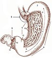


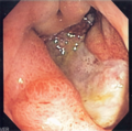

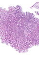
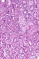
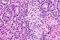
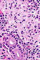


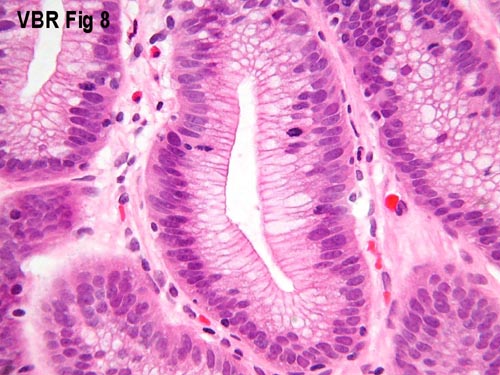