|
|
| (35 intermediate revisions by the same user not shown) |
| Line 27: |
Line 27: |
|
| |
|
| ==Pitfalls/weird stuff== | | ==Pitfalls/weird stuff== |
| *Thyroid tissue lateral to the jugular vein (often referred to as ''lateral aberrant thyroid tissue'') is generally considered metastatic thyroid carcinoma ([[papillary thyroid carcinoma]]) even if it looks benign.<ref name=pmid14452106>{{Cite journal | last1 = JOHNSON | first1 = RW. | last2 = SAHA | first2 = NC. | title = The so-called lateral aberrant thyroid. | journal = Br Med J | volume = 1 | issue = 5293 | pages = 1668-9 | month = Jun | year = 1962 | doi = | PMID = 14452106 | PMC = 1958877 }}</ref> | | *Thyroid tissue lateral to the jugular vein (often referred to as ''[[lateral aberrant thyroid tissue]]'') is generally considered metastatic thyroid carcinoma ([[papillary thyroid carcinoma]]) even if it looks benign.<ref name=pmid14452106>{{Cite journal | last1 = JOHNSON | first1 = RW. | last2 = SAHA | first2 = NC. | title = The so-called lateral aberrant thyroid. | journal = Br Med J | volume = 1 | issue = 5293 | pages = 1668-9 | month = Jun | year = 1962 | doi = | PMID = 14452106 | PMC = 1958877 }}</ref> |
| **This dictum is disputed.<ref name=pmid17319317>{{Cite journal | last1 = Escofet | first1 = X. | last2 = Khan | first2 = AZ. | last3 = Mazarani | first3 = W. | last4 = Woods | first4 = WG. | title = Lessons to be learned: a case study approach. Lateral aberrant thyroid tissue: is it always malignant? | journal = J R Soc Promot Health | volume = 127 | issue = 1 | pages = 45-6 | month = Jan | year = 2007 | doi = | PMID = 17319317 }}</ref> | | **This dictum is disputed.<ref name=pmid17319317>{{Cite journal | last1 = Escofet | first1 = X. | last2 = Khan | first2 = AZ. | last3 = Mazarani | first3 = W. | last4 = Woods | first4 = WG. | title = Lessons to be learned: a case study approach. Lateral aberrant thyroid tissue: is it always malignant? | journal = J R Soc Promot Health | volume = 127 | issue = 1 | pages = 45-6 | month = Jan | year = 2007 | doi = | PMID = 17319317 }}</ref> |
| **The level VI and VII [[lymph nodes]] are medial to the jugular. | | **The level VI and VII [[lymph nodes]] are medial to the jugular. |
| Line 62: |
Line 62: |
| *[[Follicular thyroid carcinoma|Follicular carinoma]]. | | *[[Follicular thyroid carcinoma|Follicular carinoma]]. |
| *[[Medullary thyroid carcinoma|Medullary carcinoma]]. | | *[[Medullary thyroid carcinoma|Medullary carcinoma]]. |
| *Undifferentiated (anaplastic) carcinoma. | | *[[Anaplastic thyroid carcinoma|Undifferentiated (anaplastic) carcinoma]]. |
|
| |
|
| *Poorly differentiated carcinoma. | | *[[Poorly differentiated thyroid carcinoma|Poorly differentiated carcinoma]]. |
| *[[Squamous cell carcinoma]]. | | *[[Squamous cell carcinoma]]. |
| *[[Mucoepidermoid carcinoma]]. | | *[[Mucoepidermoid carcinoma]]. |
| Line 127: |
Line 127: |
| *p63 +ve. | | *p63 +ve. |
| **-ve in clear cells. | | **-ve in clear cells. |
| *CEA +ve (polyconal).<ref name=pmid7509563>{{cite journal |author=Mizukami Y, Nonomura A, Michigishi T, ''et al.'' |title=Solid cell nests of the thyroid. A histologic and immunohistochemical study |journal=Am. J. Clin. Pathol. |volume=101 |issue=2 |pages=186–91 |year=1994 |month=February |pmid=7509563 |doi= |url=}}</ref> | | *[[CEA]] +ve (polyconal).<ref name=pmid7509563>{{cite journal |author=Mizukami Y, Nonomura A, Michigishi T, ''et al.'' |title=Solid cell nests of the thyroid. A histologic and immunohistochemical study |journal=Am. J. Clin. Pathol. |volume=101 |issue=2 |pages=186–91 |year=1994 |month=February |pmid=7509563 |doi= |url=}}</ref> |
| **+ve also in clear cells. | | **+ve also in clear cells. |
| *Chromogranin A +ve ~45% of cases.<ref name=pmid7509563/> | | *Chromogranin A +ve ~45% of cases.<ref name=pmid7509563/> |
| Line 137: |
Line 137: |
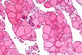
| *[[AKA]] ''[[nodular hyperplasia]]''. | | *[[AKA]] ''[[nodular hyperplasia]]''. |
| *[[AKA]] ''adenomatoid nodule''. | | *[[AKA]] ''adenomatoid nodule''. |
| | | {{Main|Thyroid gland nodular hyperplasia}} |
| ===General===
| |
| *Clinical diagnosis: ''goitre'', [[AKA]] ''sporadic goitre'', AKA ''multinodular goitre'' (MNG).
| |
| *Most common diagnosis in the thyroid.
| |
| **If you've seen a handful of thyroids you've seen this.
| |
| | |
| Notes:
| |
| *Large lesions may be clonal; however, this is clinically irrelevant.
| |
| | |
| ===Gross===
| |
| Features:
| |
| *Enlarge thyroid gland.
| |
| *+/-Distinct (well-circumscribed) nodules.
| |
| | |
| ===Microscopic===
| |
| Features:
| |
| *Follicles of variable size - '''key feature'''.
| |
| **Should be obvious at low power, i.e. with the 2.5x objective.
| |
| *+/-Nodules.
| |
| **Do not have a thick fibrous capsule.
| |
| **May have a high cellularity.
| |
| **Architecture: solid or microfollicular.<ref name=Ref_EP36>{{Ref EP|36}}</ref>
| |
| | |
| Negatives:
| |
| *No nuclear features suggestive of malignancy (at lower power).
| |
| **One should not look at high power.
| |
| *Not cellular.
| |
| | |
| DDx:
| |
| *[[Papillary thyroid carcinoma]] - esp. [[papillary thyroid carcinoma follicular variant]].
| |
| *[[Follicular thyroid adenoma]] - contained in a fibrous capsule.
| |
| *[[Follicular thyroid carcinoma]] - has fibrous capsule and invasion through it.
| |
| | |
| ===Sign out===
| |
| <pre>
| |
| HEMITHYROID, RIGHT, HEMITHYROIDECTOMY:
| |
| - NODULAR HYPERPLASIA.
| |
| - NEGATIVE FOR MALIGNANCY.
| |
| </pre>
| |
| | |
| <pre>
| |
| HEMITHYROID, RIGHT, HEMITHYROIDECTOMY:
| |
| - CELLULAR ADENOMATOID NODULE ON A BACKGROUND OF NODULAR HYPERPLASIA.
| |
| - NEGATIVE FOR MALIGNANCY.
| |
| </pre>
| |
|
| |
|
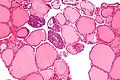
| ==Follicular thyroid adenoma== | | ==Follicular thyroid adenoma== |
| *[[AKA]] follicular adenoma, [[AKA]] thyroid follicular adenoma. | | *[[AKA]] follicular adenoma, [[AKA]] thyroid follicular adenoma. |
| ===General===
| | {{Main|Follicular thyroid adenoma}} |
| *Most common neoplasm of thyroid.<ref name=Ref_EP51>{{Ref EP|51}}</ref>
| |
| *Encapusled lesion (surrounded by fibrous capsule).
| |
| | |
| ===Gross===
| |
| *Thick capsule.
| |
| | |
| Notes:
| |
| *The entire capsule should be submitted.<ref>SR. 17 January 2011.</ref>
| |
| **A good start for most thyroid specimens with a thick capsule is 10 blocks.
| |
| | |
| ===Microsopic===
| |
| Features:
| |
| *Cellular.
| |
| *Thick capsule - '''key feature'''.
| |
| | |
| Negatives.
| |
| *No invasion of the capsule (see ''[[follicular thyroid carcinoma]]'' section).
| |
| *No nuclear features suggestive of [[papillary thyroid carcinoma]].
| |
| | |
| DDx:
| |
| *[[Thyroid gland nodular hyperplasia]] with an encapsulated nodule - not as cellular.
| |
|
| |
|
| ==Graves disease== | | ==Graves disease== |
| ===General===
| | {{Main|Graves' disease}} |
| *Often misspelled "Grave's disease".
| |
| *Autoimmune disease leading to hyperthyroidism.
| |
| *Eye problems not resolved with thyroid removal.{{fact}}
| |
| *Higher risk of [[papillary thyroid carcinoma]].
| |
| | |
| Clinical:
| |
| *TSH-receptor antibody +ve.<ref name=pmid19576193>{{Cite journal | last1 = Massart | first1 = C. | last2 = Gibassier | first2 = J. | last3 = d'Herbomez | first3 = M. | title = Clinical value of M22-based assays for TSH-receptor antibody (TRAb) in the follow-up of antithyroid drug treated Graves' disease: comparison with the second generation human TRAb assay. | journal = Clin Chim Acta | volume = 407 | issue = 1-2 | pages = 62-6 | month = Sep | year = 2009 | doi = 10.1016/j.cca.2009.06.033 | PMID = 19576193 }}</ref>
| |
| | |
| ===Gross===
| |
| Features:<ref>{{Ref EP|30}}</ref>
| |
| *Enlarged 50-150 g.
| |
| *"Beefy-red" appearance, looks like raw beef.
| |
| | |
| ===Microscopic===
| |
| Features:
| |
| *Classic:
| |
| **Hypercellular
| |
| **Patchy lymphocytes.
| |
| **Little colloid.
| |
| *Scalloping of colloid; colloid has undulating border.
| |
| **Non-specific finding.
| |
| *+/-Nuclear clearing.
| |
| *+/-Papillae (may mimic papillary thyroid carcinoma in this respect).
| |
| | |
| Notes:
| |
| *Usually has an unimpressive appearance... as it is treated, i.e. history is important.
| |
| *Nuclear clearing and papillae are usu. diffuse in Graves disease - unlike in papillary thyroid carcinoma.
| |
| | |
| Image:
| |
| *[http://library.med.utah.edu/WebPath/jpeg4/ENDO022.jpg Graves disease (med.utah.edu)].<ref>URL: [http://library.med.utah.edu/WebPath/EXAM/IMGQUIZ/enfrm.html http://library.med.utah.edu/WebPath/EXAM/IMGQUIZ/enfrm.html]. Accessed on: 4 December 2011.</ref>
| |
|
| |
|
| ==Idiopathic granulomatous thyroiditis== | | ==Idiopathic granulomatous thyroiditis== |
| Line 253: |
Line 158: |
| Clinical: | | Clinical: |
| *Tenderness.<ref name=pmid22538753>{{Cite journal | last1 = Szczepanek-Parulska | first1 = E. | last2 = Zybek | first2 = A. | last3 = Biczysko | first3 = M. | last4 = Majewski | first4 = P. | last5 = Ruchała | first5 = M. | title = What might cause pain in the thyroid gland? Report of a patient with subacute thyroiditis of atypical presentation. | journal = Endokrynol Pol | volume = 63 | issue = 2 | pages = 138-42 | month = | year = 2012 | doi = | PMID = 22538753 }}</ref> | | *Tenderness.<ref name=pmid22538753>{{Cite journal | last1 = Szczepanek-Parulska | first1 = E. | last2 = Zybek | first2 = A. | last3 = Biczysko | first3 = M. | last4 = Majewski | first4 = P. | last5 = Ruchała | first5 = M. | title = What might cause pain in the thyroid gland? Report of a patient with subacute thyroiditis of atypical presentation. | journal = Endokrynol Pol | volume = 63 | issue = 2 | pages = 138-42 | month = | year = 2012 | doi = | PMID = 22538753 }}</ref> |
| | |
| | Management: |
| | *Medical. |
| | *Rarely surgery.<ref>{{Cite journal | last1 = Volpé | first1 = R. | title = The management of subacute (DeQuervain's) thyroiditis. | journal = Thyroid | volume = 3 | issue = 3 | pages = 253-5 | month = | year = 1993 | doi = | PMID = 8257868 }}</ref> |
|
| |
|
| ===Microscopic=== | | ===Microscopic=== |
| Line 298: |
Line 207: |
| ==Riedel thyroiditis== | | ==Riedel thyroiditis== |
| *[[AKA]] ''invasive fibrous thyroiditis''.<ref name=pmid21568724>{{Cite journal | last1 = Fatourechi | first1 = MM. | last2 = Hay | first2 = ID. | last3 = McIver | first3 = B. | last4 = Sebo | first4 = TJ. | last5 = Fatourechi | first5 = V. | title = Invasive fibrous thyroiditis (Riedel thyroiditis): the Mayo Clinic experience, 1976-2008. | journal = Thyroid | volume = 21 | issue = 7 | pages = 765-72 | month = Jul | year = 2011 | doi = 10.1089/thy.2010.0453 | PMID = 21568724 }}</ref> | | *[[AKA]] ''invasive fibrous thyroiditis''.<ref name=pmid21568724>{{Cite journal | last1 = Fatourechi | first1 = MM. | last2 = Hay | first2 = ID. | last3 = McIver | first3 = B. | last4 = Sebo | first4 = TJ. | last5 = Fatourechi | first5 = V. | title = Invasive fibrous thyroiditis (Riedel thyroiditis): the Mayo Clinic experience, 1976-2008. | journal = Thyroid | volume = 21 | issue = 7 | pages = 765-72 | month = Jul | year = 2011 | doi = 10.1089/thy.2010.0453 | PMID = 21568724 }}</ref> |
| ===General===
| | {{Main|Riedel thyroiditis}} |
| Clinical features:<ref name=pmid21568724/>
| |
| *Extremely rare.
| |
| *Women > men.
| |
| *Usually smokers.
| |
| *May be associated with ''[[retroperitoneal fibrosis]]''.
| |
| *May be hypothyroid.
| |
| *+/-Obstructive symptoms.
| |
| | |
| ===Microscopic===
| |
| Features:
| |
| *Fibrosis.
| |
| *Specimen often fragmented as it was difficult to remove.
| |
| | |
| DDx:
| |
| *[[Anaplastic thyroid carcinoma|Anaplastic carcinoma]], spindle cell variant.
| |
|
| |
|
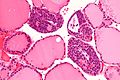
| ==Hashimoto thyroiditis== | | ==Hashimoto thyroiditis== |
| ===General===
| | {{Main|Hashimoto's thyroiditis}} |
| *'''This is a clinical diagnosis'''.
| |
| **The histomorphologic findings, generally, are '''not''' diagnostic.
| |
| | |
| Etiology:
| |
| *Autoimmune disease leading to hypothyroidism.
| |
| **Often genetic/part of a syndrome.
| |
| | |
| ====Clinical====
| |
| Serology:<ref name=pmid7813361>{{cite journal |author=Poropatich C, Marcus D, Oertel YC |title=Hashimoto's thyroiditis: fine-needle aspirations of 50 asymptomatic cases |journal=Diagn. Cytopathol. |volume=11 |issue=2 |pages=141–5 |year=1994 |pmid=7813361 |doi= |url=http://www3.interscience.wiley.com/journal/112701408/abstract?CRETRY=1&SRETRY=0}}</ref>
| |
| *Antimicrosomal (antithyroid peroxidase) +ve.
| |
| *Antithyroglobulin +ve.
| |
| | |
| Associated pathology:<ref name=pmid7813361/>
| |
| *Increased risk of B-cell lymphoma; these are classically:<ref name=pmid18018576 >{{Cite journal | last1 = Ohye | first1 = H. | last2 = Fukata | first2 = S. | last3 = Hirokawa | first3 = M. | title = [Malignant lymphoma of the thyroid]. | journal = Nihon Rinsho | volume = 65 | issue = 11 | pages = 2092-8 | month = Nov | year = 2007 | doi = | PMID = 18018576 }}</ref>
| |
| **[[MALT lymphoma]].
| |
| **[[Diffuse large B cell lymphoma]] (DLBCL).
| |
| | |
| ===Microscopic===
| |
| Features:
| |
| *Lymphocytic infiltrate - '''key feature'''.
| |
| *Nuclear clearing common.
| |
| **May confuse with [[papillary thyroid carcinoma]].
| |
| *Polymorphous lymphoplasmacytic infiltrate with germinal centres.<ref name=Ref_APBR672>{{Ref APBR|672}}</ref>
| |
| *+/-Oncocytic metaplasia.
| |
| | |
| Notes:
| |
| *Histologically often '''not''' possible to separate from "non-specific" thyroiditis.<ref name=Ref_Sternberg4_560>{{Ref Sternberg4|560}}</ref>
| |
| | |
| DDx:
| |
| *[[Lymphocytic thyroiditis]].
| |
| *[[Papillary thyroid carcinoma]].
| |
| *[[MALT lymphoma]].
| |
| *[[Diffuse large B cell lymphoma]].
| |
| | |
| ===IHC===
| |
| *Panel to exclude lymphoma may be required, e.g. CD3, CD20, CD10, BCL6, BCL2, kappa, lambda.
| |
| | |
| ===Molecular===
| |
| *Occasionally done to exclude lymphoma - see ''[[MALT lymphoma]]'' and ''[[DLBCL]]''.
| |
|
| |
|
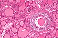
| ==C-cell hyperplasia== | | ==C-cell hyperplasia== |
| *Abbreviated ''CCH''. | | *Abbreviated ''CCH''. |
| ===General===
| | {{Main|C-cell hyperplasia}} |
| *Screening for C-cell hyperplasia/[[medullary thyroid carcinoma]] done with ''serum calcitonin level''.<ref name=pmid19726541>{{cite journal |author=Machens A, Hoffmann F, Sekulla C, Dralle H |title=Importance of gender-specific calcitonin thresholds in screening for occult sporadic medullary thyroid cancer |journal=Endocr. Relat. Cancer |volume=16 |issue=4 |pages=1291–8 |year=2009 |month=December |pmid=19726541 |doi=10.1677/ERC-09-0136 |url=http://erc.endocrinology-journals.org/cgi/content/full/16/4/1291}}</ref>
| |
| | |
| ===Gross===
| |
| *Not visible.
| |
| | |
| ===Microscopic===
| |
| Features:
| |
| *Location:<ref>URL: [http://www.cap.org/apps/docs/committees/cancer/cancer_protocols/2011/Thyroid_11protocol.pdf http://www.cap.org/apps/docs/committees/cancer/cancer_protocols/2011/Thyroid_11protocol.pdf]. Accessed on: 7 April 2012.</ref>
| |
| **Mid portion of lobe to upper third of lobe.
| |
| ***Not at the poles.
| |
| ***Not in the isthmus.
| |
| | |
| *Definitions vary.<ref>SR. 17 January 2011.</ref>
| |
| | |
| One definition - either of the following:<ref name=pmid19726541>{{cite journal |author=Machens A, Hoffmann F, Sekulla C, Dralle H |title=Importance of gender-specific calcitonin thresholds in screening for occult sporadic medullary thyroid cancer |journal=Endocr. Relat. Cancer |volume=16 |issue=4 |pages=1291–8 |year=2009 |month=December |pmid=19726541 |doi=10.1677/ERC-09-0136 |url=http://erc.endocrinology-journals.org/cgi/content/full/16/4/1291}}</ref>
| |
| #>50 C-cells per low-power field (x100).
| |
| #*This part of the definition suffers from [[LPFitis]]. The paper should have been rejected.
| |
| #Confined to the thyroid gland and no larger than 10 mm in greatest dimension.
| |
| | |
| Another definition:
| |
| *Invasion of the basement membrane with stromal reaction.
| |
| | |
| A third definition:
| |
| *"Several clusters" of more than six C cells.
| |
|
| |
|
| ====Images==== | | ==Adenolipoma of the thyroid== |
| *[http://www.nature.com/modpathol/journal/v16/n8/fig_tab/3880836f2.html CCH - crappy B&W image (nature.com)].<ref>{{Cite journal | last1 = Guyétant | first1 = S. | last2 = Josselin | first2 = N. | last3 = Savagner | first3 = F. | last4 = Rohmer | first4 = V. | last5 = Michalak | first5 = S. | last6 = Saint-André | first6 = JP. | title = C-cell hyperplasia and medullary thyroid carcinoma: clinicopathological and genetic correlations in 66 consecutive patients. | journal = Mod Pathol | volume = 16 | issue = 8 | pages = 756-63 | month = Aug | year = 2003 | doi = 10.1097/01.MP.0000081727.75778.0C | PMID = 12920219 }}</ref>
| | {{Main|Adenolipoma of the thyroid}} |
| *[http://www.nature.com/modpathol/journal/v16/n8/fig_tab/3880836f3.htm CCH - crappy B&W image (nature.com)].
| |
| *[http://www.forpath.org/workshops/0201/photos/fullsize/cas7c.jpg CCH (forpath.org)].<ref>URL: [http://www.forpath.org/workshops/0201/html/case_7.asp http://www.forpath.org/workshops/0201/html/case_7.asp]. Accessed on: 21 May 2013.</ref>
| |
| *[http://alf3.urz.unibas.ch/pathopic/e/getpic-fra.cfm?id=4849 CCH (unibas.ch)].
| |
| *[http://alf3.urz.unibas.ch/pathopic/e/getpic-fra.cfm?id=10739 Nodular CCH (unibas.ch)].
| |
|
| |
|
| =Malignant neoplasm= | | =Malignant neoplasm= |
| Line 397: |
Line 224: |
| ==Papillary thyroid carcinoma== | | ==Papillary thyroid carcinoma== |
| *Abbreviated ''PTC''. | | *Abbreviated ''PTC''. |
| ===General===
| | {{Main|Papillary thyroid carcinoma}} |
| Medical school memory device P's:
| |
| *Palpable nodes.
| |
| *Popular (most common malignant neoplasm of the thyroid).
| |
| *Prognosis is good.
| |
| *Pre-Tx iodine scan.
| |
| *Post-Sx iodine scan.
| |
| *[[Psammoma bodies]].
| |
| | |
| Notes:
| |
| *PTC is associated with radiation exposure.<ref name=Ref_Sternberg4_564>{{Ref Sternberg4|564}}</ref>
| |
| *''Papillary thyroid microcarcinoma'' is defined as a tumour with a maximal dimension of 1.0 cm or less.<ref name=pmid21267823>{{Cite journal | last1 = Sethom | first1 = A. | last2 = Riahi | first2 = I. | last3 = Riahi | first3 = K. | last4 = Akkari | first4 = K. | last5 = Benzarti | first5 = S. | last6 = Miled | first6 = I. | last7 = Chebbi | first7 = MK. | title = [Management of thyroid microcarcinoma. Report of 13 cases]. | journal = Tunis Med | volume = 89 | issue = 1 | pages = 23-5 | month = Jan | year = 2011 | doi = | PMID = 21267823 }}</ref>
| |
| | |
| ====Prognosis====
| |
| Prognosis can be predicted by ''MAICS'' score. It which includes:<ref name=pmid12016468>{{Cite journal | last1 = Hay | first1 = ID. | last2 = Thompson | first2 = GB. | last3 = Grant | first3 = CS. | last4 = Bergstralh | first4 = EJ. | last5 = Dvorak | first5 = CE. | last6 = Gorman | first6 = CA. | last7 = Maurer | first7 = MS. | last8 = McIver | first8 = B. | last9 = Mullan | first9 = BP. | title = Papillary thyroid carcinoma managed at the Mayo Clinic during six decades (1940-1999): temporal trends in initial therapy and long-term outcome in 2444 consecutively treated patients. | journal = World J Surg | volume = 26 | issue = 8 | pages = 879-85 | month = Aug | year = 2002 | doi = 10.1007/s00268-002-6612-1 | PMID = 12016468 }}</ref>
| |
| *'''M'''etastases.
| |
| *'''A'''ge.
| |
| *'''I'''nvasion of surround tissues.
| |
| *'''C'''completeness of excision.
| |
| *'''S'''ize of tumour.
| |
| | |
| ===Microscopic===
| |
| Features:
| |
| *Nuclear changes - '''key feature'''.
| |
| *#"Shrivelled nuclei"/"raisin" like nuclei, nuclei with a wavy ("textured", convoluted) nuclear membrane -- usu. easy to find.
| |
| *#[[Nuclear pseudoinclusions]] -- usu. harder to find; have high [[specificity]] (nuclear pseudoinclusions appear as a result of the very convoluted nuclear membrane wrapping around parts of the cytoplasm; true nuclear inclusions in contrast are seen only in viral infections).
| |
| *#Nuclear grooves, seen as a result of the highly "textured" nuclear membrane.
| |
| *#Nuclear clearing (only on permanent section) - also known as "Orphan Annie eyes".
| |
| *Overlap of nuclei - "cells do not respect each other's borders" (easy to see at '''key feature at low power''').
| |
| *Classically has papillae (nipple-like shape); papilla (definition): epithelium on fibrovascular core.
| |
| **Absence of papillae does not exclude diagnosis.
| |
| *[[Psammoma bodies]].
| |
| **Circular, acellular, eosinophilic whorled bodies.
| |
| **Not necessary to make diagnosis - but very specific in the context of a specimen labeled "thyroid".
| |
| **Arise from infarction & calcification of papilla tips.<ref name=Ref_Sternberg4_565>{{Ref Sternberg4|565}}</ref>
| |
| | |
| Notes:
| |
| *Psammoma bodies are awesome if you see 'em, i.e. useful for arriving at the diagnosis.
| |
| **If there are no papillae structures -- you're unlikely to see psammoma bodies.
| |
| *At low power look for cellular areas/loss of follicles.
| |
| *Nuclear clearing seen in:
| |
| **Hashimoto's and papillary thyroid carcinoma.<ref name=Ref_Sternberg4_566>{{Ref Sternberg4|566}}</ref>
| |
| **May be an artifact of [[fixation]]/processing.
| |
| *Nuclear overlapping is easy to see at lower power-- should be the tip-off to look at high power for nuclear features.
| |
| *Nuclear inclusions are quite rare and not required to make the diagnosis -- but a very convincing feature if seen.
| |
| *Papillae may be seen in Graves disease.
| |
| | |
| DDx:
| |
| *[[Lymphocytic thyroiditis]]:
| |
| **[[Graves disease]].
| |
| **[[Hashimoto thyroiditis]].
| |
| *[[Solid cell nest of thyroid]].<ref name=pmid16830963>{{Cite journal | last1 = Baloch | first1 = ZW. | last2 = LiVolsi | first2 = VA. | title = Cytologic and architectural mimics of papillary thyroid carcinoma. Diagnostic challenges in fine-needle aspiration and surgical pathology specimens. | journal = Am J Clin Pathol | volume = 125 Suppl | issue = | pages = S135-44 | month = Jun | year = 2006 | doi = | PMID = 16830963 | URL = http://ajcp.ascpjournals.org/content/supplements/125/Suppl_1/S135.full.pdf }}</ref>
| |
| | |
| ====Subtypes of papillary thyroid carcinoma====
| |
| There are many.
| |
| | |
| Poor prognosis variants:
| |
| *[[Papillary thyroid carcinoma tall cell variant|Tall cell variant]].<ref name=pmid22432054>{{Cite journal | last1 = Gonzalez-Gonzalez | first1 = R. | last2 = Bologna-Molina | first2 = R. | last3 = Carreon-Burciaga | first3 = RG. | last4 = Gómezpalacio-Gastelum | first4 = M. | last5 = Molina-Frechero | first5 = N. | last6 = Salazar-Rodríguez | first6 = S. | title = Papillary thyroid carcinoma: differential diagnosis and prognostic values of its different variants: review of the literature. | journal = ISRN Oncol | volume = 2011 | issue = | pages = 915925 | month = | year = 2011 | doi = 10.5402/2011/915925 | PMID = 22432054 | PMC = 3302055 | URL = http://www.ncbi.nlm.nih.gov/pmc/articles/pmid/22432054/?tool=pubmed }}</ref>
| |
| *[[Papillary thyroid carcinoma columnar cell variant|Columnar cell variant]].<ref name=pmid22432054/>
| |
| *[[Papillary thyroid carcinoma solid variant|Solid variant]].<ref name=pmid22432054/>
| |
| *[[Papillary thyroid carcinoma diffuse sclerosing variant|Diffuse sclerosing variant]].<ref>URL: [http://emedicine.medscape.com/article/849000-overview#a0104 http://emedicine.medscape.com/article/849000-overview#a0104]. Accessed on: 1 May 2012.</ref>
| |
| | |
| =====Papillary thyroid carcinoma tall cell variant=====
| |
| ======General======
| |
| *~10% of PTC.<ref>{{Ref Sternberg5|505}}</ref>
| |
| *Often large > 6 cm.
| |
| | |
| ======Microscopic======
| |
| Features:<ref name=pmid19373912>{{cite journal |author=Urano M, Kiriyama Y, Takakuwa Y, Kuroda M |title=Tall cell variant of papillary thyroid carcinoma: Its characteristic features demonstrated by fine-needle aspiration cytology and immunohistochemical study |journal=Diagn. Cytopathol. |volume= |issue= |pages= |year=2009 |month=April |pmid=19373912 |doi=10.1002/dc.21086 |url=}}</ref>
| |
| *50% of cells with height 2x the width.<ref name=pmid18925842>{{cite journal |author=Ghossein R, Livolsi VA |title=Papillary thyroid carcinoma tall cell variant |journal=Thyroid |volume=18 |issue=11 |pages=1179–81 |year=2008 |month=November |pmid=18925842 |doi=10.1089/thy.2008.0164 |url=}}</ref>
| |
| **There is some disagreement on these criteria;<ref name=pmid18925842/> Raphael believes the height ought to be ~3x width, for 50% of the cells.<ref>S. Raphael. 17 January 2011.</ref>
| |
| *Eosinophilic cytoplasm.
| |
| *Well-defined cell borders.
| |
| *Nucleus stratified; basal location, i.e. closer to the basement membrane.
| |
| | |
| Negative:
| |
| *Nuclei ''not'' pseudostratified, if pseudostratified consider ''columnar cell variant''.
| |
| | |
| Images:
| |
| *[http://commons.wikimedia.org/wiki/File:Papillary_thyroid_carcinoma_tall_cell_var_intermed_mag.jpg PTC tall cell variant - intermed. mag. (WC)].
| |
| *[http://commons.wikimedia.org/wiki/File:Papillary_thyroid_carcinoma_tall_cell_var_high_mag.jpg PTC tall cell variant - high mag. (WC)].
| |
| | |
| =====Papillary thyroid carcinoma columnar cell variant=====
| |
| ======General======
| |
| Epidemiology:
| |
| *Poor prognosis.
| |
| *Very rare.
| |
| | |
| ======Microscopic======
| |
| Features:<ref name=Ref_Sternberg5_506>{{Ref Sternberg5|506}}</ref>
| |
| *Elongated nuclei (similar to colorectal adenocarcinoma) - '''key feature'''.
| |
| *+/-Pseudostratification of the nuclei (like in colorectal adenocarcinoma), differentiates from ''tall cell variant''.
| |
| *Nuclear stratification - '''key feature'''.
| |
| *"Minimal" papillary features.
| |
| *"Tall cells".
| |
| *Clear-eosinophilic cytoplasm.
| |
| *Mitoses common.
| |
|
| |
| Image: [http://www3.interscience.wiley.com/cgi-bin/fulltext/75000320/nfig003a?CRETRY=1&SRETRY=0 Columnar variant PTC (wiley.com)].
| |
| =====Papillary thyroid carcinoma follicular variant=====
| |
| ======General======
| |
| *May be confused with [[follicular thyroid carcinoma|follicular carcinoma]] or [[follicular thyroid adenoma|follicular adenoma]].
| |
| *Pathologists often disagree about this diagnosis.<ref name=pmid21940284>{{Cite journal | last1 = Daniels | first1 = GH. | title = What if many follicular variant papillary thyroid carcinomas are not malignant? A review of follicular variant papillary thyroid carcinoma and a proposal for a new classification. | journal = Endocr Pract | volume = 17 | issue = 5 | pages = 768-87 | month = | year = | doi = 10.4158/EP10407.RA | PMID = 21940284 }}</ref>
| |
| | |
| ======Microscopic======
| |
| Features:<ref name=Ref_EP88>{{Ref EP|88}}</ref>
| |
| *Small tightly packed follicles - '''key feature'''.
| |
| *Hypereosinophilic colloid.
| |
| *Nuclear features of PTC.
| |
| **Large nuclei.
| |
| **Typically have less [[nuclear pseudoinclusion]]s than the conventional type.
| |
| *+/-Fibrous capsule (common).
| |
| | |
| DDx:
| |
| *[[Follicular thyroid carcinoma]] - has a fibrous capsule and invasion though it.
| |
| *[[Follicular thyroid adenoma]] - surrounded by a fibrous capsule.
| |
| *[[Adenomatoid nodule]] - round nuclei, no nuclear features of PTC.
| |
| | |
| Images:
| |
| *[http://www.surgicalpathologyatlas.com/glfusion/mediagallery/media.php?f=0&sort=0&s=2008080217023776 PTC follicular variant (surgicalpathologyatlas.com)].
| |
| *[http://www.surgicalpathologyatlas.com/glfusion/mediagallery/media.php?f=0&sort=0&s=2008080216593186 PTC follicular variant (surgicalpathologyatlas.com)].
| |
| *[http://www.thyroidcancercanada.org/userfiles/images/Follicular_slide.jpg PTC follicular variant (thyroidcancercanada.org)].<ref>URL: [http://www.thyroidcancercanada.org/types-of-thyroid-cancer.php?lang=en http://www.thyroidcancercanada.org/types-of-thyroid-cancer.php?lang=en]. Accessed on: 9 January 2013.</ref>
| |
| | |
| =====Papillary thyroid carcinoma cribriform-morular variant=====
| |
| ======General======
| |
| *Associated with [[familial adenomatous polyposis]] (FAP).<ref name=pmid18612695>{{cite journal |author=Groen EJ, Roos A, Muntinghe FL, ''et al.'' |title=Extra-intestinal manifestations of familial adenomatous polyposis |journal=Ann. Surg. Oncol. |volume=15 |issue=9 |pages=2439–50 |year=2008 |month=September |pmid=18612695 |pmc=2518080 |doi=10.1245/s10434-008-9981-3 |url=http://www.ncbi.nlm.nih.gov/pmc/articles/PMC2518080/?tool=pubmed}}</ref>
| |
| | |
| ======Microscopic======
| |
| Features:
| |
| *Cribriform architectural pattern.
| |
| *Morules - balls of tissue.
| |
| | |
| =====Papillary thyroid carcinoma diffuse sclerosing variant=====
| |
| ======General======
| |
| *Usually young adults, children.
| |
| | |
| ======Microscopic======
| |
| Features:<ref>{{Ref PBoD8|1122}}</ref>
| |
| *Papillae - usu. prominent.
| |
| *Squamous morules - '''key features'''.<ref name=pmid15233643>{{Cite journal | last1 = Hirokawa | first1 = M. | last2 = Kuma | first2 = S. | last3 = Miyauchi | first3 = A. | last4 = Qian | first4 = ZR. | last5 = Nakasono | first5 = M. | last6 = Sano | first6 = T. | last7 = Kakudo | first7 = K. | title = Morules in cribriform-morular variant of papillary thyroid carcinoma: Immunohistochemical characteristics and distinction from squamous metaplasia. | journal = APMIS | volume = 112 | issue = 4-5 | pages = 275-82 | month = | year = | doi = 10.1111/j.1600-0463.2004.apm11204-0508.x | PMID = 15233643 }}
| |
| </ref>
| |
| *Lymphocytes - abundant.
| |
| *Fibrosis.
| |
| | |
| DDx:
| |
| *Lymphocytic thyroiditis (esp. Hashimoto's thyroiditis).
| |
| | |
| =====Papillary thyroid carcinoma warthin-like variant=====
| |
| *Resemble [[Warthin tumour]].
| |
| ======Microscopic======
| |
| Features:<ref name=Ref_Sternberg5_506>{{Ref Sternberg5|506}}</ref>
| |
| *Eosinophilic cytoplasm.
| |
| *Lymphocytic thyroiditis.
| |
| *Papillae.
| |
| | |
| =====Papillary thyroid carcinoma solid variant=====
| |
| Features:<ref name=pmid22432054/>
| |
| *Some studies suggest this has a poor prognosis.
| |
| *More common in children.
| |
| *Associated with Chernobyl nuclear accident.
| |
| | |
| ======Microscopic======
| |
| Features:
| |
| *Solid sheets >50% of tumour mass.<ref name=pmid22432054/>
| |
| | |
| =====Papillary thyroid carcinoma oncocytic variant=====
| |
| Features:
| |
| *Possible association with [[autoimmune thyroiditis]].<ref name=pmid9013831>{{Cite journal | last1 = Berho | first1 = M. | last2 = Suster | first2 = S. | title = The oncocytic variant of papillary carcinoma of the thyroid: a clinicopathologic study of 15 cases. | journal = Hum Pathol | volume = 28 | issue = 1 | pages = 47-53 | month = Jan | year = 1997 | doi = | PMID = 9013831 }}</ref>
| |
| | |
| ======Microscopic======
| |
| Features:<ref name=pmid9013831/>
| |
| *Abundant oncocytic tumour cells with apical nuclei.
| |
| *Classic features of PTC:
| |
| **Grooves and and abundant pseudoinclusions.<ref name=Ref_EP86>{{Ref EP|86}}</ref>
| |
| *>70% papillary architecture.<ref name=Ref_EP86>{{Ref EP|86}}</ref>
| |
| *+/-Degenerative changes.
| |
| | |
| Note:
| |
| *CK19 +ve -- though ''not'' specific or sensitive.
| |
| | |
| ===IHC===
| |
| Thyroid versus something else:
| |
| *Thyroglobulin +ve.
| |
| *TTF-1 (thyroid transcription factor-1) +ve.
| |
| *CD15 +ve.{{fact}}
| |
| | |
| PTC versus benign:<ref>{{Cite journal | last1 = Mataraci | first1 = EA. | last2 = Ozgüven | first2 = BY. | last3 = Kabukçuoglu | first3 = F. | title = Expression of cytokeratin 19, HBME-1 and galectin-3 in neoplastic and nonneoplastic thyroid lesions. | journal = Pol J Pathol | volume = 63 | issue = 1 | pages = 58-64 | month = Mar | year = 2012 | doi = | PMID = 22535608 }}</ref>
| |
| *HBME-1 +ve (strong, diffuse).
| |
| *CK19 +ve (strong, diffuse).
| |
| *Galectin-3 +ve (strong, diffuse).
| |
| | |
| ===Molecular===
| |
| *Currently not widely used in a diagnostic context.
| |
| | |
| ====Tabular summary====
| |
| Molecular changes in papillary thyroid carcinoma as per ''Adeniran et al'':<ref name=pmid16434896>{{Cite journal | last1 = Adeniran | first1 = AJ. | last2 = Zhu | first2 = Z. | last3 = Gandhi | first3 = M. | last4 = Steward | first4 = DL. | last5 = Fidler | first5 = JP. | last6 = Giordano | first6 = TJ. | last7 = Biddinger | first7 = PW. | last8 = Nikiforov | first8 = YE. | title = Correlation between genetic alterations and microscopic features, clinical manifestations, and prognostic characteristics of thyroid papillary carcinomas. | journal = Am J Surg Pathol | volume = 30 | issue = 2 | pages = 216-22 | month = Feb | year = 2006 | doi = | PMID = 16434896 }}</ref>
| |
| {| class="wikitable sortable"
| |
| ! Molecular change
| |
| ! Frequency
| |
| ! Histology
| |
| ! Notes
| |
| |-
| |
| |BRAF point mutations
| |
| | ~ 40%
| |
| | [[papillary thyroid carcinoma tall cell variant|tall cell variant]]
| |
| | poorer prognosis, older individuals
| |
| |-
| |
| |RET/PTC rearrangments
| |
| | ~ 20%
| |
| | papillary architecture, [[psammoma bodies]]
| |
| | younger individuals
| |
| |-
| |
| |RAS point mutations
| |
| | ~ 15%
| |
| | exclusively [[papillary thyroid carcinoma follicular variant|follicular variant]]
| |
| | -
| |
| |}
| |
| | |
| ===Sign out===
| |
| <pre>
| |
| HEMITHYROID, RIGHT, COMPLETION OF TOTAL THYROIDECTOMY:
| |
| - PAPILLARY THYROID CARCINOMA, FOLLICULAR VARIANT.
| |
| -- TUMOUR SIZE: 4 MM (MAXIMAL).
| |
| -- ARCHITECTURE: FOLLICULAR.
| |
| -- CYTOMORPHOLOGY: CLASSICAL.
| |
| -- HISTOLOGIC GRADE: G1 (WELL DIFFERENTIATED).
| |
| -- NO TUMOUR CAPSULE IDENTIFIED.
| |
| -- NEGATIVE FOR LYMPHOVASCULAR INVASION.
| |
| -- NEGATIVE FOR PERINEURAL INVASION.
| |
| -- NEGATIVE FOR EXTRATHYROIDAL EXTENSION.
| |
| -- SURGICAL MARGINS NEGATIVE FOR MALIGNANCY.
| |
| </pre>
| |
| | |
| Note:
| |
| *If it is a completion thyroidectomy and the staging changes one should do a full synoptic report.
| |
| | |
| ====Microcarcinoma====
| |
| <pre>
| |
| A. LEFT HEMITHYROID, THYROIDECTOMY COMPLETION:
| |
| - PAPILLARY THYROID MICROCARCINOMA.
| |
| -- MARGINS NEGATIVE FOR MALIGNANCY.
| |
| -- TUMOUR SIZE ~ 1 MM.
| |
| -- NEGATIVE FOR LYMPHOVASCULAR INVASION.
| |
| -- NEGATIVE FOR PERINEURAL INVASION.
| |
| - PALPATION THYROIDITIS, FOCAL.
| |
| - NODULAR HYPERPLASIA.
| |
| | |
| B. LYMPH NODES, LEVEL 6 AND 7, LYMPH NODE DISSECTION:
| |
| - TWO LYMPH NODES, NEGATIVE FOR MALIGNANCY ( 0 POSITIVE / 2 ).
| |
| </pre>
| |
|
| |
|
| ==Insular carcinoma== | | ==Insular carcinoma== |
| ===General===
| | {{Main|Insular thyroid carcinoma}} |
| Features:<ref name=pmid17665497>{{cite journal |author=Rufini V, Salvatori M, Fadda G, ''et al.'' |title=Thyroid carcinomas with a variable insular component: prognostic significance of histopathologic patterns |journal=Cancer |volume=110 |issue=6 |pages=1209–17 |year=2007 |month=September |pmid=17665497 |doi=10.1002/cncr.22913 |url=}}</ref>
| |
| *Rare - approximately 5% of all thyroid carcinomas.
| |
| *Thought to be a separate tumour from papillary thyroid carcinoma and follicular thyroid carcinoma with a focal insular pattern.
| |
| *Some lump this entity with papillary carcinoma, i.e. consider it a variant of papillary thyroid carcinoma.
| |
| | |
| ===Microscopic===
| |
| Features:<ref name=pmid17665497/>
| |
| *Islands of cells - '''key feature'''.
| |
| *Scant cytoplasm.
| |
| *Nuclei monomorphic and round.
| |
| | |
| DDx:<ref>Endo. fellow. 17 September 2009.</ref>
| |
| *[[Medullary thyroid carcinoma]].
| |
| *Poorly differentiated thyroid carcinoma.
| |
|
| |
|
| ==Follicular thyroid carcinoma== | | ==Follicular thyroid carcinoma== |
| *[[AKA]] ''follicular carcinoma''. | | *[[AKA]] ''follicular carcinoma''. |
| ===Clinical===
| | {{Main|Follicular thyroid carcinoma}} |
| Medical school memory device ''4 Fs'':
| |
| *FNA NOT diagnosable.
| |
| *Far away mets (sometimes).
| |
| *Female predominant.
| |
| *Favourable prognosis.
| |
| | |
| Notes:
| |
| *Usu. has a hematologic spread.
| |
| **PTC usu. spread via lymphatics.
| |
| | |
| ===Microscopic===
| |
| Features:
| |
| *Defined by either:
| |
| *#Invasion through the capsule:
| |
| *#*Should be all the way through.<ref>SR. 17 January 2011.</ref>
| |
| *#**1/2 does not count.
| |
| *#**Fibrous reaction does not count.
| |
| *#**"Above the contour" does not count.
| |
| *#Vascular invasion (all of the following):
| |
| *##In a small vein (not a capillary), that is outside of the tumour mass.
| |
| *##Tumour adherent to the side of the vessel.
| |
| *##Tumour must be re-endothelialized.
| |
| | |
| Notes:
| |
| *'''Impossible''' to differentiate from ''[[follicular thyroid adenoma|follicular adenoma]]'' on FNA (no cytologic differences).
| |
| *Described as "over-diagnosed" ... misdiagnoses: PTC follicular variant, follicular adenoma, multinodular goitre with a thick capsule.
| |
| | |
| Images:
| |
| *[http://path.upmc.edu/cases/case653.html Follicular thyroid carcinoma - several images (upmc.edu)].
| |
|
| |
|
| ==Medullary thyroid carcinoma== | | ==Medullary thyroid carcinoma== |
| *Abbreviated ''MTC''. | | *Abbreviated ''MTC''. |
| ===General===
| | {{Main|Medullary thyroid carcinoma}} |
| Medical school memory device - 3 M's:
| |
| *[[amyloid|aMyloid]].
| |
| *Median node dissection done.
| |
| *[[MEN IIa syndrome]]/[[MEN IIb syndrome]].
| |
| **Medullary thyroid carcinoma.
| |
| **[[Pheochromocytoma]].
| |
| **[[Parathyroid adenoma]].
| |
| | |
| Epidemiology:
| |
| *Very rare.
| |
| *Poor prognosis.
| |
| *May be genetic (MEN IIa/b syndrome).
| |
| *Arises from C cells (which produce calcitonin).
| |
| | |
| Syndromic tumours - typically:<ref name=pmid21455198>{{Cite journal | last1 = Nosé | first1 = V. | title = Familial thyroid cancer: a review. | journal = Mod Pathol | volume = 24 Suppl 2 | issue = | pages = S19-33 | month = Apr | year = 2011 | doi = 10.1038/modpathol.2010.147 | PMID = 21455198 |URL = http://www.nature.com/modpathol/journal/v24/n2s/full/modpathol2010147a.html }}</ref>
| |
| *Present in 30s or 40s.
| |
| *+/-Multifocal.
| |
| *+/-Bilateral.
| |
| *[[C-cell hyperplasia]].
| |
| | |
| ===Gross===
| |
| Features:<ref name=pmid21455198/>
| |
| *Usu. well-circumscribed.
| |
| *White, gray or yellow.
| |
| *Gritty.
| |
| *Firm.
| |
|
| |
|
| Image:
| | ==Poorly differentiated thyroid carcinoma== |
| *[http://www.nature.com/modpathol/journal/v24/n2s/fig_tab/modpathol2010147f2.html MTC (nature.com)].
| | {{Main|Poorly differentiated thyroid carcinoma}} |
| | |
| ===Microscopic===
| |
| Features:
| |
| *Nuclei with "neuroendocrine features".
| |
| **Small, round nuclei.
| |
| **Coarse chromatin (''salt and pepper nuclei'').
| |
| *+/-[[Amyloid]] deposits - fluffy appearing acellular eosinophilic material in the cytoplasm.
| |
| *+/-[[C-cell hyperplasia]] - seen with familial forms of MTC.
| |
| **C cells (AKA ''parafollicular cell''): abundant cytoplasm - clear/pale.
| |
| | |
| Note:
| |
| *The amyloid is formed from ''calcitonin''.<ref name=pmid15459123>{{Cite journal | last1 = Khurana | first1 = R. | last2 = Agarwal | first2 = A. | last3 = Bajpai | first3 = VK. | last4 = Verma | first4 = N. | last5 = Sharma | first5 = AK. | last6 = Gupta | first6 = RP. | last7 = Madhusudan | first7 = KP. | title = Unraveling the amyloid associated with human medullary thyroid carcinoma. | journal = Endocrinology | volume = 145 | issue = 12 | pages = 5465-70 | month = Dec | year = 2004 | doi = 10.1210/en.2004-0780 | PMID = 15459123 }}</ref>
| |
| | |
| Images:
| |
| *www:
| |
| **[http://jcp.bmj.com/content/vol57/issue3/images/large/cp8474.f16.jpeg Medullary thyroid carcinoma (bmj.com)].
| |
| **[http://www.nature.com/ki/journal/v70/n11/fig_tab/5001888f2.html C cell hyperplasia (nature.com)].
| |
| **[http://lifesci.rutgers.edu/~babiarz/Review3/Lp6/scope8.htm C cell (rutgers.edu)].
| |
| **[http://www.anatomyatlases.org/MicroscopicAnatomy/Images/Plate287.jpg Parafollicular cells (anatomyatlases.org)].
| |
| *[[WC]]:
| |
| **[http://commons.wikimedia.org/wiki/File:Medullary_thyroid_carcinoma_-_low_mag.jpg MTC - low mag. (WC)].
| |
| **[http://commons.wikimedia.org/wiki/File:Medullary_thyroid_carcinoma_-_high_mag.jpg MTC - high mag. (WC)].
| |
| **[http://commons.wikimedia.org/wiki/File:Medullary_thyroid_carcinoma_-_2_-_high_mag.jpg MTC and amyloid - high mag. (WC)].
| |
| | |
| ===IHC===
| |
| Features:<ref>URL: [http://pathologyoutlines.com/thyroid.html#medullary http://pathologyoutlines.com/thyroid.html#medullary]. Accessed on: 17 January 2011.</ref>
| |
| *[[Calcitonin]] +ve - it arises from C cells (which produce calcitonin).
| |
| *Congo-red +ve (amyloid present) - mnemonic: ''CRAP'' -- congo red amyloid protein.
| |
| *Neuroendocrine markers.
| |
| **[[Chromogranin A]].
| |
| **[[Synaptophysin]].
| |
| *CEA +ve (often better staining than calcitonin).<ref>SB. 7 January 2010.</ref>
| |
| *Thyroglobulin usu. -ve.<ref name=pmid8454270>{{Cite journal | last1 = de Micco | first1 = C. | last2 = Chapel | first2 = F. | last3 = Dor | first3 = AM. | last4 = Garcia | first4 = S. | last5 = Ruf | first5 = J. | last6 = Carayon | first6 = P. | last7 = Henry | first7 = JF. | last8 = Lebreuil | first8 = G. | title = Thyroglobulin in medullary thyroid carcinoma: immunohistochemical study with polyclonal and monoclonal antibodies. | journal = Hum Pathol | volume = 24 | issue = 3 | pages = 256-62 | month = Mar | year = 1993 | doi = | PMID = 8454270 }}</ref>
| |
| | |
| ===EM===
| |
| *Neurosecretory granules.
| |
| **Feature seen in neuroendocrine tumours.
| |
| | |
| Images: [http://pathhsw5m54.ucsf.edu/case7/image77.html Neurosecretory granules (ucsf.edu)].
| |
|
| |
|
| ==Anaplastic thyroid carcinoma== | | ==Anaplastic thyroid carcinoma== |
| ===Epidemiology===
| | {{Main|Anaplastic thyroid carcinoma}} |
| *Very rare.
| |
| *Horrible prognosis.
| |
| *Often presents with obstruction.
| |
| *Typically there is a history of a thyroid mass.
| |
| | |
| ===Microscopic===
| |
| Features:
| |
| *Cytologically malignant:
| |
| **Huge [[NC ratio]].
| |
| **Mitoses.
| |
| *+/-[[Necrosis]].
| |
| | |
| Notes:
| |
| *May have features of other thyroid carcinomas, e.g. psammoma bodies, papillae, nuclear changes of PTC.
| |
| | |
| Image: [http://commons.wikimedia.org/wiki/File:Anaplastic_thyroid_carcinoma_low_mag.jpg Anaplastic thyroid carcinoma with a component of papillary thyroid carcinoma (WC)].
| |
| | |
| DDx:
| |
| *[[Poorly differentiated carcinoma of the thyroid|Poorly differentiated carcinoma]].
| |
| *[[Squamous cell carcinoma]].
| |
| *[[Medullary thyroid carcinoma]].
| |
| *Sarcoma.
| |
| | |
| ===IHC===
| |
| *Keratin (AE1/AE3) +ve.
| |
| *Vimentin +ve, >90%.<ref name=pmid1712540>{{cite journal |author=Ordóñez NG, El-Naggar AK, Hickey RC, Samaan NA |title=Anaplastic thyroid carcinoma. Immunocytochemical study of 32 cases |journal=Am. J. Clin. Pathol. |volume=96 |issue=1 |pages=15–24 |year=1991 |month=July |pmid=1712540 |doi= |url=}}</ref>
| |
| *Thyroglobulin - rarely +ve (~15%).<ref name=pmid1712540/>
| |
| *CEA -ve, calcitonin -ve; to r/o medullary.
| |
| *p53 +ve.
| |
| *TTF-1 +ve.
| |
|
| |
|
| ==Lymphomas of the thyroid== | | ==Lymphomas of the thyroid== |
| Line 820: |
Line 260: |
| *[[AKA]] ''hyalinizing trabecular adenoma''. | | *[[AKA]] ''hyalinizing trabecular adenoma''. |
| *Abbreviated ''HTT''. | | *Abbreviated ''HTT''. |
| ===General===
| | {{Main|Hyalinizing trabecular tumour}} |
| *Considered by some (e.g. Silvia Asa) to be a variant of [[papillary thyroid carcinoma]].<ref name=pmid11117782>{{cite journal |author=Cheung CC, Boerner SL, MacMillan CM, Ramyar L, Asa SL |title=Hyalinizing trabecular tumor of the thyroid: a variant of papillary carcinoma proved by molecular genetics |journal=Am. J. Surg. Pathol. |volume=24 |issue=12 |pages=1622–6 |year=2000 |month=December |pmid=11117782 |doi= |url=}}</ref>
| |
| *Behaviour similar to papillary thyroid carcinoma - indolent.
| |
| | |
| ===Microscopic===
| |
| Features:
| |
| *Trabecular arrangement of cells.
| |
| **May have "curved" trabeculae.
| |
| *Extracellular space has hyaline material - '''key feature'''.
| |
| *Cytoplasm mimics hyaline material in the extracellular space.
| |
| | |
| DDx:
| |
| *[[Papillary thyroid carcinoma]] (if one believes this is a separate entity).
| |
| *[[Medullary thyroid carcinoma]] - not trabecular, nuclei not [[PTC]]-like.
| |
| *[[Paraganglioma]].<ref>URL: [http://path.upmc.edu/cases/case465/dx.html http://path.upmc.edu/cases/case465/dx.html]. Accessed on: 17 January 2011.</ref>
| |
| | |
| ====Images====
| |
| <gallery>
| |
| Image: Hyalinized trabecular tumour - low mag.jpg | HHT - low mag. (WC)
| |
| Image: Hyalinized trabecular tumour - intermed mag.jpg | HHT - intermed. mag. (WC)
| |
| Image: Hyalinized trabecular tumour - high mag.jpg | HHT - high mag. (WC)
| |
| Image: Thyroid gland - high mag.jpg | Thyroid gland - high mag. (WC)
| |
| | |
| Image: Hyalinized trabecular tumour - 2 - intermed mag.jpg | HHT - intermed. mag. (WC)
| |
| Image: Hyalinized trabecular tumour - 2 - high mag.jpg | HHT - high mag. (WC)
| |
| | |
| Image: Hyalinized trabecular tumour - 3 - intermed mag.jpg | HHT - intermed. mag. (WC)
| |
| Image: Hyalinized trabecular tumour - 3 - high mag.jpg | HHT - high mag. (WC)
| |
| Image: Hyalinized trabecular tumour - 3 - very high mag.jpg | HHT - very high mag. (WC)
| |
| </gallery>
| |
| www:
| |
| *[http://archive.biomedcentral.com/1742-6413/3/17/figure/F2?highres=y HTT (biomedcentral.com)].<ref name=pmid16867191>{{Cite journal | last1 = Baloch | first1 = ZW. | last2 = Puttaswamy | first2 = K. | last3 = Brose | first3 = M. | last4 = LiVolsi | first4 = VA. | title = Lack of BRAF mutations in hyalinizing trabecular neoplasm. | journal = Cytojournal | volume = 3 | issue = | pages = 17 | month = | year = 2006 | doi = 10.1186/1742-6413-3-17 | PMID = 16867191 }}</ref>
| |
| *[http://www.ispub.com/journal/the-internet-journal-of-endocrinology/volume-2-number-1/hyalinizing-trabecular-neoplasm-of-the-thyroid-controversies-in-management.article-g01.fs.jpg HTT (ispub.com)].<ref>URL: [http://www.ispub.com/journal/the-internet-journal-of-endocrinology/volume-2-number-1/hyalinizing-trabecular-neoplasm-of-the-thyroid-controversies-in-management.html http://www.ispub.com/journal/the-internet-journal-of-endocrinology/volume-2-number-1/hyalinizing-trabecular-neoplasm-of-the-thyroid-controversies-in-management.html]. Accessed on: 1 January 2012.</ref>
| |
| | |
| ===IHC===
| |
| *Thyroglobulin +ve.
| |
| *NSE +ve.
| |
|
| |
|
| ==Hürthle cell neoplasm== | | ==Hürthle cell neoplasm== |
| *[[AKA]] ''oncocytic neoplasm''. | | *[[AKA]] ''oncocytic neoplasm''. |
| *Also spelled ''Hurthle cell neoplasm''. | | *Also spelled ''Hurthle cell neoplasm''. |
| | | {{Main|Hürthle cell neoplasm}} |
| ===General===
| |
| *Incidence: uncommon.
| |
| *This is a general category - includes:
| |
| **Hürthle cell adenoma.
| |
| **Hürthle cell carcinoma.
| |
| | |
| *Some advocate ''total thyroidectomy'' for all Hürthle cell neoplasms, as it is difficult to reliably differentiate adenomas and carcinomas.<ref name=pmid9697901>{{Cite journal | last1 = Wasvary | first1 = H. | last2 = Czako | first2 = P. | last3 = Poulik | first3 = J. | last4 = Lucas | first4 = R. | title = Unilateral lobectomy for Hurthle cell adenoma. | journal = Am Surg | volume = 64 | issue = 8 | pages = 729-32; discussion 732-3 | month = Aug | year = 1998 | doi = | PMID = 9697901 }}</ref>
| |
| *It can be understood as a special type of ''follicular neoplasm'' (including ''[[follicular thyroid adenoma]]'' and ''[[follicular thyroid carcinoma]]'').<ref name=Ref_EP104>{{Ref EP|104}}</ref>
| |
| | |
| ====Adenoma vs. carcinoma====
| |
| Suggestive for carcinoma:<ref name=pmid9697901/>
| |
| *Male.
| |
| *>4 cm
| |
| **Adenomas usu. <3 cm.
| |
| Definite for carcinoma:<ref name=pmid9697901/>
| |
| *Lymphovascular invasion.
| |
| *Capsular invasion.
| |
| | |
| ===Gross===
| |
| *Yellow.
| |
| *Encapsulated.
| |
| | |
| ===Microscopic===
| |
| Features:<ref name=Ref_EP104>{{Ref EP|104}}</ref>
| |
| *Oncocytes >= 75% of cells:
| |
| **Abundant granular, eosinophilic cytoplasm.
| |
| **Round regular nucleus +/- prominent nucleolus.
| |
| *+/-Degenerative changes.
| |
| | |
| Negatives:
| |
| *Lack nuclear features of [[papillary thyroid carcinoma]].
| |
| *Lack features of [[medullary thyroid carcinoma]].
| |
| | |
| DDx:<ref name=pmid18684023>{{cite journal |author=Montone KT, Baloch ZW, LiVolsi VA |title=The thyroid Hürthle (oncocytic) cell and its associated pathologic conditions: a surgical pathology and cytopathology review |journal=Arch. Pathol. Lab. Med. |volume=132 |issue=8 |pages=1241–50 |year=2008 |month=August |pmid=18684023 |doi= |url=}}</ref>
| |
| *Papillary thyroid carcinoma oncocytic variant.
| |
| *Medullary thyroid carcinoma oncocytic variant.
| |
| *Others.
| |
|
| |
|
| ==Minocycline associated thyroid pigmentation== | | ==Minocycline associated thyroid pigmentation== |
| Line 905: |
Line 272: |
| ===General=== | | ===General=== |
| *Benign pigmentation of the thyroid due to ''minocycline'', an antibiotic. | | *Benign pigmentation of the thyroid due to ''minocycline'', an antibiotic. |
| **Reported at other sites, e.g. [[heart valves]],<ref name=pmid10615019/> coronary arteries. | | **Reported at other sites, e.g. [[heart valves]],<ref name=pmid10615019/> [[skin]],<ref name=pmid19595269>{{cite journal |author=Geria AN, Tajirian AL, Kihiczak G, Schwartz RA |title=Minocycline-induced skin pigmentation: an update |journal=Acta Dermatovenerol Croat |volume=17 |issue=2 |pages=123–6 |year=2009 |pmid=19595269 |doi= |url=}}</ref> coronary arteries. |
|
| |
|
| ===Gross=== | | ===Gross=== |
| Line 925: |
Line 292: |
| *Pigment described as ''lipofuscin-like''.<ref name=pmid6435454>{{Cite journal | last1 = Gordon | first1 = G. | last2 = Sparano | first2 = BM. | last3 = Kramer | first3 = AW. | last4 = Kelly | first4 = RG. | last5 = Iatropoulos | first5 = MJ. | title = Thyroid gland pigmentation and minocycline therapy. | journal = Am J Pathol | volume = 117 | issue = 1 | pages = 98-109 | month = Oct | year = 1984 | doi = | PMID = 6435454 | PMC = 1900569 }}</ref> | | *Pigment described as ''lipofuscin-like''.<ref name=pmid6435454>{{Cite journal | last1 = Gordon | first1 = G. | last2 = Sparano | first2 = BM. | last3 = Kramer | first3 = AW. | last4 = Kelly | first4 = RG. | last5 = Iatropoulos | first5 = MJ. | title = Thyroid gland pigmentation and minocycline therapy. | journal = Am J Pathol | volume = 117 | issue = 1 | pages = 98-109 | month = Oct | year = 1984 | doi = | PMID = 6435454 | PMC = 1900569 }}</ref> |
|
| |
|
| Images: | | ====Images==== |
| *[http://flylib.com/books/2/953/1/html/2/44%20-%20Thyroid_files/DA11C44FF7.png Pigmentation due to minocycline (flylib.com)].<ref>URL: [http://flylib.com/books/en/2.953.1.50/1/ http://flylib.com/books/en/2.953.1.50/1/]. Accessed on: 11 March 2012.</ref>
| |
| *[http://www.archivesofpathology.org/doi/full/10.1043/1543-2165(2004)128%3C355:PQCTIP%3E2.0.CO;2 Minocycline thyroid - gross and microscopic (archivesofpathology.org)].<ref name=pmid14987144>{{Cite journal | last1 = Raghavan | first1 = R. | last2 = Snyder | first2 = WH. | last3 = Sharma | first3 = S. | title = Pathologic quiz case: tumor in pigmented thyroid gland in a young man. Papillary thyroid carcinoma in a minocycline-induced, diffusely pigmented thyroid gland. | journal = Arch Pathol Lab Med | volume = 128 | issue = 3 | pages = 355-6 | month = Mar | year = 2004 | doi = 10.1043/1543-2165(2004)128355:PQCTIP2.0.CO;2 | PMID = 14987144 }}</ref> | | *[http://www.archivesofpathology.org/doi/full/10.1043/1543-2165(2004)128%3C355:PQCTIP%3E2.0.CO;2 Minocycline thyroid - gross and microscopic (archivesofpathology.org)].<ref name=pmid14987144>{{Cite journal | last1 = Raghavan | first1 = R. | last2 = Snyder | first2 = WH. | last3 = Sharma | first3 = S. | title = Pathologic quiz case: tumor in pigmented thyroid gland in a young man. Papillary thyroid carcinoma in a minocycline-induced, diffusely pigmented thyroid gland. | journal = Arch Pathol Lab Med | volume = 128 | issue = 3 | pages = 355-6 | month = Mar | year = 2004 | doi = 10.1043/1543-2165(2004)128355:PQCTIP2.0.CO;2 | PMID = 14987144 }}</ref> |
|
| |
|
| ===Stains=== | | ===Stains=== |
| *Fontana-Masson stain +ve.<ref name=pmid10615019>{{Cite journal | last1 = Sant'Ambrogio | first1 = S. | last2 = Connelly | first2 = J. | last3 = DiMaio | first3 = D. | title = Minocycline pigmentation of heart valves. | journal = Cardiovasc Pathol | volume = 8 | issue = 6 | pages = 329-32 | month = | year = | doi = | PMID = 10615019 }}</ref> | | *[[Fontana-Masson stain]] +ve.<ref name=pmid10615019>{{Cite journal | last1 = Sant'Ambrogio | first1 = S. | last2 = Connelly | first2 = J. | last3 = DiMaio | first3 = D. | title = Minocycline pigmentation of heart valves. | journal = Cardiovasc Pathol | volume = 8 | issue = 6 | pages = 329-32 | month = | year = | doi = | PMID = 10615019 }}</ref> |
| | |
| | ==Sclerosing mucoepidermoid carcinoma with eosinophilia== |
| | {{Main|Sclerosing mucoepidermoid carcinoma with eosinophilia}} |
|
| |
|
| =See also= | | =See also= |


