Difference between revisions of "Case 115"
Jump to navigation
Jump to search
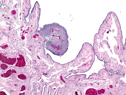
Low magnification. H&E stain.
(→IHC: add image :-)) |
|||
| (2 intermediate revisions by the same user not shown) | |||
| Line 115: | Line 115: | ||
| cd56= | | cd56= | ||
| cd57= | | cd57= | ||
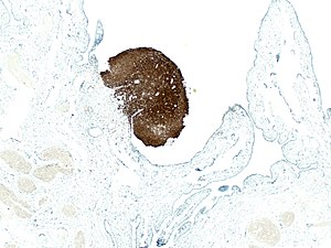
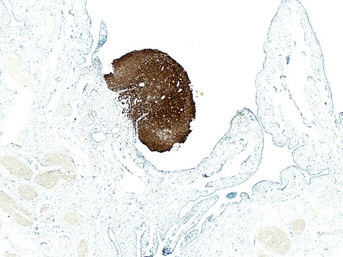
| cd68= | | cd68= <br>[[Image:Nodular histiocytic hyperplasia in fallopian tube 4.jpg|link=|thumb|CD68 IHC]] | ||
| cd7= | | cd7= | ||
| cd79a= | | cd79a= | ||
| Line 273: | Line 273: | ||
===Diagnosis=== | ===Diagnosis=== | ||
{{hidden|Diagnosis|<center>NODULAR HISTIOCYTIC HYPERPLASIA IN THE FALLOPIAN TUBE | {{hidden|Diagnosis|<center>NODULAR HISTIOCYTIC HYPERPLASIA IN THE FALLOPIAN TUBE</center> | ||
<br> | <br> | ||
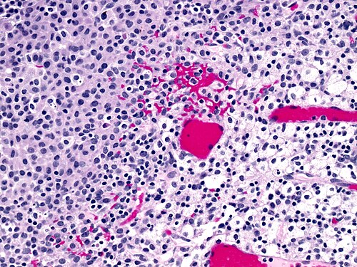
Originally called nodular histiocytic and mesothelial hyperplasia, it was reported in [[hernia sac]]s and was believed to be of predominant mesothelial cells. The advent of [[immunohistochemistry]] indicated that the vast majority of the cells are histiocytes. In the gynecologic tract it is reported in the [[endometrium]] also, where it can simulate neoplasia.<ref name=pmid24300536>{{Cite journal | last1 = Parkash | first1 = V. | last2 = Domfeh | first2 = AB. | last3 = Fadare | first3 = O. | title = Nodular histiocytic aggregates in the endometrium: a report of 7 cases. | journal = Int J Gynecol Pathol | volume = 33 | issue = 1 | pages = 52-7 | month = Jan | year = 2014 | doi = 10.1097/PGP.0b013e3182894365 | PMID = 24300536 }}</ref> | |||
===References=== | |||
{{Reflist|1}} | |||
}} | |||
<br> | |||
===Other cases=== | ===Other cases=== | ||
{{Cases navigation}} | {{Cases navigation}} | ||
| Line 282: | Line 289: | ||
[[Category:Cases]] | [[Category:Cases]] | ||
[[Category:Cases in gynecologic pathology]] | [[Category:Cases in gynecologic pathology]] | ||
- | [[Category:Cases in gynecologic pathology - senior]] | ||
[[Category:Cases difficulty 4]] <!-- difficulty 1-7 -- should roughly correspond to the PGY level --> | [[Category:Cases difficulty 4]] <!-- difficulty 1-7 -- should roughly correspond to the PGY level --> | ||
Latest revision as of 03:52, 27 September 2015
Provided clinical history
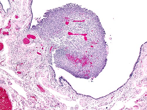
54 year old woman, incidental lesion discovered at time of risk reducing salpingooophorectomy
Site
Fallopian tube
Primary image

Intermediate magnification
|
|---|
 |
High magnification
|
|---|
 |
High magnification
|
|---|
 |
Differential diagnosis
Differential diagnosis
|
|---|
|
|
Additional tests
More history
More history
|
|---|
|
|
Ask a colleague
Ask a colleague
|
|---|
|
|
Stains
|
|
|
|
|
|
|---|
IHC
|
|
|
|
|
|
|---|
Molecular testing
Chromosomal translocations
|
|
|
|
|---|
Other molecular tests
|
|
|
|
|---|
Diagnosis
Diagnosis
|
|---|
|
References
|
