P16
Jump to navigation
Jump to search

p16 is a commonly used immunostain. It can be considered a surrogate marker for HPV infection. p16, like most other "p" stains, is a nuclear stain. The antibody target is a cell cycle protein, cyclin-dependent kinase inhibitor 2A, sometimes denoted p16INK4a.
| P16 | |
|---|---|
| Immunostain in short | |
 HSIL showing the characteristic p16 staining. (WC/Nephron) | |
| Similar stains | HPV |
| Use | HSIL versus LSIL, HPV associated SCC versus non-HPV associated SCC |
| Subspeciality | gynecologic pathology, head and neck pathology |
| Normal staining pattern | nuclear and cytoplasmic |
| Positive | endometrial tubal metaplasia, cervical SCC, HPV-associated head and neck SCC, serous carcinoma of the endometrium |

Endocervical AIS showing the characteristic p16 staining.
Pattern
- Nuclear stain +/- cytoplasmic staining.
Use
- Squamous lesions of the uterine cervix - see HSIL.
- Head and neck squamous cell carcinoma, specifically human papillomavirus-associated head and neck squamous cell carcinoma.
- Increased expression in well-differentiated liposarcoma.[1]
- Limited use in melanocytic lesions.[2]
Head and neck squamous cell carcinoma
p16 testing is useful in:
- Lymph node metastases with an unknown primary - positivity suggests an oropharyngeal primary.
- Oropharyngeal carcinomas.
Note:
- Like elsewhere, i.e. other anatomical sites, p16 is an imperfect surrogate marker for the presence of HPV.[3]
- Non-oropharyngeal sites (oral cavity, larynx, and hypopharynx) are not well-studied; however, it is known that p16 positivity is much less common in there.[3]
Images
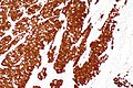
c/w HPV-assoc. SCC - p16 - intermed. mag. (WC/Nephron)
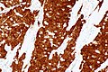
c/w HPV-assoc. SCC - p16 - high mag. (WC/Nephron)


Penile cancer
- p16 is considered surrogate for HPV and block-type positivity is considered indicative of HPV.[4]
- A meta-analysis from 2018 shows that various p16 staining criteria have been used in the past.[5]
The p16 staining is patchy (~10% of tumour) and moderate.  The 2022 WHO Classification describes "block-type positivity" as being relevant; as such, the case is interpreted as p16 NEGATIVE.

HPV testing will be requested and the findings reported in an addendum.
Positive
- Squamous cell carcinoma - esp. cervical SCC, anal SCC, penile SCC, HPV-associated head and neck SCC.
- High grade urothelial carcinoma ~86% of cases by PCR.[6]
- Serous carcinoma of the endometrium - should be strong.[7]
- High-grade squamous intraepithelial lesion - full thickness, strong.
- A subset of LSIL stains with p16; however, it is not full thickness - see HSIL article.
- Small cell carcinoma of the lung - most cases (95 of 101 cases in one series[8]).
Note:
- Positive staining (in the head and neck pathology context) is defined as (strong block) positive staining in >75% (or >50%) of lesional cells.[9]
- Staining varies somewhat by the p16 clone used.
Benign
- p16 endometrial tubal metaplasia.[10]
Focal staining
- Endometriosis - focal/weak staining may be seen.[11][12]
Negative
- Lung squamous cell carcinoma[13] - 21% positive (7/33).[14]
References
- ↑ Thway, K.; Flora, R.; Shah, C.; Olmos, D.; Fisher, C. (Mar 2012). "Diagnostic utility of p16, CDK4, and MDM2 as an immunohistochemical panel in distinguishing well-differentiated and dedifferentiated liposarcomas from other adipocytic tumors.". Am J Surg Pathol 36 (3): 462-9. doi:10.1097/PAS.0b013e3182417330. PMID 22301498.
- ↑ Koh, SS.; Cassarino, DS. (Jul 2018). "Immunohistochemical Expression of p16 in Melanocytic Lesions: An Updated Review and Meta-analysis.". Arch Pathol Lab Med 142 (7): 815-828. doi:10.5858/arpa.2017-0435-RA. PMID 29939777.
- ↑ 3.0 3.1 Stephen, JK.; Divine, G.; Chen, KM.; Chitale, D.; Havard, S.; Worsham, MJ. (2013). "Significance of p16 in Site-specific HPV Positive and HPV Negative Head and Neck Squamous Cell Carcinoma.". Cancer Clin Oncol 2 (1): 51-61. doi:10.5539/cco.v2n1p51. PMID 23935769.
- ↑ Menon S, Moch H, Berney DM, Cree IA, Srigley JR, Tsuzuki T, Compérat E, Hartmann A, Netto G, Rubin MA, Gill AJ, Turajlic S, Tan PH, Raspollini MR, Tickoo SK, Amin MB (March 2023). "WHO 2022 classification of penile and scrotal cancers: updates and evolution". Histopathology 82 (4): 508–520. doi:10.1111/his.14824. PMID 36221864.
- ↑ Sand FL, Rasmussen CL, Frederiksen MH, Andersen KK, Kjaer SK (October 2018). "Prognostic Significance of HPV and p16 Status in Men Diagnosed with Penile Cancer: A Systematic Review and Meta-analysis". Cancer Epidemiol Biomarkers Prev 27 (10): 1123–1132. doi:10.1158/1055-9965.EPI-18-0322. PMID 29987099.
- ↑ Piaton, E.; Casalegno, JS.; Advenier, AS.; Decaussin-Petrucci, M.; Mege-Lechevallier, F.; Ruffion, A.; Mekki, Y. (Oct 2014). "p16(INK4a) overexpression is not linked to oncogenic human papillomaviruses in patients with high-grade urothelial cancer cells.". Cancer Cytopathol 122 (10): 760-9. doi:10.1002/cncy.21462. PMID 25069600.
- ↑ Chiesa-Vottero, AG.; Malpica, A.; Deavers, MT.; Broaddus, R.; Nuovo, GJ.; Silva, EG. (Jul 2007). "Immunohistochemical overexpression of p16 and p53 in uterine serous carcinoma and ovarian high-grade serous carcinoma.". Int J Gynecol Pathol 26 (3): 328-33. doi:10.1097/01.pgp.0000235065.31301.3e. PMID 17581420.
- ↑ Švajdler M, Mezencev R, Ondič O, Šašková B, Mukenšnábl P, Michal M (April 2018). "P16 is a useful supplemental diagnostic marker of pulmonary small cell carcinoma in small biopsies and cytology specimens". Ann Diagn Pathol 33: 23–29. doi:10.1016/j.anndiagpath.2017.11.008. PMID 29566943.
- ↑ Shelton J, Purgina BM, Cipriani NA, Dupont WD, Plummer D, Lewis JS (September 2017). "p16 immunohistochemistry in oropharyngeal squamous cell carcinoma: a comparison of antibody clones using patient outcomes and high-risk human papillomavirus RNA status". Mod Pathol 30 (9): 1194–1203. doi:10.1038/modpathol.2017.31. PMID 28621317.
- ↑ Horree, N.; Heintz, AP.; Sie-Go, DM.; van Diest, PJ. (2007). "p16 is consistently expressed in endometrial tubal metaplasia.". Cell Oncol 29 (1): 37-45. PMID 17429140.
- ↑ Stewart, CJ.; Bharat, C. (Feb 2016). "Clinicopathological and immunohistological features of polypoid endometriosis.". Histopathology 68 (3): 398-404. doi:10.1111/his.12755. PMID 26095917.
- ↑ O'Neill, CJ.; McCluggage, WG. (Jan 2006). "p16 expression in the female genital tract and its value in diagnosis.". Adv Anat Pathol 13 (1): 8-15. doi:10.1097/01.pap.0000201828.92719.f3. PMID 16462152.
- ↑ Pereira, TC.; Share, SM.; Magalhães, AV.; Silverman, JF. (Jan 2011). "Can we tell the site of origin of metastatic squamous cell carcinoma? An immunohistochemical tissue microarray study of 194 cases.". Appl Immunohistochem Mol Morphol 19 (1): 10-4. doi:10.1097/PAI.0b013e3181ecaf1c. PMID 20823766.
- ↑ Wang, CW.; Wu, TI.; Yu, CT.; Wu, YC.; Teng, YH.; Chin, SY.; Lai, CH.; Chen, TC. (May 2009). "Usefulness of p16 for differentiating primary pulmonary squamous cell carcinoma from cervical squamous cell carcinoma metastatic to the lung.". Am J Clin Pathol 131 (5): 715-22. doi:10.1309/AJCPTPBC6V5KUITM. PMID 19369633.