Ductal carcinoma in situ
Ductal carcinoma in situ, abbreviated DCIS, in a common type of non-invasive breast carcinoma.
General
- Diagnosis based on nuclear abnormalities and/or architecture.
- Low-grade DCIS does not have a malignant cytology.
- It is typically picked-up during radiologic screening.
Microscopic
Features:
- Architectural changes:
- Equal spacing of cells - "cookie cutter" look.
- Cells line-up along lumen/glandular spaces - form "Roman briges".
- Architecture suggestive of DCIS - see Subtypes of DCIS.
- Nuclear changes:
- Nuclear enlargement - at least 2-3x size of RBC - key feature.
- Compared to RBCs to grade DCIS - see Grading DCIS.
- Compare sizes of nuclei if you cannot find RBCs.
- Compared to RBCs to grade DCIS - see Grading DCIS.
- Nuclear pleomorphism - important feature.
- Nuclear enlargement - at least 2-3x size of RBC - key feature.
- +/-Mitoses.
Note:
- Apocrine changes of cytoplasm -- several sets of criteria exist -- any of the following:
Subtypes of DCIS
The subtypes are based on architecture.
Note:
- Comedonecrosis used to be considered a separate subtype. Necrosis is seen most often in the context of solid ductal carcinoma in situ.
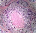
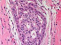
Solid ductal carcinoma in situ
Features:
- Sheet of cells fills the duct
- No spaces between cells.
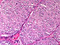
Breast - Ductal carcinoma in situ - Solid variant- Intermediate grade - Medium power (SKB)
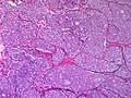
Breast - Ductal carcinoma in situ - Solid variant- Intermediate grade - Low power (SKB)
Breast - Ductal carcinoma in situ - Solid variant - Medium power (SKB)
Breast - Ductal carcinoma in situ - Solid variant - Comedonecrosis (SKB)
Breast - Ductal carcinoma in situ - Solid variant - Comedonecrosis (SKB)


DDx:
- LCIS.
- May show dyscohesion
- More monomorphic population of cells
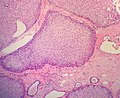
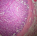
Cribriform ductal carcinoma in situ
Features:
- Honeycomb-like appearance: circular holes.
- "Cookie cutter" appearance/"punched-out" appearance/"Roman bridges" -- cells surround the circular holes.
Breast - Ductal carcinoma in situ - cribriform varient - medium power (SKB)
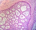
Breast - Ductal carcinoma in situ - cribriform varient - medium power (SKB)


DDx:
- Collagenous spherulosis.
- Adenoid cystic carcinoma of the breast.
- Invasive cribriform carcinoma of the breast
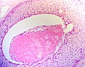
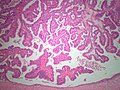
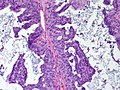
Papillary ductal carcinoma in situ
Features:
- Papillae with fibrovascular cores.
- Papillae lack a myoepithelial layer
- Papillae are lined by atypical cells.
- Papillae within a ductal space lined by myoepithelial cells.
Breast - Ductal carcinoma in situ - Papillary variant - low power (SKB)
Breast - Ductal carcinoma in situ - Papillary variant - Medium power (SKB)


DDX:
- Intraductal papilloma
- Ductal carcinoma in situ arising within an intraductal papilloma
- Intracystic papillary breast carcinoma
- Invasive papillary breast carcinoma
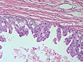
Micropapillary ductal carcinoma in situ
Features:
- Small papillae without fibrovascular cores.
- Have "drum stick" shape.
DDx:
Breast - Ductal carcinoma in situ - micropapillary variant - Medium power - (SKB)
Breast - Ductal carcinoma in situ - micropapillary variant - High power - (SKB)
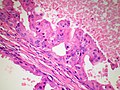
Breast - Ductal carcinoma in situ - Micropapillary type with apocrine features - High power - (SKB)


Grading DCIS
Graded 1-3 (low-high)[3] - compare lesional nuclei to one another.
- Grade 1:
- Nuclei 2-3x size of RBC.
- No necrosis.
- Grade 2:
- Nuclei 2-3x size of RBC.
- +/-Necrosis.
- Grade 3:
- Nuclei >3x size of RBC.
- Necrosis usually present.
Notes:
- It is often hard to find RBCs when you want 'em. DCIS is pleomorphic.
- If no RBCs are present to compare with compare the nuclei to one another.
- If you see nuclei >3x larger than their neigbour you're ready to call DCIS Grade 3.
Size criteria for low-grade DCIS
ADH is diagnosed if the lesion is small - specifically:[4][5]
- < Two membrane-bound spaces.
- < 2 mm extent. ‡
The treatment is similar; ADH and DCIS are both excised.
The differences are:
- DCIS is cancer, i.e. this has life insurance implications.
- Radiation treatment - DCIS is irradiated; ADH does not get radiation.
Notes:
- ‡ 3 mm is used in papillary lesions.[citation needed]
Micrometastasis in DCIS
See also
References
- ↑ URL: http://surgpathcriteria.stanford.edu/breast/dcis/apocrinedcis.html. Accessed on: 4 August 2011.
- ↑ 2.0 2.1 O'Malley, FP.; Bane, A. (Jan 2008). "An update on apocrine lesions of the breast.". Histopathology 52 (1): 3-10. doi:10.1111/j.1365-2559.2007.02888.x. PMID 18171412.
- ↑ URL: http://surgpathcriteria.stanford.edu/breast/dcis/. Accessed on: 4 August 2011.
- ↑ O'Malley, Frances P.; Pinder, Sarah E. (2006). Breast Pathology: A Volume in Foundations in Diagnostic Pathology series (1st ed.). Churchill Livingstone. pp. 168. ISBN 978-0443066801.
- ↑ Tadrous, Paul.J. Diagnostic Criteria Handbook in Histopathology: A Surgical Pathology Vade Mecum (1st ed.). Wiley. pp. 258. ISBN 978-0470519035.
- ↑ Lara, JF.; Young, SM.; Velilla, RE.; Santoro, EJ.; Templeton, SF. (Nov 2003). "The relevance of occult axillary micrometastasis in ductal carcinoma in situ: a clinicopathologic study with long-term follow-up.". Cancer 98 (10): 2105-13. doi:10.1002/cncr.11761. PMID 14601079.
- ↑ Broekhuizen, LN.; Wijsman, JH.; Peterse, JL.; Rutgers, EJ. (Jun 2006). "The incidence and significance of micrometastases in lymph nodes of patients with ductal carcinoma in situ and T1a carcinoma of the breast.". Eur J Surg Oncol 32 (5): 502-6. doi:10.1016/j.ejso.2006.02.006. PMID 16569492.