Small cell carcinoma of the lung
Jump to navigation
Jump to search
| Small cell carcinoma of the lung | |
|---|---|
| Diagnosis in short | |
_by_core_needle_biopsy.jpg) Lung small cell carcinoma. H&E stain. | |
|
| |
| LM | stippled chromatin, high NC ratio with scant basophilic cytoplasm, typically small cells (~2x RBC diameter), +/-nuclear moulding, nuclei with smudgy appearance (Azzopardi phenomenon), necrosis, mitoses |
| Subtypes | large cell neuroendocrine carcinoma (LCNEC) |
| LM DDx | poorly differentiated adenocarcinoma of the lung, atypical carcinoid, lung carcinoid, metastatic small cell carcinoma, lymphoma, other small round blue cell tumours |
| Stains | chromogranin +ve, synaptophysin +ve, CD56 +ve, NSE +ve, TTF-1 +ve |
| Staging | lung cancer staging |
| Site | lung - see lung tumours |
|
| |
| Clinical history | smoking - usually a long history, heavy |
| Signs | +/-hemoptysis |
| Prevalence | not common |
| Radiology | lung mass, usu. central location |
| Prognosis | poor |
| Clin. DDx | other lung tumours (squamous cell carcinoma of the lung), metastatic tumours |
| Treatment | medical (chemotherapy) |
Small cell carcinoma of the lung, also small cell lung carcinoma (abbreviated SCLC)[1] is an aggressive malignant tumour of the lung. It is strongly associated with smoking.
Small cell carcinoma in general is dealt with in the small cell carcinoma article.
General
- Strong association with smoking.
- Typically treated with chemotherapy.
- Poor prognosis.
On a spectrum of lesions (benign to malignant):[1]
- Tumourlet.
- Carcinoid.
- Atypical carcinoid.
- Small cell carinoma/large cell neuroendocrine carcinoma (LCNEC).
Precursor lesion - uncommonly seen:
- Pulmonary neuroendocrine cell hyperplasia.[1]
Clinical:
- +/-Hemoptysis.
Gross
- Central location (close to large airways) - typical.
- Necrosis.
Images
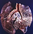
Small cell carcinoma of the lung - centre of image. (WC/Rosen)
.jpg)
Microscopic
Features:
- Stippled chromatin.
- High NC ratio, scant basophilic cytoplasm.
- Typically small cells ~2x RBC diameter.
- +/-Nuclear moulding.
- Nuclei with a smudgy appearance (Azzopardi phenomenon).
- Necrosis.
- Mitoses.
Notes:
- There should be no nucleolus.
DDx:
- Poorly differentiated adenocarcinoma of the lung.
- Metastatic small cell carcinoma.
- Lymphoma.
- Atypical carcinoid.
- Basaloid squamous cell carcinoma of the lung.[2]
- Other small round blue cell tumours.
Subtypes
- Large cell neuroendocrine carcinoma (LCNEC).
Images
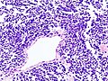
SCLC - low mag. (WC)
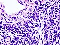
SCLC - high mag. (WC)
_by_core_needle_biopsy.jpg)
IHC
Sign out
Lung, Left Lower Lobe, Core Biopsy:
- SMALL CELL CARCINOMA.
Block letters
LOWER LOBE OF LUNG, LEFT, CORE BIOPSY:
- SMALL CELL CARCINOMA.
See also
References
- ↑ 1.0 1.1 1.2 Travis, WD. (Oct 2010). "Advances in neuroendocrine lung tumors.". Ann Oncol 21 Suppl 7: vii65-71. doi:10.1093/annonc/mdq380. PMID 20943645.
- ↑ Maleki, Z. (Mar 2011). "Diagnostic issues with cytopathologic interpretation of lung neoplasms displaying high-grade basaloid or neuroendocrine morphology.". Diagn Cytopathol 39 (3): 159-67. doi:10.1002/dc.21351. PMID 21319315.
- ↑ Wu, M.; Szporn, AH.; Zhang, D.; Wasserman, P.; Gan, L.; Miller, L.; Burstein, DE. (Oct 2005). "Cytology applications of p63 and TTF-1 immunostaining in differential diagnosis of lung cancers.". Diagn Cytopathol 33 (4): 223-7. doi:10.1002/dc.20337. PMID 16138374.
- ↑ 4.0 4.1 Gyure, KA.; Morrison, AL. (Jun 2000). "Cytokeratin 7 and 20 expression in choroid plexus tumors: utility in differentiating these neoplasms from metastatic carcinomas.". Mod Pathol 13 (6): 638-43. doi:10.1038/modpathol.3880111. PMID 10874668.