Difference between revisions of "Non-malignant skin disease"
(→Syringoma: more) |
|||
| (443 intermediate revisions by the same user not shown) | |||
| Line 1: | Line 1: | ||
'''Non-malignant skin disease''' is relatively common. The pathology may or may not be specific. Some diseases require clinical information to diagnose. | '''Non-malignant skin disease''' is relatively common. The pathology may or may not be specific. Some diseases require clinical information to diagnose. | ||
An introduction to dermatopathology is in the ''[[dermatopathology]]'' article. | An introduction to dermatopathology is in the ''[[dermatopathology]]'' article. [[Nevi]] (moles) and other melanocytic lesions are dealt with in the article ''[[melanocytic lesions]]''. Inflammatory skin conditions are dealt with in ''[[inflammatory skin disorders]]''. | ||
== | =Other= | ||
==Lichen simplex chronicus== | |||
{{Main|Lichen simplex chronicus}} | |||
''' | ==Prurigo nodularis== | ||
*Abbreviated ''PN''. | |||
*[[AKA]] ''chronic prurigo'' and ''picker nodule''.<ref name=Ref_Derm26>{{Ref Derm|26}}</ref> | |||
===General=== | |||
*Can be thought of as a localized/focal version of [[lichen simplex chronicus]] (LSC). | |||
==Lichen | ===Gross=== | ||
*Dome-shaped/raised - papular (<1 cm) ''or'' nodular (>1 cm).<ref>URL: [http://www.pediatrics.wisc.edu/education/derm/text.html http://www.pediatrics.wisc.edu/education/derm/text.html]. Accessed on: 23 August 2012.</ref> | |||
===Microscopic=== | |||
*See ''[[lichen simplex chronicus]]''. | |||
DDx: | |||
*[[Lichen simplex chronicus]] - a more diffuse process, not a raised lesion. | |||
===Sign out=== | |||
<pre> | |||
SKIN LESION, LEFT CHIN, BIOPSY: | |||
- PRURIGO NODULARIS. | |||
</pre> | |||
====Micro==== | |||
The sections show a raised lesion with compact hyperkeratosis and irregular acanthosis. Spongiosis is seen focally. There is minimal hypergranulosis. | |||
There is no thinning of the suprapapillary plate and no dilated superficial blood vessels. There is no interface activity. | |||
=Very common= | |||
==Dermatomycosis== | |||
:''Dermatophytosis'' redirects here. | |||
===General=== | ===General=== | ||
* | *[[microorganisms|Fungal infection]] of skin. | ||
* | |||
Note: | |||
*''Dermatophytosis'' (ring worm) is a type of dermatomycosis. | |||
===Microscopic=== | |||
Features: | |||
*Microorganisms - '''key feature'''. | |||
**Often hyphae (candida) - like twigs of a tree... branching. | |||
***May be very fragmented in section ~ size of a neutrophil. | |||
*Perivascular inflammation, esp. neutrophils. | |||
*[[Exocytosis]] - blood cell infiltrate the epidermis. | |||
====Images==== | |||
<gallery> | |||
Image:Dermatomycosis_-_intermed_mag.jpg | Dermatomycosis - intermed. mag. (WC) | |||
Image:Dermatomycosis_-_high_mag.jpg | Dermatomycosis - high mag. (WC) | |||
Image:Dermatomycosis_-_gms_-_low_mag.jpg | Dermatomycosis - GMS stain - low mag. (WC) | |||
Image:Dermatomycosis_-_gms_-_high_mag.jpg | Dermatomycosis - GMS stain - high mag. (WC) | |||
</gallery> | |||
www: | |||
*[http://missinglink.ucsf.edu/lm/DermatologyGlossary/img/Dermatology%20Glossary/Glossary%20Histo%20Images/tinea_pas.jpg Dermatophytosis (ucsf.edu)].<ref>URL: [http://missinglink.ucsf.edu/lm/DermatologyGlossary/tinea.html http://missinglink.ucsf.edu/lm/DermatologyGlossary/tinea.html]. Accessed on: 25 February 2013.</ref> | |||
===Stains=== | |||
*[[GMS stain]]. | |||
*[[PAS-D stain]]. | |||
===Sign out=== | |||
<pre> | |||
SKIN, BIOPSY: | |||
- SKIN WITH SUPERFICIAL FUNGAL ORGANISMS CONSISTENT WITH CANDIDA. | |||
- REACTIVE CHANGES OF THE EPITHELIUM. | |||
</pre> | |||
====Micro==== | |||
The sections show skin with a neutrophilic infiltrate in the superficial epidermis. PAS-D staining demonstrates fungal organisms with a morphology suggestive of candida. | |||
The epithelium has parakeratosis, acanthosis and spongiosis. No mitotic activity is appreciated. The keratinocytes are moderately enlarged and have evident nucleoli. | |||
==Cicatrix== | |||
{{Main|Dermal scar}} | |||
==Fibroepithelial polyp== | |||
{{Main|Fibroepithelial polyp}} | |||
==Actinic keratosis== | |||
{{Main|Actinic keratosis}} | |||
== | ==Actinic cheilitis== | ||
*[[ | ===General=== | ||
*[[Actinic keratosis]] of the lip.<ref name=pmid3305604>{{Cite journal | last1 = Picascia | first1 = DD. | last2 = Robinson | first2 = JK. | title = Actinic cheilitis: a review of the etiology, differential diagnosis, and treatment. | journal = J Am Acad Dermatol | volume = 17 | issue = 2 Pt 1 | pages = 255-64 | month = Aug | year = 1987 | doi = | PMID = 3305604 }}</ref> | |||
===Microscopic=== | ===Microscopic=== | ||
:See ''[[actinic keratosis]]''. | |||
== | ===Sign out=== | ||
<pre> | |||
LESION, LOWER LIP, BIOPSY: | |||
- ACTINIC CHEILITIS. | |||
- SOLAR ELASTOSIS. | |||
</pre> | |||
====Micro==== | |||
The sections show skin with moderate basal nuclear hyperchromasia and atypia, and parakeratosis. The squamous epithelium has maturation to the surface. There is no inflammation at the dermal-epidermal interface. Solar elastosis is present. | |||
==Seborrheic keratosis== | |||
{{Main|Seborrheic keratosis}} | |||
==Pilomatricoma== | |||
{{Main|Pilomatricoma}} | |||
==Dermatofibroma== | |||
{{Main|Dermatofibroma}} | |||
==Ezcema== | |||
===General=== | ===General=== | ||
* | *A nebulous thingy. | ||
*Very common. | |||
DDx: | |||
* | *Contact allergy. | ||
*[[Drug reaction]]. | |||
*Food allergy. | |||
===Microscopic=== | ===Microscopic=== | ||
Features:<ref | Features:<ref>{{Ref PBoD8|1188}}</ref> | ||
*Spongiosis (epidermal edema); keratinocytes spacing increased - '''key feature'''. | |||
*+/-Interdermal vesicles. | |||
*+/-Eosinophils (may suggest Rx reaction). | |||
*Spongiosis (epidermal | *Perivascular lymphocytes. | ||
* | |||
* | |||
* | |||
== | ==Acne vulgaris== | ||
===General=== | ===General=== | ||
* | *Extremely common - esp. among adolescents. | ||
* | *Very rarely seen by pathologists. | ||
Treatments: | |||
* | *Antibiotic (minocycline). | ||
*Isotretinoin [[AKA]] all-trans retinoic acid (ATRA). | |||
===Gross=== | ===Gross=== | ||
* | *Papules, pustules, nodules or cysts. | ||
**White, black or erythematous. | |||
Images: | |||
*[http:// | *[http://commons.wikimedia.org/wiki/File:Blackheads.JPG Blackheads (WC)]. | ||
===Microscopic=== | ===Microscopic=== | ||
Features: | Features:<ref>{{Ref Derm|76}}</ref> | ||
* | *Folliculitis:<ref name=Ref_Derm77>{{Ref Derm|77}}</ref> | ||
**[[Neutrophil]]s around hair follicle and infiltrate into it - including the follicular canal. | |||
*Epidermal invagination ''or'' cyst at site of a hair follicle - contains: | |||
**Sebum. | |||
**+/-Bacteria (''Propionibacterium acnes'') and inflammatory cells - typically neurophils. | |||
Subtyped into: | |||
#Open comedones ("blackheads") - no extension to epidermal surface. | |||
#Closed comedones ("whiteheads") - to epidermal surface have wide opening. | |||
DDx - acneiform disorder:<ref name=Ref_Derm77>{{Ref Derm|77}}</ref> | |||
*[[Rosacea]]. | |||
*Infective folliculitis. | |||
*Perioral dermatitis. | |||
*Acne vulgaris. | |||
Image | Image: | ||
*[http://www.dermnet.com/images/Acne-Histology/picture/4069 Acne (dermnet.com)]. | |||
*[http://www. | |||
== | ==Solar elastosis== | ||
*[[AKA]] ''actinic elastosis''.<ref>URL: [http://www.dermnetnz.org/dermal-infiltrative/solar-elastosis.html http://www.dermnetnz.org/dermal-infiltrative/solar-elastosis.html]. Accessed on: 27 March 2013.</ref> | |||
===General=== | ===General=== | ||
* | *Very common. | ||
* | *Caused by sun exposure - specifically UV light.<ref name=pmid20802019>{{Cite journal | last1 = Thomas | first1 = NE. | last2 = Kricker | first2 = A. | last3 = From | first3 = L. | last4 = Busam | first4 = K. | last5 = Millikan | first5 = RC. | last6 = Ritchey | first6 = ME. | last7 = Armstrong | first7 = BK. | last8 = Lee-Taylor | first8 = J. | last9 = Marrett | first9 = LD. | title = Associations of cumulative sun exposure and phenotypic characteristics with histologic solar elastosis. | journal = Cancer Epidemiol Biomarkers Prev | volume = 19 | issue = 11 | pages = 2932-41 | month = Nov | year = 2010 | doi = 10.1158/1055-9965.EPI-10-0686 | PMID = 20802019 }}</ref> | ||
**Severity correlated with cumulative exposure to UV light..<ref name=pmid17204514/> | |||
*Often co-localized with skin cancers - as UV light is risk factor for skin cancers.<ref name=pmid17204514 >{{Cite journal | last1 = Karagas | first1 = MR. | last2 = Zens | first2 = MS. | last3 = Nelson | first3 = HH. | last4 = Mabuchi | first4 = K. | last5 = Perry | first5 = AE. | last6 = Stukel | first6 = TA. | last7 = Mott | first7 = LA. | last8 = Andrew | first8 = AS. | last9 = Applebaum | first9 = KM. | title = Measures of cumulative exposure from a standardized sun exposure history questionnaire: a comparison with histologic assessment of solar skin damage. | journal = Am J Epidemiol | volume = 165 | issue = 6 | pages = 719-26 | month = Mar | year = 2007 | doi = 10.1093/aje/kwk055 | PMID = 17204514 }}</ref> | |||
*Benign. | |||
===Microscopic=== | ===Microscopic=== | ||
Features: | Features: | ||
* | *Grey, spaghetti-like material in the superficial dermis. | ||
* | |||
* | DDx: | ||
* | *[[Actinic keratosis]]. | ||
*[[Basal cell carcinoma]]. | |||
*[[Squamous cell carcinoma of the skin|Squamous cell carcinoma]]. | |||
Note: | |||
*The DDx above is things associated with sun damaged skin. | |||
*Dermal mucin (as my be seen in [[lupus erythematosus]]) is a possible mimic - but it isn't spaghetti-like and the "background" (an [[interface dermatitis]]) is different. | |||
== | ====Images==== | ||
* | <gallery> | ||
Image:Solar_elastosis_-_intermed_mag.jpg | Solar elastosis - intermed. mag. (WC) | |||
Image:Solar_elastosis_-_high_mag.jpg | Solar elastosis - high mag. (WC) | |||
</gallery> | |||
www: | |||
*[http://dermpathexpert.com/id88.html Solar elastosis - several images (dermpathexpert.com)]. | |||
===Sign out=== | |||
<pre> | |||
SKIN, RIGHT CHEEK, RE-EXCISION: | |||
- DERMAL SCAR. | |||
- EXTENSIVE SOLAR ELASTOSIS. | |||
</pre> | |||
=====Prominent blood vessels===== | |||
<pre> | |||
SKIN LESION, LEFT CHEEK, BIOPSY: | |||
- SKIN WITH SOLAR ELASTOSIS AND PROMINENT SMALL BLOOD VESSELS. | |||
</pre> | |||
<pre> | |||
SUPERIOR SHOULDER, LEFT, PUNCH BIOPSY: | |||
- BENIGN SKIN WITH MODERATE SOLAR ELASTOSIS, PROMINENT SMALL BLOOD VESSELS AND | |||
SCATTERED PERIVASCULAR LYMPHOCYTES AND PLASMA CELLS. | |||
- NEGATIVE FOR BASAL CELL CARCINOMA. | |||
- NEGATIVE FOR ACTINIC KERATOSIS. | |||
</pre> | |||
====Micro==== | |||
The sections show hair bearing skin with solar elastosis and numerous small dilated blood vessels. The dermis is mildly fibrotic. Compact keratin is present. | |||
The epidermis matures to the surface. A granular layer is present. There is no basal | |||
epidermal atypia. No melanocytic nests are identified. There is no palisading of the basal | |||
cells. Rare scattered lymphocytes are in the dermis. | |||
== | =Very common - viral= | ||
==Verruca vulgaris== | |||
{{Main|Verruca vulgaris}} | |||
==Verruca plana== | |||
===General=== | |||
*Common. | |||
*Usu. hands and face.<ref name=Ref_WMSP480>{{Ref WMSP|480}}</ref> | |||
===Microscopic=== | ===Microscopic=== | ||
Features:<ref> | Features:<ref name=Ref_WMSP480>{{Ref WMSP|480}}</ref> | ||
* | *Orthokeratosis with basketweave pattern. | ||
* | *Hypergranulosis. | ||
* | *Viral keratohyaline. | ||
* | *Koilocytes. | ||
* | *[[Acanthosis]] - yet flat surface and base. | ||
=== | Notes: | ||
* | *It differs from [[verruca vulgaris]]... (1) orthokeratosis, (2) flat surface and base. | ||
====Images==== | |||
* | www: | ||
*[http://www.fujita-hu.ac.jp/~tsutsumi/case/case180.htm Verruca plana - several images (fujita-hu.ac.jp)]. | |||
*[http://www.dermatopathonline.com/verruca%20plana2.html Verruca plana - several good images (dermatopathonline.com)]. | |||
== | =Less common= | ||
==Chronic folliculitis== | |||
:''Folliculitis'' redirect here. | |||
===General=== | ===General=== | ||
* | *Common. | ||
* | *Infrequently biopsied. | ||
===Gross=== | |||
* | *Erythema.<ref>URL: [http://www.webmd.com/skin-problems-and-treatments/tc/folliculitis-topic-overview http://www.webmd.com/skin-problems-and-treatments/tc/folliculitis-topic-overview]. Accessed on: 7 November 2012.</ref> | ||
DDx gross: | |||
* | *[[Melanocytic lesion]]s. | ||
===Microscopic=== | ===Microscopic=== | ||
Features: | Features: | ||
* | *Inflammation around the hair follicle - '''key feature'''. | ||
* | **Lymphocytes - usu. predominant. | ||
* | *+/-Chronic changes: | ||
* | **[[Acanthosis]]. | ||
** | **Hyperkeratosis. | ||
** | **Hypergranulosis. | ||
* | |||
DDx: | |||
*[[Acne vulgaris]]. | |||
===Sign out=== | |||
<pre> | |||
SKIN LESION, UPPER ARM, BIOPSY: | |||
- CHRONIC FOLLICULITIS WITH SECONDARY SURFACE CHANGES. | |||
</pre> | |||
====Micro==== | |||
The sections show hair-bearing skin with abundant lymphocytes around and within the hair follicle wall. | |||
The non-hair follicle epidermis has acanthosis, hypergranulosis and compact hyperkeratosis. There is no inflammatory cell infiltrate in the non-hair follicle epidermis or at the non-hair follicle interface. | |||
There are no granulomas. | |||
==Clear cell acanthoma== | |||
{{Main|Clear cell acanthoma}} | |||
== | ==Chondrodermatitis nodularis chronica helicis== | ||
* | *[[AKA]] ''chondrodermatitis nodularis helicis''. | ||
* | *Abbreviated ''CNCH''. | ||
* | *[[AKA]] ''Winkler disease''.<ref>URL: [http://www.head-face-med.com/content/4/1/2 http://www.head-face-med.com/content/4/1/2]. Accessed on: 16 January 2014.</ref> | ||
{{Main|Chondrodermatitis nodularis chronica helicis}} | |||
==Cutaneous calcinosis== | |||
*[ | *[[AKA]] ''calcinosis cutis''. | ||
{{Main|Cutaneous calcinosis}} | |||
== | ==Dilated pore of Winer== | ||
===General=== | ===General=== | ||
*Benign | *Benign. | ||
* | *Looks like a zit. | ||
===Microscopic=== | ===Microscopic=== | ||
Features: | Features:<ref name=Ref_WMSP486>{{Ref WMSP|486}}</ref> | ||
* | *Dilated hair follicle with keratin. | ||
*[[Acanthosis]]. | |||
*Budding of epidermis (into dermis). | |||
DDx: | |||
*[[Pilar sheath acanthoma]]. | |||
Image: | |||
*[http://dermpathexpert.com/id90.html Dilated pore of Winer (dermpathexpert.com)]. | |||
==Lichenoid keratosis== | |||
*[[AKA]] ''lichen planus-like keratosis''. | |||
{{Main|Lichenoid keratosis}} | |||
==Granuloma annulare== | |||
{{Main|Granuloma annulare}} | |||
== | ==Necrobiosis lipoidica== | ||
{{Main|Necrobiosis lipoidica}} | |||
=== | ==Keloid== | ||
{{Main|Keloid}} | |||
== | ==Angiofibroma== | ||
:See also: ''[[nasopharyngeal angiofibroma]]''. | |||
:Should '''not''' be confused with ''[[angiokeratoma]]''. | |||
{{Main|Angiofibroma}} | |||
==Benign fibrous papule== | |||
*[ | *[[AKA]] ''fibrous papule''. | ||
===General=== | ===General=== | ||
* | *An ''[[angiofibroma]]'' on the face that is solitary.<ref name=Ref_Derm505>{{Ref Derm|505}}</ref><ref name=pmid18032900>{{Cite journal | last1 = Jacyk | first1 = WK. | last2 = Rütten | first2 = A. | last3 = Requena | first3 = L. | title = Fibrous papule of the face with granular cells. | journal = Dermatology | volume = 216 | issue = 1 | pages = 56-9 | month = | year = 2008 | doi = 10.1159/000109359 | PMID = 18032900 }}</ref> | ||
* | |||
*Usually | ===Gross=== | ||
*Solitary lesion of the face - '''important'''.<ref name=Ref_Derm505>{{Ref Derm|505}}</ref> | |||
**Usually on the nose.<ref name=pmid18032900/> | |||
===Microscopic=== | ===Microscopic=== | ||
Features:<ref name=Ref_WMSP492>{{Ref WMSP|492}}</ref> | Features:<ref name=Ref_WMSP492>{{Ref WMSP|492}}</ref> | ||
* | *Dome-shaped. | ||
* | *Fibrotic dermis. | ||
** | **Enlarged fibroblasts. | ||
* | *Dilated small vessels. | ||
*+/- | *+/-Multinucleated stromal cells.<ref name=pmid543528>{{Cite journal | last1 = Ragaz | first1 = A. | last2 = Berezowsky | first2 = V. | title = Fibrous papule of the face. A study of five cases by electron microscopy. | journal = Am J Dermatopathol | volume = 1 | issue = 4 | pages = 353-6 | month = | year = 1979 | doi = | PMID = 543528 }}</ref> | ||
* | *+/-Stellate cells.<ref name=pmid543528/> | ||
DDx: | |||
*[ | *[[Angiofibroma]] - not solitary or not on the nose. | ||
*[http://www. | |||
Note: | |||
*Several variants exist.<ref name=pmid18032900>{{Cite journal | last1 = Jacyk | first1 = WK. | last2 = Rütten | first2 = A. | last3 = Requena | first3 = L. | title = Fibrous papule of the face with granular cells. | journal = Dermatology | volume = 216 | issue = 1 | pages = 56-9 | month = | year = 2008 | doi = 10.1159/000109359 | PMID = 18032900 }}</ref> | |||
===Images=== | |||
*[http://www.dermaamin.com/site/histopathology-of-the-skin/58-f/1739-fibrous-papule-angiofibroma-.html Fibrous papule (dermaamin.com)]. | |||
===Sign out=== | |||
<pre> | |||
SKIN LESION, CHIN, BIOPSY: | |||
- BENIGN FIBROUS PAPULE. | |||
</pre> | |||
==Molluscum contagiosum== | |||
{{Main|Molluscum contagiosum}} | |||
==Superficial dermal infiltrates== | ==Superficial dermal infiltrates== | ||
Discussed in detail by Alsaad and Ghazarian.<ref name=pmid16311340>{{Cite journal | last1 = Alsaad | first1 = KO. | last2 = Ghazarian | first2 = D. | title = My approach to superficial inflammatory dermatoses. | journal = J Clin Pathol | volume = 58 | issue = 12 | pages = 1233-41 | month = Dec | year = 2005 | doi = 10.1136/jcp.2005.027151 | PMID = 16311340 }}</ref> | Discussed in detail by Alsaad and Ghazarian.<ref name=pmid16311340>{{Cite journal | last1 = Alsaad | first1 = KO. | last2 = Ghazarian | first2 = D. | title = My approach to superficial inflammatory dermatoses. | journal = J Clin Pathol | volume = 58 | issue = 12 | pages = 1233-41 | month = Dec | year = 2005 | doi = 10.1136/jcp.2005.027151 | PMID = 16311340 }}</ref> | ||
===Dermal perivascular lymphoeosinophilic infiltration | ===Dermal perivascular lymphoeosinophilic infiltration=== | ||
*Microscopic appearance is just what it is called: | *Abbreviated ''DPLI''. | ||
Microscopic appearance is just what it is called: | |||
*Lymphocytes and eosinophils around the vessels in the superficial dermis. | |||
DDx:<ref name=pmid16311340/> | DDx:<ref name=pmid16311340/> | ||
*Insect bite - classically wedge-shaped.<ref name=Ref_PBoD1269>{{Ref PBoD|1269}}</ref> | *Insect bite - classically wedge-shaped.<ref name=Ref_PBoD1269>{{Ref PBoD|1269}}</ref> | ||
*Drug | *[[Drug reaction]]. | ||
*Urticarial | *Urticarial reaction. | ||
*Prevesicular early stage of [[bullous pemphigoid]]. | *Prevesicular early stage of [[bullous pemphigoid]]. | ||
*[[HIV]] related dermatoses. | *[[HIV]] related dermatoses. | ||
| Line 301: | Line 400: | ||
Gross: | Gross: | ||
*Brown or blue-grey patch in the lumbosacral area. | *Brown or blue-grey patch in the lumbosacral area. | ||
==Mastocytosis== | |||
{{Main|Mastocytosis}} | |||
==Ichthyosis== | |||
===General=== | |||
*Comes in different flavours. | |||
*Usu. inherited... thus a pediatric condition. | |||
===Gross=== | |||
*Fish scale-like appearance. | |||
Image: | |||
*[http://commons.wikimedia.org/wiki/File:Ichthyosis_1.jpg Ichtyosis (WC)]. | |||
===Microscopic=== | |||
Features:<ref name=Ref_PBoD8|1185>{{Ref PBoD8|1185}}</ref> | |||
*Thick stratum corneum without basket-weave pattern. | |||
==Palmar fibromatosis== | ==Palmar fibromatosis== | ||
*[[AKA]] ''Dupuytren's contracture''. | |||
*[[AKA]] ''Dupuytren disease''. | |||
{{Main|Palmar fibromatosis}} | |||
==Angiomyoma== | |||
*Do '''not''' confuse with ''[[aggressive angiomyxoma|angiomyxoma]]''. | |||
===General=== | ===General=== | ||
*[ | *Benign. | ||
*Female > male.<ref name=pmid3201831 >{{cite journal |author=Katenkamp D, Kosmehl H, Langbein L |title=[Angiomyoma. A pathologo-anatomic analysis of 229 cases] |language=German |journal=Zentralbl Allg Pathol |volume=134 |issue=4-5 |pages=423–33 |year=1988 |pmid=3201831 |doi= |url=}}</ref> | |||
Clinical:<ref>URL: [http://www. | ===Microscopic=== | ||
* | Features: | ||
* | *Well-circumscribed lesion with [[fascicular]] architecture. | ||
*Spindle cells/epithelioid cell with moderate eosinophilic (pink) cytoplasm. | |||
*Thick-walled blood vessels. (???) | |||
Images: | |||
*[http://commons.wikimedia.org/wiki/File:Cutaneous_angiomyoma_%281%29.jpg Angiomymoma (WC)]. | |||
*[http://commons.wikimedia.org/wiki/File:Cutaneous_angiomyoma_%282%29.jpg Angiomymoma (WC)]. | |||
==Angiokeratoma== | |||
{{Main|Angiokeratoma}} | |||
==Inverted follicular keratosis== | |||
*Abbreviated ''IFK''.<ref name=pmid11411260>{{Cite journal | last1 = Shih | first1 = CC. | last2 = Yu | first2 = HS. | last3 = Tung | first3 = YC. | last4 = Tsai | first4 = KB. | last5 = Cheng | first5 = ST. | title = Inverted follicular keratosis. | journal = Kaohsiung J Med Sci | volume = 17 | issue = 1 | pages = 50-4 | month = Jan | year = 2001 | doi = | PMID = 11411260 }}</ref> | |||
===General=== | |||
*Benign skin lesion. | |||
*Central face - middle age.<ref name=Ref_Derm387>{{Ref Derm|387}}</ref> | |||
*Uncommon. | |||
*May be considered a variant of ''[[seborrheic keratosis]]'' that is predominantly endophytic.<ref name=Ref_Derm341>{{Ref Derm|341}}</ref> | |||
Clinical DDx:<ref name=Ref_Derm387>{{Ref Derm|387}}</ref><ref>URL: [http://www.ncbi.nlm.nih.gov/pmc/articles/PMC475744/ http://www.ncbi.nlm.nih.gov/pmc/articles/PMC475744/]. Accessed on: 11 May 2010.</ref> | |||
*[[BCC]]. | |||
*[[Wart]]. | |||
===Microscopic=== | ===Microscopic=== | ||
Features:<ref> | Features:<ref name=Ref_Derm387>{{Ref Derm|387}}</ref> | ||
* | *Keratinocyte of cytologically benign proliferation. | ||
** | *"Squamous eddies" (whorls of keratin). | ||
* | *Coarse keratohyaline granules. | ||
* | |||
DDx: | |||
*[[Squamous cell carcinoma of the skin]]. | |||
*[[Trichilemmoma]]. | |||
*[[Seborrheic keratosis]] - has an exophytic component. | |||
Images: | Images: | ||
*[http://www. | *[http://www.flickr.com/photos/euthman/3059309003/ Inverted follicular keratosis - low mag. (flickr.com)]. | ||
*[http://www.flickr.com/photos/euthman/3060145702/ Inverted follicular keratosis - high mag. (flickr.com)]. | |||
*[http://mckeedermpath.com/SPOT%20DIAGNOSIS%20CASE%20474.html Inverted follicular keratosis - several images (mckeedermpath.com)]. | |||
===Sign out=== | |||
<pre> | |||
SKIN LESION, FACE, BIOPSY: | |||
- INVERTED FOLLICULAR KERATOSIS. | |||
</pre> | |||
====Micro==== | |||
The sections show skin with acanthosis, pseudohorn cysts, and focal basal epidermal pigmentation. There is no basal nuclear atypia, no mitoses and there are no melanocytic nests. There is minimal dermal inflammation. There is no apparent solar elastosis. | |||
==Focal cutaneous mucinosis== | |||
===General=== | |||
*Benign. | |||
*May be associated with systemic disease.<ref>{{Cite journal | last1 = Gandhi | first1 = V. | last2 = Dogra | first2 = D. | last3 = Pandhi | first3 = RK. | title = Cutaneous focal mucinosis. | journal = Indian J Dermatol Venereol Leprol | volume = 62 | issue = 4 | pages = 260-1 | month = | year = | doi = | PMID = 20948074 }}</ref> | |||
===Microscopic=== | |||
Features: | |||
*Light blue whispy material in the dermis - '''key feature'''. | |||
DDx: | |||
*Dermal edema. | |||
*[[Digital mucous cyst]]. | |||
==Panniculitis== | |||
{{Main|Panniculitis}} | |||
This is dealt with in the ''[[panniculitis]]'' article. | |||
DDx for panniculitis: | |||
*[[Erythema nodosum]]. | |||
*[[Erythema induratum]]. | |||
*Self-inflicted trauma. | |||
*[[Systemic lupus erythematosus]]. | |||
*Weird stuff. | |||
=Rare= | |||
==Necrotizing fasciitis== | |||
:'''''Not''' to be confused with [[nodular fasciitis]]''. | |||
*[[AKA]] ''flesh-eating disease''. | |||
{{Main|Necrotizing fasciitis}} | |||
==Porokeratosis== | |||
{{Main|Porokeratosis}} | |||
==Nevus sebaceous== | |||
*[[AKA]] ''nevus sebaceous of Jadassohn''. | |||
{{Main|Nevus sebaceous}} | |||
==Nevus lipomatosus superficialis== | |||
*Abbreviated ''NLS''. | |||
*[[AKA]] ''nevus lipomatosus cutaneous superficialis'', abbreviated ''NLCS''. | |||
*[[AKA]] ''nevus lipomatosus''.<ref name=pmid15677959>{{Cite journal | last1 = Kaw | first1 = P. | last2 = Carlson | first2 = A. | last3 = Meyer | first3 = DR. | title = Nevus lipomatosus (pedunculated lipofibroma) of the eyelid. | journal = Ophthal Plast Reconstr Surg | volume = 21 | issue = 1 | pages = 74-6 | month = Jan | year = 2005 | doi = | PMID = 15677959 }}</ref> | |||
{{Main|Nevus lipomatosus superficialis}} | |||
=Bullous disease= | |||
{{main|Bullous disease}} | {{main|Bullous disease}} | ||
=Cysts= | |||
{{main|Dermal cysts}} | {{main|Dermal cysts}} | ||
=See also= | |||
*[[Dermatopathology introduction]]. | *[[Dermatopathology introduction]]. | ||
*[[Malignant skin disease]]. | *[[Malignant skin disease]]. | ||
=References= | |||
{{reflist|2}} | {{reflist|2}} | ||
[[Category:Non-malignant skin disease]] | [[Category:Non-malignant skin disease]] | ||
[[Category:Dermatopathology]] | [[Category:Dermatopathology]] | ||
Revision as of 16:53, 2 June 2017
Non-malignant skin disease is relatively common. The pathology may or may not be specific. Some diseases require clinical information to diagnose.
An introduction to dermatopathology is in the dermatopathology article. Nevi (moles) and other melanocytic lesions are dealt with in the article melanocytic lesions. Inflammatory skin conditions are dealt with in inflammatory skin disorders.
Other
Lichen simplex chronicus
Prurigo nodularis
General
- Can be thought of as a localized/focal version of lichen simplex chronicus (LSC).
Gross
- Dome-shaped/raised - papular (<1 cm) or nodular (>1 cm).[2]
Microscopic
DDx:
- Lichen simplex chronicus - a more diffuse process, not a raised lesion.
Sign out
SKIN LESION, LEFT CHIN, BIOPSY:
- PRURIGO NODULARIS.
Micro
The sections show a raised lesion with compact hyperkeratosis and irregular acanthosis. Spongiosis is seen focally. There is minimal hypergranulosis.
There is no thinning of the suprapapillary plate and no dilated superficial blood vessels. There is no interface activity.
Very common
Dermatomycosis
- Dermatophytosis redirects here.
General
- Fungal infection of skin.
Note:
- Dermatophytosis (ring worm) is a type of dermatomycosis.
Microscopic
Features:
- Microorganisms - key feature.
- Often hyphae (candida) - like twigs of a tree... branching.
- May be very fragmented in section ~ size of a neutrophil.
- Often hyphae (candida) - like twigs of a tree... branching.
- Perivascular inflammation, esp. neutrophils.
- Exocytosis - blood cell infiltrate the epidermis.
Images
Dermatomycosis - intermed. mag. (WC)
Dermatomycosis - high mag. (WC)
Dermatomycosis - GMS stain - low mag. (WC)
Dermatomycosis - GMS stain - high mag. (WC)


www:
Stains
Sign out
SKIN, BIOPSY:
- SKIN WITH SUPERFICIAL FUNGAL ORGANISMS CONSISTENT WITH CANDIDA.
- REACTIVE CHANGES OF THE EPITHELIUM.
Micro
The sections show skin with a neutrophilic infiltrate in the superficial epidermis. PAS-D staining demonstrates fungal organisms with a morphology suggestive of candida.
The epithelium has parakeratosis, acanthosis and spongiosis. No mitotic activity is appreciated. The keratinocytes are moderately enlarged and have evident nucleoli.
Cicatrix
Fibroepithelial polyp
Actinic keratosis
Actinic cheilitis
General
- Actinic keratosis of the lip.[4]
Microscopic
- See actinic keratosis.
Sign out
LESION, LOWER LIP, BIOPSY:
- ACTINIC CHEILITIS.
- SOLAR ELASTOSIS.
Micro
The sections show skin with moderate basal nuclear hyperchromasia and atypia, and parakeratosis. The squamous epithelium has maturation to the surface. There is no inflammation at the dermal-epidermal interface. Solar elastosis is present.
Seborrheic keratosis
Pilomatricoma
Dermatofibroma
Ezcema
General
- A nebulous thingy.
- Very common.
DDx:
- Contact allergy.
- Drug reaction.
- Food allergy.
Microscopic
Features:[5]
- Spongiosis (epidermal edema); keratinocytes spacing increased - key feature.
- +/-Interdermal vesicles.
- +/-Eosinophils (may suggest Rx reaction).
- Perivascular lymphocytes.
Acne vulgaris
General
- Extremely common - esp. among adolescents.
- Very rarely seen by pathologists.
Treatments:
- Antibiotic (minocycline).
- Isotretinoin AKA all-trans retinoic acid (ATRA).
Gross
- Papules, pustules, nodules or cysts.
- White, black or erythematous.
Images:
Microscopic
Features:[6]
- Folliculitis:[7]
- Neutrophils around hair follicle and infiltrate into it - including the follicular canal.
- Epidermal invagination or cyst at site of a hair follicle - contains:
- Sebum.
- +/-Bacteria (Propionibacterium acnes) and inflammatory cells - typically neurophils.
Subtyped into:
- Open comedones ("blackheads") - no extension to epidermal surface.
- Closed comedones ("whiteheads") - to epidermal surface have wide opening.
DDx - acneiform disorder:[7]
- Rosacea.
- Infective folliculitis.
- Perioral dermatitis.
- Acne vulgaris.
Image:
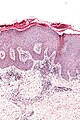
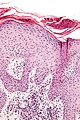
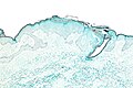
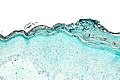
Solar elastosis
General
- Very common.
- Caused by sun exposure - specifically UV light.[9]
- Severity correlated with cumulative exposure to UV light..[10]
- Often co-localized with skin cancers - as UV light is risk factor for skin cancers.[10]
- Benign.
Microscopic
Features:
- Grey, spaghetti-like material in the superficial dermis.
DDx:
Note:
- The DDx above is things associated with sun damaged skin.
- Dermal mucin (as my be seen in lupus erythematosus) is a possible mimic - but it isn't spaghetti-like and the "background" (an interface dermatitis) is different.
Images
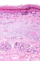
Solar elastosis - intermed. mag. (WC)
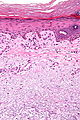
Solar elastosis - high mag. (WC)


www:
Sign out
SKIN, RIGHT CHEEK, RE-EXCISION:
- DERMAL SCAR.
- EXTENSIVE SOLAR ELASTOSIS.
Prominent blood vessels
SKIN LESION, LEFT CHEEK, BIOPSY:
- SKIN WITH SOLAR ELASTOSIS AND PROMINENT SMALL BLOOD VESSELS.
SUPERIOR SHOULDER, LEFT, PUNCH BIOPSY:
- BENIGN SKIN WITH MODERATE SOLAR ELASTOSIS, PROMINENT SMALL BLOOD VESSELS AND
  SCATTERED PERIVASCULAR LYMPHOCYTES AND PLASMA CELLS.
- NEGATIVE FOR BASAL CELL CARCINOMA.
- NEGATIVE FOR ACTINIC KERATOSIS.
Micro
The sections show hair bearing skin with solar elastosis and numerous small dilated blood vessels. The dermis is mildly fibrotic. Compact keratin is present.
The epidermis matures to the surface. A granular layer is present. There is no basal epidermal atypia. No melanocytic nests are identified. There is no palisading of the basal cells. Rare scattered lymphocytes are in the dermis.
Very common - viral
Verruca vulgaris
Verruca plana
General
- Common.
- Usu. hands and face.[11]
Microscopic
Features:[11]
- Orthokeratosis with basketweave pattern.
- Hypergranulosis.
- Viral keratohyaline.
- Koilocytes.
- Acanthosis - yet flat surface and base.
Notes:
- It differs from verruca vulgaris... (1) orthokeratosis, (2) flat surface and base.
Images
www:
- Verruca plana - several images (fujita-hu.ac.jp).
- Verruca plana - several good images (dermatopathonline.com).
Less common
Chronic folliculitis
- Folliculitis redirect here.
General
- Common.
- Infrequently biopsied.
Gross
- Erythema.[12]
DDx gross:
Microscopic
Features:
- Inflammation around the hair follicle - key feature.
- Lymphocytes - usu. predominant.
- +/-Chronic changes:
- Acanthosis.
- Hyperkeratosis.
- Hypergranulosis.
DDx:
Sign out
SKIN LESION, UPPER ARM, BIOPSY:
- CHRONIC FOLLICULITIS WITH SECONDARY SURFACE CHANGES.
Micro
The sections show hair-bearing skin with abundant lymphocytes around and within the hair follicle wall.
The non-hair follicle epidermis has acanthosis, hypergranulosis and compact hyperkeratosis. There is no inflammatory cell infiltrate in the non-hair follicle epidermis or at the non-hair follicle interface.
There are no granulomas.
Clear cell acanthoma
Chondrodermatitis nodularis chronica helicis
Cutaneous calcinosis
- AKA calcinosis cutis.
Dilated pore of Winer
General
- Benign.
- Looks like a zit.
Microscopic
Features:[14]
- Dilated hair follicle with keratin.
- Acanthosis.
- Budding of epidermis (into dermis).
DDx:
Image:
Lichenoid keratosis
- AKA lichen planus-like keratosis.
Granuloma annulare
Necrobiosis lipoidica
Keloid
Angiofibroma
- See also: nasopharyngeal angiofibroma.
- Should not be confused with angiokeratoma.
Benign fibrous papule
- AKA fibrous papule.
General
- An angiofibroma on the face that is solitary.[15][16]
Gross
Microscopic
Features:[17]
- Dome-shaped.
- Fibrotic dermis.
- Enlarged fibroblasts.
- Dilated small vessels.
- +/-Multinucleated stromal cells.[18]
- +/-Stellate cells.[18]
DDx:
- Angiofibroma - not solitary or not on the nose.
Note:
- Several variants exist.[16]
Images
Sign out
SKIN LESION, CHIN, BIOPSY:
- BENIGN FIBROUS PAPULE.
Molluscum contagiosum
Superficial dermal infiltrates
Discussed in detail by Alsaad and Ghazarian.[19]
Dermal perivascular lymphoeosinophilic infiltration
- Abbreviated DPLI.
Microscopic appearance is just what it is called:
- Lymphocytes and eosinophils around the vessels in the superficial dermis.
DDx:[19]
- Insect bite - classically wedge-shaped.[20]
- Drug reaction.
- Urticarial reaction.
- Prevesicular early stage of bullous pemphigoid.
- HIV related dermatoses.
Notes:
- May superficially resemble cutaneous lymphoma.[20]
Images:
Congenital dermal melanocytosis
- AKA Mongolian spots.
- Classically seen in asian children.
Gross:
- Brown or blue-grey patch in the lumbosacral area.
Mastocytosis
Ichthyosis
General
- Comes in different flavours.
- Usu. inherited... thus a pediatric condition.
Gross
- Fish scale-like appearance.
Image:
Microscopic
Features:[21]
- Thick stratum corneum without basket-weave pattern.
Palmar fibromatosis
Angiomyoma
- Do not confuse with angiomyxoma.
General
- Benign.
- Female > male.[22]
Microscopic
Features:
- Well-circumscribed lesion with fascicular architecture.
- Spindle cells/epithelioid cell with moderate eosinophilic (pink) cytoplasm.
- Thick-walled blood vessels. (???)
Images:
Angiokeratoma
Inverted follicular keratosis
- Abbreviated IFK.[23]
General
- Benign skin lesion.
- Central face - middle age.[24]
- Uncommon.
- May be considered a variant of seborrheic keratosis that is predominantly endophytic.[25]
Microscopic
Features:[24]
- Keratinocyte of cytologically benign proliferation.
- "Squamous eddies" (whorls of keratin).
- Coarse keratohyaline granules.
DDx:
- Squamous cell carcinoma of the skin.
- Trichilemmoma.
- Seborrheic keratosis - has an exophytic component.
Images:
- Inverted follicular keratosis - low mag. (flickr.com).
- Inverted follicular keratosis - high mag. (flickr.com).
- Inverted follicular keratosis - several images (mckeedermpath.com).
Sign out
SKIN LESION, FACE, BIOPSY:
- INVERTED FOLLICULAR KERATOSIS.
Micro
The sections show skin with acanthosis, pseudohorn cysts, and focal basal epidermal pigmentation. There is no basal nuclear atypia, no mitoses and there are no melanocytic nests. There is minimal dermal inflammation. There is no apparent solar elastosis.
Focal cutaneous mucinosis
General
- Benign.
- May be associated with systemic disease.[27]
Microscopic
Features:
- Light blue whispy material in the dermis - key feature.
DDx:
- Dermal edema.
- Digital mucous cyst.
Panniculitis
This is dealt with in the panniculitis article.
DDx for panniculitis:
- Erythema nodosum.
- Erythema induratum.
- Self-inflicted trauma.
- Systemic lupus erythematosus.
- Weird stuff.
Rare
Necrotizing fasciitis
- Not to be confused with nodular fasciitis.
- AKA flesh-eating disease.
Porokeratosis
Nevus sebaceous
- AKA nevus sebaceous of Jadassohn.
Nevus lipomatosus superficialis
- Abbreviated NLS.
- AKA nevus lipomatosus cutaneous superficialis, abbreviated NLCS.
- AKA nevus lipomatosus.[28]
Bullous disease
Cysts
See also
References
- ↑ Busam, Klaus J. (2009). Dermatopathology: A Volume in the Foundations in Diagnostic Pathology Series (1st ed.). Saunders. pp. 26. ISBN 978-0443066542.
- ↑ URL: http://www.pediatrics.wisc.edu/education/derm/text.html. Accessed on: 23 August 2012.
- ↑ URL: http://missinglink.ucsf.edu/lm/DermatologyGlossary/tinea.html. Accessed on: 25 February 2013.
- ↑ Picascia, DD.; Robinson, JK. (Aug 1987). "Actinic cheilitis: a review of the etiology, differential diagnosis, and treatment.". J Am Acad Dermatol 17 (2 Pt 1): 255-64. PMID 3305604.
- ↑ Kumar, Vinay; Abbas, Abul K.; Fausto, Nelson; Aster, Jon (2009). Robbins and Cotran pathologic basis of disease (8th ed.). Elsevier Saunders. pp. 1188. ISBN 978-1416031215.
- ↑ Busam, Klaus J. (2009). Dermatopathology: A Volume in the Foundations in Diagnostic Pathology Series (1st ed.). Saunders. pp. 76. ISBN 978-0443066542.
- ↑ 7.0 7.1 Busam, Klaus J. (2009). Dermatopathology: A Volume in the Foundations in Diagnostic Pathology Series (1st ed.). Saunders. pp. 77. ISBN 978-0443066542.
- ↑ URL: http://www.dermnetnz.org/dermal-infiltrative/solar-elastosis.html. Accessed on: 27 March 2013.
- ↑ Thomas, NE.; Kricker, A.; From, L.; Busam, K.; Millikan, RC.; Ritchey, ME.; Armstrong, BK.; Lee-Taylor, J. et al. (Nov 2010). "Associations of cumulative sun exposure and phenotypic characteristics with histologic solar elastosis.". Cancer Epidemiol Biomarkers Prev 19 (11): 2932-41. doi:10.1158/1055-9965.EPI-10-0686. PMID 20802019.
- ↑ 10.0 10.1 Karagas, MR.; Zens, MS.; Nelson, HH.; Mabuchi, K.; Perry, AE.; Stukel, TA.; Mott, LA.; Andrew, AS. et al. (Mar 2007). "Measures of cumulative exposure from a standardized sun exposure history questionnaire: a comparison with histologic assessment of solar skin damage.". Am J Epidemiol 165 (6): 719-26. doi:10.1093/aje/kwk055. PMID 17204514.
- ↑ 11.0 11.1 Humphrey, Peter A; Dehner, Louis P; Pfeifer, John D (2008). The Washington Manual of Surgical Pathology (1st ed.). Lippincott Williams & Wilkins. pp. 480. ISBN 978-0781765275.
- ↑ URL: http://www.webmd.com/skin-problems-and-treatments/tc/folliculitis-topic-overview. Accessed on: 7 November 2012.
- ↑ URL: http://www.head-face-med.com/content/4/1/2. Accessed on: 16 January 2014.
- ↑ Humphrey, Peter A; Dehner, Louis P; Pfeifer, John D (2008). The Washington Manual of Surgical Pathology (1st ed.). Lippincott Williams & Wilkins. pp. 486. ISBN 978-0781765275.
- ↑ 15.0 15.1 Busam, Klaus J. (2009). Dermatopathology: A Volume in the Foundations in Diagnostic Pathology Series (1st ed.). Saunders. pp. 505. ISBN 978-0443066542.
- ↑ 16.0 16.1 16.2 Jacyk, WK.; Rütten, A.; Requena, L. (2008). "Fibrous papule of the face with granular cells.". Dermatology 216 (1): 56-9. doi:10.1159/000109359. PMID 18032900.
- ↑ Humphrey, Peter A; Dehner, Louis P; Pfeifer, John D (2008). The Washington Manual of Surgical Pathology (1st ed.). Lippincott Williams & Wilkins. pp. 492. ISBN 978-0781765275.
- ↑ 18.0 18.1 Ragaz, A.; Berezowsky, V. (1979). "Fibrous papule of the face. A study of five cases by electron microscopy.". Am J Dermatopathol 1 (4): 353-6. PMID 543528.
- ↑ 19.0 19.1 Alsaad, KO.; Ghazarian, D. (Dec 2005). "My approach to superficial inflammatory dermatoses.". J Clin Pathol 58 (12): 1233-41. doi:10.1136/jcp.2005.027151. PMID 16311340.
- ↑ 20.0 20.1 Cotran, Ramzi S.; Kumar, Vinay; Fausto, Nelson; Nelso Fausto; Robbins, Stanley L.; Abbas, Abul K. (2005). Robbins and Cotran pathologic basis of disease (7th ed.). St. Louis, Mo: Elsevier Saunders. pp. 1269. ISBN 0-7216-0187-1.
- ↑ Kumar, Vinay; Abbas, Abul K.; Fausto, Nelson; Aster, Jon (2009). Robbins and Cotran pathologic basis of disease (8th ed.). Elsevier Saunders. pp. 1185. ISBN 978-1416031215.
- ↑ Katenkamp D, Kosmehl H, Langbein L (1988). "[Angiomyoma. A pathologo-anatomic analysis of 229 cases]" (in German). Zentralbl Allg Pathol 134 (4-5): 423–33. PMID 3201831.
- ↑ Shih, CC.; Yu, HS.; Tung, YC.; Tsai, KB.; Cheng, ST. (Jan 2001). "Inverted follicular keratosis.". Kaohsiung J Med Sci 17 (1): 50-4. PMID 11411260.
- ↑ 24.0 24.1 24.2 Busam, Klaus J. (2009). Dermatopathology: A Volume in the Foundations in Diagnostic Pathology Series (1st ed.). Saunders. pp. 387. ISBN 978-0443066542.
- ↑ Busam, Klaus J. (2009). Dermatopathology: A Volume in the Foundations in Diagnostic Pathology Series (1st ed.). Saunders. pp. 341. ISBN 978-0443066542.
- ↑ URL: http://www.ncbi.nlm.nih.gov/pmc/articles/PMC475744/. Accessed on: 11 May 2010.
- ↑ Gandhi, V.; Dogra, D.; Pandhi, RK.. "Cutaneous focal mucinosis.". Indian J Dermatol Venereol Leprol 62 (4): 260-1. PMID 20948074.
- ↑ Kaw, P.; Carlson, A.; Meyer, DR. (Jan 2005). "Nevus lipomatosus (pedunculated lipofibroma) of the eyelid.". Ophthal Plast Reconstr Surg 21 (1): 74-6. PMID 15677959.