Difference between revisions of "Mixed germ cell tumour"
Jump to navigation
Jump to search
(redirect w/ cat.) |
(→IHC) |
||
| (23 intermediate revisions by the same user not shown) | |||
| Line 1: | Line 1: | ||
# | {{ Infobox diagnosis | ||
| Name = {{PAGENAME}} | |||
| Image = Mixed_germ_cell_tumour_-_intermed_mag.jpg | |||
| Width = | |||
| Caption = Mixed germ cell tumour. [[H&E stain]]. | |||
| Micro = depends on the components | |||
| Subtypes = | |||
| LMDDx = other [[germ cell tumours]] | |||
| Stains = | |||
| IHC = variable | |||
| EM = | |||
| Molecular = | |||
| IF = | |||
| Gross = heterogeneous appearance, typically solid and cystic | |||
| Grossing = [[orchiectomy grossing]] | |||
| Staging = [[testicular cancer staging]] | |||
| Site = [[ovary]], [[testis]], [[mediastinum]], other | |||
| Assdx = | |||
| Syndromes = | |||
| Clinicalhx = | |||
| Signs = mass lesion | |||
| Symptoms = | |||
| Prevalence = most common germ cell tumour | |||
| Bloodwork = +/-[[AFP]] elevated, +/-beta-hCG elevated, +/-LDH elevated | |||
| Rads = | |||
| Endoscopy = | |||
| Prognosis = worse than [[seminoma]]/[[dysgerminoma]] | |||
| Other = | |||
| ClinDDx = gonads: [[germ cell tumours]], other tumours | |||
}} | |||
'''Mixed germ cell tumour''', abbreviated '''MGCT''', is a lesion composed of different [[germ cell tumours]]. Most germ cell tumours are mixed. | |||
==General== | |||
*60% of GCTs are mixed. † | |||
Common combinations: | |||
# Teratoma + embryonal carcinoma + endodermal sinus tumour (yolk sac tumour) (TEE). | |||
# Seminoma + embryonal (SE). | |||
# Teratoma + embryonal +(TE). | |||
Memory device: ''TEE'' + all combinations have embryonal carcinoma. | |||
Note: | |||
*† Numbers vary between sources. One series suggests it is almost 70%.<ref name=pmid15017200>{{Cite journal | last1 = Mosharafa | first1 = AA. | last2 = Foster | first2 = RS. | last3 = Leibovich | first3 = BC. | last4 = Ulbright | first4 = TM. | last5 = Bihrle | first5 = R. | last6 = Einhorn | first6 = LH. | last7 = Donohue | first7 = JP. | title = Histology in mixed germ cell tumors. Is there a favorite pairing? | journal = J Urol | volume = 171 | issue = 4 | pages = 1471-3 | month = Apr | year = 2004 | doi = 10.1097/01.ju.0000116841.30826.85 | PMID = 15017200 }}</ref> | |||
*There has been in increase in MGCTs over the past 20 years that is probably due to changes how in how [[germ cell tumours|GCT]]s are classified.<ref name=pmid21623833>{{Cite journal | last1 = Trabert | first1 = B. | last2 = Stang | first2 = A. | last3 = Cook | first3 = MB. | last4 = Rusner | first4 = C. | last5 = McGlynn | first5 = KA. | title = Impact of classification of mixed germ-cell tumours on incidence trends of non-seminoma. | journal = Int J Androl | volume = 34 | issue = 4 Pt 2 | pages = e274-7 | month = Aug | year = 2011 | doi = 10.1111/j.1365-2605.2011.01187.x | PMID = 21623833 }}</ref> | |||
==Gross== | |||
*Heterogeneous appearance - distinctive regions that look different from one another. | |||
*Typically solid and cystic. | |||
===Images=== | |||
<gallery> | |||
Image:Mixed_Germ_Cell_Tumor_of_Testis_(3260625567).jpg | Mixed germ cell tumour. (WC/euthman) | |||
</gallery> | |||
==Microscopic== | |||
Features: | |||
*Depends on the components. | |||
*Classic appearances: | |||
**[[Seminoma]]: fried egg-like" cells with lymphocytes. | |||
**[[Yolk sac tumour]]: edematous appearing/paucicellular regions, Schiller-Duval bodies. | |||
**[[Embryonal carcinoma]]: moderate-to-marked [[nuclear atypia]] with overlapping nuclei and usu. necrosis. | |||
**[[Teratoma]]: cysts with GI like epithelium, cysts with squamous epithelium & keratin (skin), immature cartilage, others. | |||
**[[Choriocarcinoma]]: hemorrhagic, multinucleated cells (syncytiotrophoblasts) and cells with pale cytoplasm (cytotrophoblasts). | |||
Notes: | |||
*If one cannot identify the component... it is probably yolk sac as this has so many different patterns. | |||
===Images=== | |||
<gallery> | |||
Image:Mixed_germ_cell_tumour_-_intermed_mag.jpg | Mixed GCT - intermed mag. (WC/Nephron) | |||
Image:Mixed germ cell tumour - high mag.jpg | Mixed GCT - high mag. (WC/Nephron) | |||
</gallery> | |||
www: | |||
*[http://path.upmc.edu/cases/case192/micro.html Mixed germ cell tumour - several images (upmc.edu)]. | |||
*[http://path.upmc.edu/cases/case356.html Mixed germ cell tumour - several cases (upmc.edu)]. | |||
==IHC== | |||
*Immunostains are useful for differentiating components, e.g. [[yolk sac tumour]] versus [[embryonal carcinoma]]. | |||
Looking for elements | |||
*Beta-hCG +ve - if syncytiotrophoblasts are present. | |||
*[[AFP]] +ve (or Glypican 3 +ve) - a yolk sac tumour component is present. | |||
*GFAP +ve - if neuroepithelium is present. | |||
A panel: | |||
*CD30 +ve -- [[embryonal carcinoma]]. | |||
*OCT4 +ve -- [[seminoma]]. | |||
*D2-40 +ve -- seminoma, useful for [[LVI]]. | |||
*[[Pankeratin]] +ve -- embryonal carcinoma. | |||
*CEA-M. | |||
*[[EMA]] +ve -- metastatic carcinoma.<ref>{{Cite journal | last1 = Shek | first1 = TW. | last2 = Yuen | first2 = ST. | last3 = Luk | first3 = IS. | last4 = Wong | first4 = MP. | title = Germ cell tumour as a diagnostic pitfall of metastatic carcinoma. | journal = J Clin Pathol | volume = 49 | issue = 3 | pages = 223-5 | month = Mar | year = 1996 | doi = | PMID = 8675733 }}</ref> | |||
*[[Vimentin]]. | |||
*[[Glypican 3]] +ve -- [[yolk sac tumour]]. | |||
**Others: A1A +ve -- yolk sac tumour, AFP +ve -- yolk sac tumour. | |||
==Sign out== | |||
<pre> | |||
TESTIS, RIGHT, ORCHIECTOMY: | |||
- MALIGNANT MIXED GERM CELL TUMOUR, pT1 pNx: | |||
-- 80% OF TUMOUR TERATOMA. | |||
-- 20% OF TUMOUR SEMINOMA. | |||
-- PLEASE SEE TUMOUR SUMMARY. | |||
</pre> | |||
==See also== | |||
*[[Germ cell tumours]]. | |||
*[[Testis]]. | |||
*[[Ovarian tumours]]. | |||
==References== | |||
{{Reflist|1}} | |||
[[Category:Diagnosis]] | [[Category:Diagnosis]] | ||
[[Category:Germ cell tumours]] | |||
[[Category:Genitourinary pathology]] | |||
[[Category:Gynecologic pathology]] | |||
Latest revision as of 02:21, 2 August 2016
| Mixed germ cell tumour | |
|---|---|
| Diagnosis in short | |
 Mixed germ cell tumour. H&E stain. | |
|
| |
| LM | depends on the components |
| LM DDx | other germ cell tumours |
| IHC | variable |
| Gross | heterogeneous appearance, typically solid and cystic |
| Grossing notes | orchiectomy grossing |
| Staging | testicular cancer staging |
| Site | ovary, testis, mediastinum, other |
|
| |
| Signs | mass lesion |
| Prevalence | most common germ cell tumour |
| Blood work | +/-AFP elevated, +/-beta-hCG elevated, +/-LDH elevated |
| Prognosis | worse than seminoma/dysgerminoma |
| Clin. DDx | gonads: germ cell tumours, other tumours |
Mixed germ cell tumour, abbreviated MGCT, is a lesion composed of different germ cell tumours. Most germ cell tumours are mixed.
General
- 60% of GCTs are mixed. †
Common combinations:
- Teratoma + embryonal carcinoma + endodermal sinus tumour (yolk sac tumour) (TEE).
- Seminoma + embryonal (SE).
- Teratoma + embryonal +(TE).
Memory device: TEE + all combinations have embryonal carcinoma.
Note:
- † Numbers vary between sources. One series suggests it is almost 70%.[1]
- There has been in increase in MGCTs over the past 20 years that is probably due to changes how in how GCTs are classified.[2]
Gross
- Heterogeneous appearance - distinctive regions that look different from one another.
- Typically solid and cystic.
Images
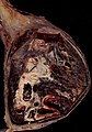
Mixed germ cell tumour. (WC/euthman)
.jpg)
Microscopic
Features:
- Depends on the components.
- Classic appearances:
- Seminoma: fried egg-like" cells with lymphocytes.
- Yolk sac tumour: edematous appearing/paucicellular regions, Schiller-Duval bodies.
- Embryonal carcinoma: moderate-to-marked nuclear atypia with overlapping nuclei and usu. necrosis.
- Teratoma: cysts with GI like epithelium, cysts with squamous epithelium & keratin (skin), immature cartilage, others.
- Choriocarcinoma: hemorrhagic, multinucleated cells (syncytiotrophoblasts) and cells with pale cytoplasm (cytotrophoblasts).
Notes:
- If one cannot identify the component... it is probably yolk sac as this has so many different patterns.
Images
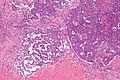
Mixed GCT - intermed mag. (WC/Nephron)
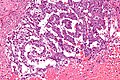
Mixed GCT - high mag. (WC/Nephron)

www:
- Mixed germ cell tumour - several images (upmc.edu).
- Mixed germ cell tumour - several cases (upmc.edu).
IHC
- Immunostains are useful for differentiating components, e.g. yolk sac tumour versus embryonal carcinoma.
Looking for elements
- Beta-hCG +ve - if syncytiotrophoblasts are present.
- AFP +ve (or Glypican 3 +ve) - a yolk sac tumour component is present.
- GFAP +ve - if neuroepithelium is present.
A panel:
- CD30 +ve -- embryonal carcinoma.
- OCT4 +ve -- seminoma.
- D2-40 +ve -- seminoma, useful for LVI.
- Pankeratin +ve -- embryonal carcinoma.
- CEA-M.
- EMA +ve -- metastatic carcinoma.[3]
- Vimentin.
- Glypican 3 +ve -- yolk sac tumour.
- Others: A1A +ve -- yolk sac tumour, AFP +ve -- yolk sac tumour.
Sign out
TESTIS, RIGHT, ORCHIECTOMY:
- MALIGNANT MIXED GERM CELL TUMOUR, pT1 pNx:
-- 80% OF TUMOUR TERATOMA.
-- 20% OF TUMOUR SEMINOMA.
-- PLEASE SEE TUMOUR SUMMARY.
See also
References
- ↑ Mosharafa, AA.; Foster, RS.; Leibovich, BC.; Ulbright, TM.; Bihrle, R.; Einhorn, LH.; Donohue, JP. (Apr 2004). "Histology in mixed germ cell tumors. Is there a favorite pairing?". J Urol 171 (4): 1471-3. doi:10.1097/01.ju.0000116841.30826.85. PMID 15017200.
- ↑ Trabert, B.; Stang, A.; Cook, MB.; Rusner, C.; McGlynn, KA. (Aug 2011). "Impact of classification of mixed germ-cell tumours on incidence trends of non-seminoma.". Int J Androl 34 (4 Pt 2): e274-7. doi:10.1111/j.1365-2605.2011.01187.x. PMID 21623833.
- ↑ Shek, TW.; Yuen, ST.; Luk, IS.; Wong, MP. (Mar 1996). "Germ cell tumour as a diagnostic pitfall of metastatic carcinoma.". J Clin Pathol 49 (3): 223-5. PMID 8675733.