Difference between revisions of "Uterine leiomyoma"
Jump to navigation
Jump to search
(→Microscopic: tweak) |
|||
| (4 intermediate revisions by the same user not shown) | |||
| Line 1: | Line 1: | ||
{{ Infobox diagnosis | |||
| Name = {{PAGENAME}} | |||
| Image = Histopathology of uterine leiomyoma.jpg | |||
| Width = | |||
| Caption = Uterine leiomyoma. [[H&E stain]]. | |||
| Synonyms = uterine fibroid | |||
| Micro = spindle cells arranged in fascicles, usually without atypia | |||
| Subtypes = atypical leiomyoma (symplastic leiomyoma), lipoleiomyoma, cellular leiomyoma, others | |||
| LMDDx = [[leiomyosarcoma]], [[STUMP]], [[dermatomyofibroma]], [[adenomatoid tumour]] | |||
| Stains = | |||
| IHC = | |||
| EM = | |||
| Molecular = | |||
| IF = | |||
| Gross = sharply circumscribed lesion, gray-white, whorled appearance | |||
| Grossing = [[Hysterectomy for fibroids grossing]] | |||
| Staging = | |||
| Site = | |||
| Assdx = | |||
| Syndromes = | |||
| Clinicalhx = | |||
| Signs = | |||
| Symptoms = | |||
| Prevalence = very common | |||
| Bloodwork = | |||
| Rads = | |||
| Endoscopy = | |||
| Prognosis = benign | |||
| Other = | |||
| ClinDDx = other [[uterine tumours]] | |||
| Tx = surgical (myomectomy, hysterectomy) or medical | |||
}} | |||
'''Uterine leiomyoma''', commonly '''fibroid''', is a very common benign smooth muscle tumour of the [[uterus]]. | '''Uterine leiomyoma''', commonly '''fibroid''', is a very common benign smooth muscle tumour of the [[uterus]]. | ||
| Line 21: | Line 53: | ||
* [[Necrosis]]. | * [[Necrosis]]. | ||
==Microscopic== | |||
Features: | Features: | ||
* Spindle cells arranged in fascicles. | * Spindle cells arranged in fascicles. | ||
| Line 33: | Line 65: | ||
* Few mitoses. | * Few mitoses. | ||
DDx: | |||
*[ | *[[Leiomyosarcoma]]. | ||
*[[Smooth muscle tumour of uncertain malignant potential]] (STUMP). | |||
*[[Adenomatoid tumour]]. | |||
===Variants=== | ===Variants=== | ||
<!-- variants https://www.ajronline.org/doi/full/10.2214/AJR.14.13946 --> | |||
*Lipoleiomyoma - with adipose tissue. | *Lipoleiomyoma - with adipose tissue. | ||
**Image: [http://commons.wikimedia.org/wiki/File:Lipoleiomyoma1.jpg Lipoleiomyoma - low mag. (WC)]. | **Image: [http://commons.wikimedia.org/wiki/File:Lipoleiomyoma1.jpg Lipoleiomyoma - low mag. (WC)]. | ||
Latest revision as of 17:26, 15 September 2022
| Uterine leiomyoma | |
|---|---|
| Diagnosis in short | |
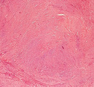
 Uterine leiomyoma. H&E stain. | |
|
| |
| Synonyms | uterine fibroid |
|
| |
| LM | spindle cells arranged in fascicles, usually without atypia |
| Subtypes | atypical leiomyoma (symplastic leiomyoma), lipoleiomyoma, cellular leiomyoma, others |
| LM DDx | leiomyosarcoma, STUMP, dermatomyofibroma, adenomatoid tumour |
| Gross | sharply circumscribed lesion, gray-white, whorled appearance |
| Grossing notes | Hysterectomy for fibroids grossing |
| Prevalence | very common |
| Prognosis | benign |
| Clin. DDx | other uterine tumours |
| Treatment | surgical (myomectomy, hysterectomy) or medical |
Uterine leiomyoma, commonly fibroid, is a very common benign smooth muscle tumour of the uterus.
The more general topic of leiomyoma is covered in the article leiomyoma.
General
- Extremely common... 40% of women by age 40.
- Benign.
- Can be a cause of abnormal uterine bleeding (commonly abbreviated AUB).
- Large & multiple associated with infertility.
- May be treated medically with a selective progesterone receptor modulator, e.g. ulipristal (Fibristal).[1]
Gross
Feature:
- Sharply circumscribed.
- Gray-white.
- Whorled appearance.
Factor that raise concern for leiomyosarcoma:
- Haemorrhage.
- Cystic degeneration.
- Necrosis.
Microscopic
Features:
- Spindle cells arranged in fascicles.
- Fascicular appearance: adjacent groups of cells have their long axis perpendicular to one another; looks somewhat like a braided hair that was cut.
- Whorled arrangement of cells.
Negatives:
- Necrosis (low power) - suggestive of leiomyosarcoma.
- Hypercellularity.
- Nuclear atypia seen at low power.
- Few mitoses.
DDx:
Variants
- Lipoleiomyoma - with adipose tissue.
- Image: Lipoleiomyoma - low mag. (WC).
- Hypercellular leiomyoma - hypercellularity associated with more mutations.[2]
- Atypical leiomyoma (AKA symplastic leiomyoma) - leiomyoma with nuclear atypia.
- Image: Atypical leiomyoma (WC).
- Benign metastasizing leiomyoma.[3]
- This is just what it sounds like. Some believe these are low grade leiomyosarcomas.
IHC
Work-up of suspicious leiomyomas:[4]
Others:
Sign out
Uterine Cervix, Uterus, Bilateral Tubes and IUD, Total Hysterectomy and Bilateral Salpingectomy:
- Uterine leiomyomas.
- Mild atherosclerosis.
- Inactive endometrium.
- Intrauterine device (IUD) - gross only.
- Uterine cervix within normal limits.
- Left uterine tube with small paratubal cyst, negative for
  significant pathology.
- Right uterine tube with paratubal cyst, negative for significant pathology.
- NEGATIVE for malignancy.
Block letters
UTERUS WITH CERVIX, UTERINE TUBES AND LEFT OVARY, TOTAL HYSTERECTOMY, BILATERAL SALPINGECTOMY
AND LEFT OOPHRECTOMY:
- LEIOMYOMATA WITH FOCAL CALCIFICATION AND HYALINE CHANGE.
- SECRETORY PHASE ENDOMETRIUM.
- LEFT OVARY WITHIN NORMAL LIMITS.
- UTERINE TUBES WITHIN NORMAL LIMITS.
- UTERINE CERVIX WITHIN NORMAL LIMITS.
Myomectomy
UTERINE MASSES ("FIBROIDS"), MYOMECTOMY:
- LEIOMYOMATA.
UTERINE MASS, HYSTEROSCOPIC MYOMECTOMY:
- BENIGN SMOOTH MUSCLE FRAGMENTS COMPATIBLE WITH LEIOMYOMA.
- SECRETORY PHASE ENDOMETRIUM.
Micro
The sections show bland spindle cells within a fascicular architecture. Hyaline change is present. No necrosis is seen. Mild proliferative activity is seen (~ 2 mitoses/10 HPFs, 1 HPF ~0.2376 mm*mm). No cytologic atypia is apparent.
See also
References
- ↑ Delev, DP.. "Ulipristal acetate--a review of the new therapeutic indications and future prospects.". Folia Med (Plovdiv) 55 (3-4): 5-10. PMID 24712276.
- ↑ Pandis, N.; Heim, S.; Willén, H.; Bardi, G.; Flodérus, U-M.; Mandahl, N.; Mitelman, F. (Jan 1991). "Histologic—cytogenetic correlations in uterine leiomyomas.". International Journal of Gynecological Cancer 1 (4): 163-68. http://www3.interscience.wiley.com/journal/119360394/abstract.
- ↑ Patton, KT.; Cheng, L.; Papavero, V.; Blum, MG.; Yeldandi, AV.; Adley, BP.; Luan, C.; Diaz, LK. et al. (Jan 2006). "Benign metastasizing leiomyoma: clonality, telomere length and clinicopathologic analysis.". Mod Pathol 19 (1): 130-40. doi:10.1038/modpathol.3800504. PMID 16357844. http://www.nature.com/modpathol/journal/v19/n1/full/3800504a.html.
- ↑ STC. 25 February 2009.
- ↑ 5.0 5.1 Zhu, XQ.; Shi, YF.; Cheng, XD.; Zhao, CL.; Wu, YZ. (Jan 2004). "Immunohistochemical markers in differential diagnosis of endometrial stromal sarcoma and cellular leiomyoma.". Gynecol Oncol 92 (1): 71-9. PMID 14751141.
- ↑ Gannon, BR.; Manduch, M.; Childs, TJ. (Jan 2008). "Differential Immunoreactivity of p16 in leiomyosarcomas and leiomyoma variants.". Int J Gynecol Pathol 27 (1): 68-73. doi:10.1097/pgp.0b013e3180ca954f. PMID 18156978.