Synaptophysin
Jump to navigation
Jump to search


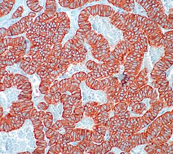
Synaptophysin staining in Atypical carcinoid.
Synaptophysin is the principal structural element of the walls of synaptic vesicles and thus a neuroendocrine marker.
Normal tissues
- Note: resists autolytic degradation (can be used 5 days post mortem).
- Marker of late onset (neuronal maturation).
Positive staining
- Cerebral cortex
- Hippocampus
- Corpus striatum,
- Globus pallidus
- Substantia nigra
- Cerebellum system
- Olfactory bulb
- Pineal gland
- Adrenal gland
- Langerhans islets
Tumours
- Ganglioglioma.
- Medulloblastoma +/-ve.
- Pituitary adenoma.
- Neurocytoma.
- Paraganglioma.
- Dysembryoplastic neuroepithelial tumour.
- Atypical carcinoid.