Plasma cell neoplasms
Jump to navigation
Jump to search
| Plasma cell neoplasms | |
|---|---|
| Diagnosis in short | |
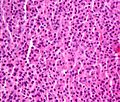
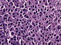
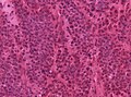
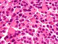
 Plasma cell neoplasm. H&E stain | |
|
| |
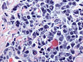
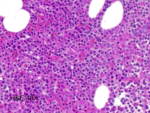
| LM | dyscohesive plasmacytoid cells (abundant cytoplasm, eccentrically placed nucleus), often prominent perinuclear hof, +/-nucleoli, +/-Russell bodies, +/-Dutcher bodies |
| LM DDx | lymphoplasmacytic lymphoma, neuroendocrine carcinoma, poorly differentiated carcinoma, others |
| IHC | CD138 +ve, CD56 +ve, CD45 -ve/+ve, CD79a +ve |
| Site | bone - bone marrow |
|
| |
| Associated Dx | renal failure, myeloma cast nephropathy |
| Blood work | +/-anemia, +/-hypercalcemia |
| Radiology | +/-lytic bone lesions |
| Prognosis | poor |
| Clin. DDx | metastatic disease |
Plasma cell neoplasms arise from plasma cells. They are encountered by anatomical pathologists on occasion.
Plasma cell myeloma, and plasmacytoma (solitary myeloma)[1] redirect to this article.
General
- Malignancy derived from the plasma cells.
- Prognosis: poor.
- Common primary bone tumour in adults.
Clinical:[2]
- Bence Jones protein (urine).
- Abnormal protein electrophoresis (monoclonal gammopathy, dysproteinemia, paraproteinemia).
Note:
- Plasmacytoma = histology of multiple myeloma; to diagnose multiple myeloma other (non-pathology) criteria are needed.
Classified by site:
- Medullary.
- Extramedullary - usu. upper aerodigestive tract.[3]
Multiple myeloma
Diagnosis requires the following:[4]
- Clonal plasma cells. Must >10% if on bone marrow biopsy.
- Monoclonal protein, i.e. paraprotein, in serum or urine.
- End-organ damage thought to be due to the neoplasm - mnemonic CARL:
- Calcium (in the serum) is elevated.
- Anemia.
- Renal failure.
- Lytic bone lesions.
Note:
- CRAB (calclium, renal failure, anemia, bony lesions) is another mnemonic.[5]
Microscopic
Features (plasmacytoma):
- Abundant eosinophilic cytoplasm.
- Eccentrically placed nucleus.
- Usually with "clock face" morphology.
- "Clock face" morphology = chromatin clumps around the edge of the nucleus, like the numbers on a clock face.
- May have nucleoli.
- Usually with "clock face" morphology.
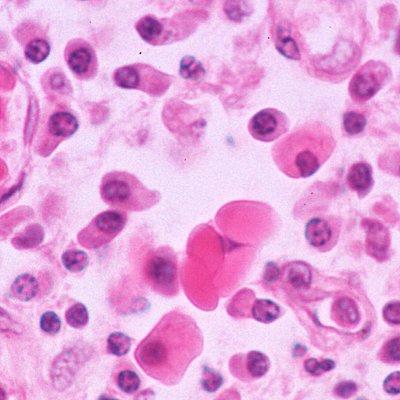
- Russell bodies:
- Eosinophilic, large (10-15 micrometres), homogenous immunoglobulin-containing inclusions.
- Dutcher bodies - intranuclear crystalline rods.
- Dutcher bodies are PAS stain +ve.[6]
- Prominent perinuclear hof - cytoplasmic crescent shaped lucency adjacent to the nuclear membrane (due to large Golgi apparatus); nucleus has a "bib".
DDx:
- Neuroendocrine carcinoma - nucleus often has a plasmacytoid (plasma cell-like) appearance.
- Lymphoplasmacytic lymphoma (AKA Waldenström's macroglobulinemia).
- Syphilis.
Images
Case
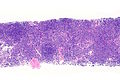
PCN - low mag.
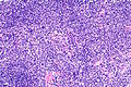
PCN - intermed. mag.
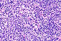
PCN - high mag.
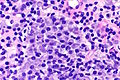
PCN - very high mag.


Other cases
Plasmacytoma. (WC)
Skull base plasmacytoma, higher magnification (WC/jensflorian)
Plasmacytoma smear (WC/jensflorian)
Leptomeningeal plasmacytoma, frozen section (WC/jensflorian)


www
Russell bodies
Russell bodies. (WC)

www
Dutcher bodies
Dutcher bodies and Russell bodies. (WC/Gabriel Caponetti)

IHC
- CD138 +ve.
- CD56 +ve.[8]
- Also +ve in NK/T cell lymphomas.
- Kappa -- usu. slightly stronger than lambda.
- Lambda.
- CD57.
- Also +ve in T-cell large granular lymphocytic leukemia.[9]
- CD38 +ve.[10]
Others:[11]
- CD79a +ve.
- CD45 -ve/+ve.
- CD10 -ve.
- CD5 -ve.
- Cyclin D1 -ve/+ve.
A panel:
- CD3, CD20, CD56, CD117, CD138, IG-kappa (plasma), IG-lambda (plasma).
Molecular
- t(4;14)(p16.3;q32.3) / IGH–MMSET.[12]
- Associated with poor prognosis.[13]
- 13q deletion.
- Worse prognosis.[1]
- 17q deletion.
- Worse prognosis.[1]
See also
References
- ↑ 1.0 1.1 1.2 Mitchell, Richard; Kumar, Vinay; Fausto, Nelson; Abbas, Abul K.; Aster, Jon (2011). Pocket Companion to Robbins & Cotran Pathologic Basis of Disease (8th ed.). Elsevier Saunders. pp. 324. ISBN 978-1416054542.
- ↑ Mitchell, Richard; Kumar, Vinay; Fausto, Nelson; Abbas, Abul K.; Aster, Jon (2011). Pocket Companion to Robbins & Cotran Pathologic Basis of Disease (8th ed.). Elsevier Saunders. pp. 323. ISBN 978-1416054542.
- ↑ Alexiou, C.; Kau, RJ.; Dietzfelbinger, H.; Kremer, M.; Spiess, JC.; Schratzenstaller, B.; Arnold, W. (Jun 1999). "Extramedullary plasmacytoma: tumor occurrence and therapeutic concepts.". Cancer 85 (11): 2305-14. PMID 10357398.
- ↑ Kyle RA, Rajkumar SV (January 2009). "Criteria for diagnosis, staging, risk stratification and response assessment of multiple myeloma". Leukemia 23 (1): 3–9. doi:10.1038/leu.2008.291. PMC 2627786. PMID 18971951. http://www.nature.com/leu/journal/v23/n1/full/leu2008291a.html.
- ↑ "Criteria for the classification of monoclonal gammopathies, multiple myeloma and related disorders: a report of the International Myeloma Working Group.". Br J Haematol 121 (5): 749-57. Jun 2003. PMID 12780789.
- ↑ URL: http://www.thefreelibrary.com/Dutcher+bodies+in+chronic+synovitis-a083551789. Accessed on: 4 August 2010.
- ↑ URL: http://path.upmc.edu/cases/case515.html. Accessed on: 25 January 2012.
- ↑ URL: http://www.ncbi.nlm.nih.gov/omim/116930. Accessed on: 31 August 2010.
- ↑ URL: http://www.nature.com/bmt/journal/v33/n1/full/1704298a.html. Accessed on: 31 August 2010.
- ↑ Tamamori, T.; Nakayama, F.; Sugimoto, H.; Fenxiang, J.; Iwatsuki, K.; Takigawa, M. (Oct 1993). "Extramedullary plasmacytoma: cytological and genotypic studies.". Br J Dermatol 129 (4): 468-72. PMID 8217765.
- ↑ URL: http://e-immunohistochemistry.info/web/Plasmacytoma_plasma_cell_myeloma.htm. Accessed on: 9 November 2015.
- ↑ Chesi, M.; Nardini, E.; Lim, RS.; Smith, KD.; Kuehl, WM.; Bergsagel, PL. (Nov 1998). "The t(4;14) translocation in myeloma dysregulates both FGFR3 and a novel gene, MMSET, resulting in IgH/MMSET hybrid transcripts.". Blood 92 (9): 3025-34. PMID 9787135.
- ↑ Keats, JJ.; Reiman, T.; Maxwell, CA.; Taylor, BJ.; Larratt, LM.; Mant, MJ.; Belch, AR.; Pilarski, LM. (Feb 2003). "In multiple myeloma, t(4;14)(p16;q32) is an adverse prognostic factor irrespective of FGFR3 expression.". Blood 101 (4): 1520-9. doi:10.1182/blood-2002-06-1675. PMID 12393535.