Uterine cervix
The uterine cervix, also simply cervix, is the gateway to the uterine corpus. It is not infrequently afflicted by cancer -- squamous cell carcinoma. Prior to routine pap tests it was a leading cause of cancer death in women in the Western world.
Polyps associated with the cervix are discussed the cervical polyp article.
Cytopathology of the cervix is dealt with in the gynecologic cytopathology article.
Introduction
- Consists of non-keratinized squamous epithelium and simple columnar epithelium.
- The area of overlap (between squamous & columnar) is known as the "transformation zone".[1]
- Also known as "transition zone".
- Most cervix cancer is squamous cell carcinoma.
Normal histology
Endocervical glands
Cervical glands normally have round nuclei and vaguely resemble the colonic mucosa.
- If the nuclei are columnar think cancer! This is like in the colon-- columnar nuclei = badness.
Memory device: The Cs (Cervix & Colon) are similar.
Where to start
- Identify epithelium - exocervical (stratified squamous), endocervical (simple columnar), both.
- If there is both exocervix and endocervix --> transition zone.
- Identify possible squamous lesions.
- Identify possible endocervical lesions.
Benign (common)
Nabothian cyst
Features:
- Simple endocervical cyst.
- Lined by endocervical epithelial cells.
- Columnar morphology with large clear, apical vacuoles.
- Lined by endocervical epithelial cells.
Image:
Tunnel cluster
General
- Benign.[2]
- Not the same as microglandular hyperplasia.[3]
Microscopic
- Well-circumscribed lesion consisting of:
- Benign endocervical glands.
- Dilated & filled with mucin or (less commonly) eosinophilic secretions.
- Lining epithelium compressed/flattened (attenuated).
- Gland architecture: branching, tortuous.
- Scant intervening stroma.
Notes:
- Usually no nuclear atypia and no mitotic activity.
- Important only as one could possibly mistake it as minimal deviation adenocarcinoma, AKA adenoma malignum.[6]
Images:
Microglandular hyperplasia
- Not to be confused with microglandular adenosis.
- Abbreviated MGH.
General
- Associated with OCP use.[3]
Microscopic
Features:[3]
- Cytologically benign - important.
- Usu. cuboidal morphology.
- Typically clear cytoplasm.
- Crowded small glands (classic), reticular or solid.
DDx:
- Adenocarcinoma in situ of the cervix.
Image:
Wolffian duct hyperplasia
General
- Benign.
Microscopic
Features:
- Abundant small tubules with a simple cuboidal epithelium.
- Round small bland nucleus.
Stains
- PAS-D+ve (cytoplasm).
Squamous metaplasia of the uterine cervix
General
- Benign process: columnar cells -> squamoid cells.
- Biologic response to irritation and/or inflammation.
Microscopic
Features:
- Nuclei are uniform size and round.
- Nucleoli present.
- +/-Intercellular bridges (due to edema) - common.
- Uniform cell spacing, i.e. no crowding.
Negatives:
- No mitoses (think cancer/CIN if you see 'em).
- Usually no hyperchromatism (think cancer/CIN if you see it).
Notes:
- NC ratio high - possible to confuse CIN III.
DDx:
IHC
- p16 +ve - in SCC; a poor man's test for HPV.
- Ki-67 - stains a large number of cells; proliferation marker.
Non-invasive
Cervical intraepithelial neoplasia
- Abbreviated CIN.
General
- Refers to changes in squamous epithelium.
Grades (squamous intraepithelial neoplasia):
- CIN I = mild dysplasia.
- CIN II = moderate dysplasia.
- CIN III = severe dysplasia.
Bethesda system:
- LSIL (low-grade squamous intraepithelial lesion) = CIN I.
- HSIL (high-grade squamous intraepithelial lesion) = CIN II, CIN III.
Treatment
LEEP = Loop Electrosurgical Excision Procedure (LEEP) Procedure.
- Used for squamous lesions -- pathologist typically gets several pieces.
Cone
- Used for endocervical lesions, i.e. adenocarcinoma in situ (AIS).
- Pathologist gets a ring or donut-shaped piece of tissue.
Microscopic
Features:
- CIN I = cytoplasmic halos (koilocytic atypia), atypical cells close to basement membrane only.
- CIN II = increased nuclear-cytoplasmic ratio, loss of polarity, incr. mitoses, hyperchromasia.
- If there are large nuclei... you should seen 'em on low power, i.e. 25x.
- CIN III = same changes as in CIN II + outer third (or full thickness).
Ref.:[9]
Notes:
- Hyperchromasia is a very useful feature for identifying CIN (particularly at low power, i.e. 25x).
- Koilocytes are the key feature of CIN I.
- Koilocytes are not considered to be part of a CIN II lesion or CIN III lesion.
- Large irregular nuclei are not required for CIN II... but you should think about it.
- Some mild changes at the squamo-columnar junction are expected.
- Look for the location of mitoses...
- If there is a mitosis in the inner third (of the epithelial layer) = at least CIN I.
- If there is a mitosis in the middle third (of the epithelial layer) = at least CIN II.
- If there is a mitosis in the outer third = CIN III.
- Nucleoli are usually NOT present in CIN.[10]
- Nucleoli are common in reactive changes.[11]
- The most probably place for CIN is the posterior cervix (6 o'clock position) - risk is marginally increased.[12]
Koilocytes versus benign squamous
Koilocytes:
- Perinuclear clearing.
- Nuclear changes.
- Size similar (or larger) to those in the basal layer of the epithelium.
- Nuclear enlargement should be evident on low power, i.e. 25x. [13]
- Central location - nucleus should be smack in the middle of the cell.
Notes:
- Both perinuclear clearing and nuclear changes are essential.
- Benign cells have a small nucleus that is peripheral.
Endocervical adenocarcinoma in situ
- For the cytology see Gynecologic_cytopathology#Endocervical_adenocarcinoma_in_situ
- AKA adenocarcinoma in situ, abbreviated AIS.
General
- Usually to HPV.
- May be found together with squamous neoplasias of the cervix.
- AIS of the cervix is much less common than squamous dysplasia of the cervix/SCC of the cervix.
Microscopic
Features:[3]
- Nuclear changes - key feature:
- Variable nuclear stratification.
- Nuclear crowding/pseudostratification.
- Nuclear enlargement.
- Often cigar-shaped nuclei.
- Coarse chromatin.
- Small nucleolus or nucleoli.
- Variable nuclear stratification.
- +/-Mitoses.
- +/-Reduced cytoplasmic mucin.
- Preservation of glandular architecture.
- Normal gland spacing - lack of complexity.
- Normal gland depth (subjective).
DDx:
Cancer
Squamous cell carcinoma of the uterine cervix
- AKA cervical squamous cell carcinoma.
General
- Most common type of cervical cancer.
Risk factors:
- Low socioeconomic status.
- Smoking.
- Early first intercourse.
- High risk partners.
- Human papillomavirus (HPV) infection, esp. "high risk HPV".
- HPV 16 closely assoc. with SCC.[14]
Microscopic
Features:
- Penetration of basement membrane.
- May be challenging to determine.
- Nuclear atypia.
SCC of the cervix versus CIN III: Invasive cancer look for:
- Eosinophilia.
- Extra large nuclei, i.e. nuclei 5x normal size.
- Stromal inflammation (lymphocytes, plasma cells).
- Long rete ridges.
- Numerous beeds/blobs of epithelial cells that seem unlikely to be rete ridges.
- Desmoplastic stroma - increased cellularity, spindle cell morphology.
Grading:[15]
- Well-differentiated (keratinizing).
- Moderately differentiated (nonkeratinizing).
- Poorly differentiated.
DDx:
- Squamous metaplasia of the uterine cervix - if you can trace the squamous cells from a gland to the surface it is less likely to be invasive cancer.[16]
- CIN III.
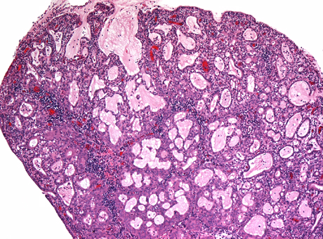
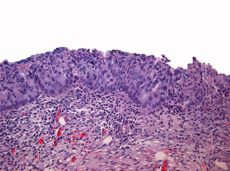
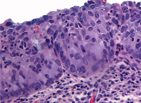
Adenocarcinoma of the uterine cervix
General
- Adenocarcinoma of the cervix is much less common than squamous dysplasia of the cervix/SCC of the cervix.
- Arises from the endocervical glands.
Microscopic
Features:
- Stromal changes - "desmoplastic stroma/desmoplastic reaction".
- Fibrosis/streaming cells.
- Gland fusion.
- Glands too deep -- very fuzzy criterion.
Notes:
- AIS changes - similar to colonic dysplasia.
- AIS may occur together with CIN.
- Not infrequently they (AIS, CIN) occur together - both are due, indirectly, to HPV infection.
- May be difficult to be certain of invasion.
DDx:
- Endocervical adenocarcinoma in situ.
- Metastatic adenocarcinoma.
Images:
- Cervical adenocarcinoma - low mag. (WC).
- Cervical adenocarcinoma - intermed. mag. (WC).
- Cervical adenocarcinoma - high mag. (WC).
IHC
Uterus vs. cervix:[17]
- Cervix (typically): CEA +ve, p16 +ve.
- ER -ve, PR -ve, vimentin -ve.
- Uterus (typically): vimentin +ve, ER +ve, PR +ve.
- CEA -ve, p16 -ve.
Uncommon non-invasive
Stratified mucin-producing intraepithelial lesions of the cervix
- Abbreviated SMILE (Stratified Mucin-producing Intraepithelial LEsion).
General
- Rare.
- Often accompanied by cervical intraepithelial neoplasia and adenocarcinoma in situ.[18]
Microscopic
Features:[18]
- Stratified epithelium with:
- Nuclear atypia.
- Cytoplasmic clearing or vacuoles in lesions - through-out.
DDx:
Images:
IHC
Features:
- Ki-67 high.
- Keratin 14 -ve.
- p63 +ve/-ve -- only basal if positive.
Uncommon types of cervical cancer
There are a number of uncommon type of cervical cancer.
Adenosquamous carcinoma
Features:
- Morphologic features of both squamous carcinoma and adenocarcinoma:
- Adenocarcinoma: gland forming or mucin vacuoles.
- Squamous carcinoma: abundant eosinophilic cytoplasm, central nucleus.
Image: Adenosquamous carcinoma - high mag. (WC).
Clear cell carcinoma of the uterine cervix
- AKA cervical clear cell carcinoma.
General
- Associated with diethylstilbestrol exposure in utero.[20]
Microscopic
Features:
- Like clear cell carcinoma elsewhere:
- +/-Clear cytoplasm.
- Cells have large free/luminal surface area (hobnailing pattern) and small non-free surface.
- Moderate-to-severe nuclear pleomorphism.
Small cell carcinoma of the cervix
- Like small cell carcinoma elsewhere.
DDx:
IHC
- HPV +ve.
Adenoid basal carcinoma
- See also: Basal cell carcinoma.
General
- Good prognosis.[21]
Microscopic
Features:[21]
- Nests of cells with basaloid rim and squamoid center.
- Basaloid cells look benign.
Image:
Glassy cell carcinoma
General
- Rare.
- Rapid growth, poor prognosis.[22]
Microscopic
Features:[23]
- Epithelioid cells in sheets or cords.
- Round/oval nucleus.
- One or more prominent nucleoli.
- Abundant finely vacuolated eosinophilic to amphophilic cytoplasm.
- Distinct cell borders.
- Inflammation - esp. eosinophils.[24]
DDx:
Images:
- WC:
- www:
Stains
Villoglandular adenocarcinoma of the cervix
- AKA well-differentiated papillary villoglandular adenocarcinoma,[26] AKA villoglandular papillary adenocarcinoma, AKA well-differentiated villoglandular adenocarcinoma.
General
- Rare.
- Younger patients and relatively good prognosis.[27]
- Associated with HPV.
- May also arise from the endometrium.[28]
Microscopic
Features:[29]
- Papillary structures (nipple-like shapes with a fibrovascular core) that are long.
- Nobody defines "long".
- Perhaps - long >3:1 length:width.
- Nobody defines "long".
- Covered by columnar (or cuboidal) epithelium.
- Intracellular mucin (focal).
DDx:
- Serous carcinoma of the cervix.
Images:
- www:
- WC:
Mucoepidermoid carcinoma
General
- Controversial - not in the WHO.[30]
Microscopic
Features:[30]
- Like salivary gland tumour.
Molecular
Like the salivary gland tumour:
- t(11;19) CRTC1/MAML2.[30]
See also
References
- ↑ URL: http://www.med-ed.virginia.edu/Courses/path/gyn/cervix1.cfm. Accessed on: 12 May 2010.
- ↑ Nucci, MR. (Oct 2002). "Symposium part III: tumor-like glandular lesions of the uterine cervix.". Int J Gynecol Pathol 21 (4): 347-59. PMID 12352183.
- ↑ 3.0 3.1 3.2 3.3 Zaino, RJ. (Mar 2000). "Glandular lesions of the uterine cervix.". Mod Pathol 13 (3): 261-74. doi:10.1038/modpathol.3880047. PMID 10757337. Cite error: Invalid
<ref>tag; name "pmid10757337" defined multiple times with different content - ↑ URL: http://pathologyoutlines.com/cervix.html#tunnelclusters. Accessed on: 27 February 2011.
- ↑ URL: http://surgpath4u.com/caseviewer.php?case_no=477. Accessed on: 5 September 2011.
- ↑ Gilks CB, Young RH, Aguirre P, DeLellis RA, Scully RE (September 1989). "Adenoma malignum (minimal deviation adenocarcinoma) of the uterine cervix. A clinicopathological and immunohistochemical analysis of 26 cases". Am. J. Surg. Pathol. 13 (9): 717–29. PMID 2764221.
- ↑ URL: http://sunnybrook.ca/content/?page=Dept_LabS_APath_GynPath_ImgAt_Cvx_neo_micro. Accessed on: 25 February 2012.
- ↑ Roteli-Martins CM, Derchain SF, Martinez EZ, Siqueira SA, Alves VA, Syrjänen KJ (2001). "Morphological diagnosis of HPV lesions and cervical intraepithelial neoplasia (CIN) is highly reproducible". Clin Exp Obstet Gynecol 28 (2): 78–80. PMID 11491378.
- ↑ Cotran, Ramzi S.; Kumar, Vinay; Fausto, Nelson; Nelso Fausto; Robbins, Stanley L.; Abbas, Abul K. (2005). Robbins and Cotran pathologic basis of disease (7th ed.). St. Louis, Mo: Elsevier Saunders. pp. 1075-6. ISBN 0-7216-0187-1.
- ↑ STC. January 2009.
- ↑ STC. January 2009.
- ↑ Pretorius, RG.; Zhang, X.; Belinson, JL.; Zhang, WH.; Ren, SD.; Bao, YP.; Qiao, YL. (Jan 2006). "Distribution of cervical intraepithelial neoplasia 2, 3 and cancer on the uterine cervix.". J Low Genit Tract Dis 10 (1): 45-50. PMID 16378031.
- ↑ V. Dube 2008.
- ↑ De Boer, MA.; Peters, LA.; Aziz, MF.; Siregar, B.; Cornain, S.; Vrede, MA.; Jordanova, ES.; Fleuren, GJ. (Apr 2005). "Human papillomavirus type 18 variants: histopathology and E6/E7 polymorphisms in three countries.". Int J Cancer 114 (3): 422-5. doi:10.1002/ijc.20727. PMID 15551313.
- ↑ Cotran, Ramzi S.; Kumar, Vinay; Fausto, Nelson; Nelso Fausto; Robbins, Stanley L.; Abbas, Abul K. (2005). Robbins and Cotran pathologic basis of disease (7th ed.). St. Louis, Mo: Elsevier Saunders. pp. 1077. ISBN 0-7216-0187-1.
- ↑ http://www.nature.com/modpathol/journal/v15/n3/pdf/3880520a.pdf
- ↑ LAE. 15 January 2009.
- ↑ 18.0 18.1 18.2 Park, JJ.; Sun, D.; Quade, BJ.; Flynn, C.; Sheets, EE.; Yang, A.; McKeon, F.; Crum, CP. (Oct 2000). "Stratified mucin-producing intraepithelial lesions of the cervix: adenosquamous or columnar cell neoplasia?". Am J Surg Pathol 24 (10): 1414-9. PMID 11023104.
- ↑ URL: http://sunnybrook.ca/content/?page=Dept_LabS_APath_GynPath_ImgAt_Cvx_mal_ais_smile. Accessed on: 30 March 2012.
- ↑ van Dijck, JA.; Doorduijn, Y.; Bulten, JH.; Verloop, J.; Massuger, LF.; Kiemeney, BA. (2009). "[Vaginal and cervical cancer due to diethylstilbestrol (DES); end epidemic]". Ned Tijdschr Geneeskd 153: A366. PMID 19857300.
- ↑ 21.0 21.1 Senzaki H, Osaki T, Uemura Y, et al. (December 1997). "Adenoid basal carcinoma of the uterine cervix: immunohistochemical study and literature review". Jpn. J. Clin. Oncol. 27 (6): 437–41. PMID 9438010. http://jjco.oxfordjournals.org/cgi/content/full/27/6/437.
- ↑ Nasu, K.; Takai, N.; Narahara, H. (Jun 2009). "Multimodal treatment for glassy cell carcinoma of the uterine cervix.". J Obstet Gynaecol Res 35 (3): 584-7. doi:10.1111/j.1447-0756.2008.00968.x. PMID 19527406.
- ↑ Reis-Filho, JS.; Fillus Neto, J.; Schonemann, E.; Sanderson, A.; Schmitt, FC.. "Glassy cell carcinoma of the uterine cervix. Report of a case with cytohistologic and immunohistochemical study.". Acta Cytol 45 (3): 407-10. PMID 11393075.
- ↑ URL: http://www.webpathology.com/image.asp?n=2&Case=561. Accessed on: 4 September 2011.
- ↑ Deshpande, AH.; Kotwal, MN.; Bobhate, SK.. "Glassy cell carcinoma of the uterine cervix a rare histology. Report of three cases with a review of the literature.". Indian J Cancer 41 (2): 92-5. PMID 15318016.
- ↑ Fadare, O.; Zheng, W. (Nov 2005). "Well-differentiated papillary villoglandular adenocarcinoma of the uterine cervix with a focal high-grade component: is there a need for reassessment?". Virchows Arch 447 (5): 883-7. doi:10.1007/s00428-005-0030-3. PMID 16088403.
- ↑ Korach, J.; Machtinger, R.; Perri, T.; Vicus, D.; Segal, J.; Fridman, E.; Ben-Baruch, G. (2009). "Villoglandular papillary adenocarcinoma of the uterine cervix: a diagnostic challenge.". Acta Obstet Gynecol Scand 88 (3): 355-8. doi:10.1080/00016340902730359. PMID 19172445.
- ↑ Zaino, RJ.; Kurman, RJ.; Brunetto, VL.; Morrow, CP.; Bentley, RC.; Cappellari, JO.; Bitterman, P. (Nov 1998). "Villoglandular adenocarcinoma of the endometrium: a clinicopathologic study of 61 cases: a gynecologic oncology group study.". Am J Surg Pathol 22 (11): 1379-85. PMID 9808130.
- ↑ Nucci, Marisa R.; Oliva, Esther (2009). Gynecologic Pathology: A Volume in Foundations in Diagnostic Pathology Series (1st ed.). Churchill Livingstone. pp. 180-1. ISBN 978-0443069208.
- ↑ 30.0 30.1 30.2 Lennerz, JK.; Perry, A.; Mills, JC.; Huettner, PC.; Pfeifer, JD. (Jun 2009). "Mucoepidermoid carcinoma of the cervix: another tumor with the t(11;19)-associated CRTC1-MAML2 gene fusion.". Am J Surg Pathol 33 (6): 835-43. doi:10.1097/PAS.0b013e318190cf5b. PMID 19092631.