Squamous metaplasia of the uterine cervix
Jump to navigation
Jump to search
| Squamous metaplasia of the uterine cervix | |
|---|---|
| Diagnosis in short | |
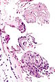
 Squamous metaplasia and endocervical epithelium. H&E stain. | |
|
| |
| LM | uniform cell spacing (no "crowding"), nuclei are uniform size and round, +/-nucleoli, distinct cell borders +/- intercellular bridges visible (common), adjacent/closely associated with endocervical epithelium - classically sits on surface (immature metaplasia), mitoses - rare, usually no nuclear hyperchromasia |
| LM DDx | HSIL, reactive squamous epithelium of the uterine cervix, squamous cell carcinoma of the uterine cervix |
| IHC | p16 patchy +ve (not diffuse), Ki-67 low & predominantly basal |
| Site | uterine cervix |
|
| |
| Signs | acetowhite lesion |
| Symptoms | none |
| Prevalence | common |
| Prognosis | benign |
| Clin. DDx | squamous intraepithelial lesion of the uterine cervix |
Squamous metaplasia of the uterine cervix is a common change of the uterine cervix that may resemble squamous intraepithelial lesion.
It may be abbreviated SMC.
General
- Benign process: columnar cells -> squamoid cells.
- Biologic response to irritation and/or inflammation.
Gross
Microscopic
Features:
- Uniform cell spacing - no crowding - key feature.
- Nuclei are uniform size and round.
- Nucleoli present.
- Distinct cell borders
- +/-Intercellular bridges (due to edema) - common.
- Adjacent/closely associated with columnar epithelium.
- Columnar epithelium superficial in immature metaplasia.
Negatives:
- No mitoses (think cancer/CIN if you see 'em).
- Usually no hyperchromatism (think cancer/CIN if you see it).
Notes:
- NC ratio high - possible to confuse with CIN III.
- May have goblet cells - uncommon.[2]
DDx:
- CIN II - esp. for immature squamous metaplasia.
- CIN III.
- Squamous cell carcinoma of the uterine cervix.
Images
Endocx and SM - high mag.
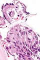
Endocx and SM - very high mag.

www:
- Squamous metaplasia - cervix (sciencephoto.com).
- Squamous metaplasia - bronchus (WC).
- Squamous metaplasia - cytology (techriver.net).[3]
IHC
- p16 weak-to-moderate patchy +ve -- checkerboard-like; not full thickness.
- Strong diffuse full thickness positivity in HSIL and SCC.
- Ki-67 - low proliferative rate.
Sign out
ECC
UTERINE ENDOCERVIX, CURETTAGE:
- SQUAMOUS METAPLASTIC EPITHELIUM.
- VERY SCANT STRIPPED ENDOCERVICAL EPITHELIUM.
Cervical biopsy
UTERINE CERVIX, BIOPSY:
- SQUAMOUS METAPLASTIC EPITHELIUM.
- SCANT BENIGN ENDOCERVICAL GLANDS.
UTERINE CERVIX, BIOPSY:
- SQUAMOUS METAPLASTIC EPITHELIUM.
- SCANT BENIGN ENDOCERVICAL GLANDS.
- NEGATIVE FOR DYSPLASIA AND NEGATIVE FOR MALIGNANCY.
Micro
The sections show stratified squamous epithelium. The cells are equally spaced and spaces are seen between the cells (edema).
The nuclei are not significantly enlarged (<3x resting lymphocyte diameter). No nuclear halos are apparent. The nuclear membranes are regular. Mild inflammation is present. Nucleoli are present focally.
No endocervical cells are identified.
See also
References
- ↑ Li, W.; Venkataraman, S.; Gustafsson, U.; Oyama, JC.; Ferris, DG.; Lieberman, RW.. "Using acetowhite opacity index for detecting cervical intraepithelial neoplasia.". J Biomed Opt 14 (1): 014020. doi:10.1117/1.3079810. PMID 19256708.
- ↑ Sivridis, E.; Karpathiou, G.; Malamou-Mitsi, V.; Giatromanolaki, A. (2010). "Intestinal-type metaplasia in the original squamous epithelium of the cervix.". Eur J Gynaecol Oncol 31 (3): 319-22. PMID 21077478.
- ↑ URL: http://nih.techriver.net/view.php?patientId=1. Accessed on: 17 September 2015.