Difference between revisions of "Synaptophysin"
Jump to navigation
Jump to search
Jensflorian (talk | contribs) (created) |
Jensflorian (talk | contribs) (→Tumours: +more added) |
||
| (2 intermediate revisions by the same user not shown) | |||
| Line 3: | Line 3: | ||
==Normal tissues== | ==Normal tissues== | ||
* | * Note: resists autolytic degradation (can be used 5 days post mortem). | ||
* Marker of late onset (neuronal maturation). | |||
* | |||
===Positive staining=== | |||
* Cerebral cortex | |||
* Hippocampus | |||
* Corpus striatum, | |||
* Globus pallidus | |||
* Substantia nigra | |||
* Cerebellum system | |||
* Olfactory bulb | |||
* Pineal gland | |||
* Adrenal gland | |||
* Langerhans islets | |||
==Tumours== | ==Tumours== | ||
* | * [[Ganglioglioma]]. | ||
* [[Medulloblastoma]] +/-ve. | |||
* [[Pituitary adenoma]]. | |||
* [[Neurocytoma]]. | |||
* [[Paraganglioma]]. | |||
* [[Dysembryoplastic neuroepithelial tumour]]. | |||
* [[Atypical carcinoid]]. | |||
===Negative staining=== | |||
==See also== | ==See also== | ||
Latest revision as of 12:02, 8 April 2019

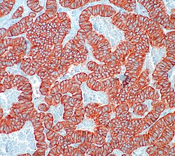
Synaptophysin staining in Atypical carcinoid.
Synaptophysin is the principal structural element of the walls of synaptic vesicles and thus a neuroendocrine marker.
Normal tissues
- Note: resists autolytic degradation (can be used 5 days post mortem).
- Marker of late onset (neuronal maturation).
Positive staining
- Cerebral cortex
- Hippocampus
- Corpus striatum,
- Globus pallidus
- Substantia nigra
- Cerebellum system
- Olfactory bulb
- Pineal gland
- Adrenal gland
- Langerhans islets
Tumours
- Ganglioglioma.
- Medulloblastoma +/-ve.
- Pituitary adenoma.
- Neurocytoma.
- Paraganglioma.
- Dysembryoplastic neuroepithelial tumour.
- Atypical carcinoid.