Difference between revisions of "Medullary thyroid carcinoma"
m (→Images) |
|||
| (27 intermediate revisions by 2 users not shown) | |||
| Line 8: | Line 8: | ||
| Subtypes = | | Subtypes = | ||
| LMDDx = | | LMDDx = | ||
| Stains = | | Stains = congo red +ve (amyloid deposits) | ||
| IHC = | | IHC = calcitonin +ve, [[CEA]] +ve, chromogranin A +ve, synaptophysin +ve, thyroglobulin -ve (usually) | ||
| EM = | | EM = | ||
| Molecular = | | Molecular = | ||
| Line 15: | Line 15: | ||
| Gross = usu. well-circumscribed, white, gray or yellow, gritty, firm | | Gross = usu. well-circumscribed, white, gray or yellow, gritty, firm | ||
| Grossing = | | Grossing = | ||
| Staging = [[thyroid cancer staging]] | |||
| Site = [[thyroid gland]] | | Site = [[thyroid gland]] | ||
| Assdx = | | Assdx = [[C-cell hyperplasia]] | ||
| Syndromes = [[multiple endocrine neoplasia IIa]], [[multiple endocrine neoplasia IIb]] | | Syndromes = [[multiple endocrine neoplasia IIa]], [[multiple endocrine neoplasia IIb]] | ||
| Clinicalhx = | | Clinicalhx = | ||
| Line 22: | Line 23: | ||
| Symptoms = | | Symptoms = | ||
| Prevalence = uncommon | | Prevalence = uncommon | ||
| Bloodwork = | | Bloodwork = +/-serum calcitonin elevated | ||
| Rads = | | Rads = | ||
| Endoscopy = | | Endoscopy = | ||
| Line 46: | Line 47: | ||
*May be genetic (MEN IIa/b syndrome). | *May be genetic (MEN IIa/b syndrome). | ||
*Arises from C cells (which produce calcitonin). | *Arises from C cells (which produce calcitonin). | ||
Sporadic tumours | |||
*~80% | |||
*Slightly older age at presentation (~45) | |||
*Tend to be solitary | |||
Syndromic tumours - typically:<ref name=pmid21455198>{{Cite journal | last1 = Nosé | first1 = V. | title = Familial thyroid cancer: a review. | journal = Mod Pathol | volume = 24 Suppl 2 | issue = | pages = S19-33 | month = Apr | year = 2011 | doi = 10.1038/modpathol.2010.147 | PMID = 21455198 |URL = http://www.nature.com/modpathol/journal/v24/n2s/full/modpathol2010147a.html }}</ref> | Syndromic tumours - typically:<ref name=pmid21455198>{{Cite journal | last1 = Nosé | first1 = V. | title = Familial thyroid cancer: a review. | journal = Mod Pathol | volume = 24 Suppl 2 | issue = | pages = S19-33 | month = Apr | year = 2011 | doi = 10.1038/modpathol.2010.147 | PMID = 21455198 |URL = http://www.nature.com/modpathol/journal/v24/n2s/full/modpathol2010147a.html }}</ref> | ||
| Line 52: | Line 58: | ||
*+/-Bilateral. | *+/-Bilateral. | ||
*[[C-cell hyperplasia]]. | *[[C-cell hyperplasia]]. | ||
Serology: | |||
*Serum calcitonin classically elevated.<ref name=pmid24470736>{{Cite journal | last1 = Vainas | first1 = I. | last2 = Marthopoulos | first2 = A. | last3 = Chrisoulidou | first3 = A. | last4 = Raptou | first4 = K. | last5 = Tziomalos | first5 = K. | last6 = Pazaitou-Panayiotou | first6 = K. | title = Calcitonin stimulation tests for the early diagnosis and follow-up of patients with C cell disease: a descriptive analysis. | journal = Hippokratia | volume = 17 | issue = 3 | pages = 246-51 | month = Jul | year = 2013 | doi = | PMID = 24470736 }}</ref> | |||
*CEA may also be elevated. | |||
==Gross== | ==Gross== | ||
| Line 64: | Line 74: | ||
==Microscopic== | ==Microscopic== | ||
Architecture - various | |||
*Nested with delicate vascular septa | |||
*Trabecular | |||
*Tubular/glandular | |||
*Pseudo-papillary | |||
Cells | |||
*Polygonal to spindle to small cells | |||
*Amphophilic, somewhat granular cytoplasm | |||
*Cells may have a more bizarre appearance | |||
*Cells may appear to be 'falling apart'due to interstitial oedema. | |||
Stroma | |||
*+/-[[Amyloid]] deposits - fluffy appearing acellular eosinophilic material in the cytoplasm. | |||
*Stroma is vascular and can show haemorrhage, hyalinised collagen, oedema or metaplastic bone | |||
*Coarse calcification | |||
*True psammoma bodies may be present | |||
Nuclei | |||
*Nuclei with "neuroendocrine features". | *Nuclei with "neuroendocrine features". | ||
**Small, round nuclei. | **Small, round nuclei. | ||
**Coarse chromatin (''salt and pepper nuclei''). | **Coarse chromatin (''salt and pepper nuclei''). | ||
Surrounding Thyroid | |||
*+/-[[C-cell hyperplasia]] - seen with familial forms of MTC. | *+/-[[C-cell hyperplasia]] - seen with familial forms of MTC. | ||
**C cells (AKA ''parafollicular cell''): abundant cytoplasm - clear/pale. | **C cells (AKA ''parafollicular cell''): abundant cytoplasm - clear/pale. | ||
| Line 76: | Line 105: | ||
DDx: | DDx: | ||
*[[Anaplastic thyroid carcinoma]]. | *Other thyroid tumours: | ||
*[[Papillary thyroid carcinoma]]. | **[[Anaplastic thyroid carcinoma]]. | ||
**[[Papillary thyroid carcinoma]]. | |||
**[[Hurthle cell carcinoma]] | |||
***The oncocytic variant of medullary carcinoma can be confused with [[Hurthle cell carcinoma]]. Clues to suggest medullary carcinoma: | |||
****Cytoplasm is amphophilic as opposed to eosinophilic | |||
****Nests of tumour cells separated by fibrous septa | |||
*[[C-cell hyperplasia]]. | |||
**Invasive medullary carcinoma shows fibrosis around tumor cells and stains more weakly for calcitonin. | |||
*Other neuroendocrine tumours - primary or metastatic: | |||
**[[Paraganglioma]] - negative for keratin, calcitonin and [[CEA]]. | |||
**[[Carcinoid]] - negative for calcitonin. | |||
*Metastatic [[melanoma]]. | |||
**Pigment. | |||
**Melanoma markers positive, calcitonin and CEA negative. | |||
===Images=== | ===Images=== | ||
www: | www: | ||
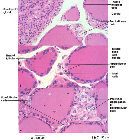
*[http://www.anatomyatlases.org/MicroscopicAnatomy/Images/Plate287.jpg Parafollicular cells (anatomyatlases.org)]. | *[http://www.anatomyatlases.org/MicroscopicAnatomy/Images/Plate287.jpg Parafollicular cells (anatomyatlases.org)]. | ||
<gallery> | <gallery> | ||
| Line 89: | Line 128: | ||
Image:Medullary_thyroid_carcinoma_-_high_mag.jpg | MTC - high mag. (WC) | Image:Medullary_thyroid_carcinoma_-_high_mag.jpg | MTC - high mag. (WC) | ||
Image:Medullary_thyroid_carcinoma_-_2_-_high_mag.jpg | MTC and amyloid - high mag. (WC) | Image:Medullary_thyroid_carcinoma_-_2_-_high_mag.jpg | MTC and amyloid - high mag. (WC) | ||
Image:Thyroid MedullaryCarcinoma 216 PA.JPG|Thyroid - Medullary carcinoma - low power (SKB) | |||
Image:Thyroid MedullaryCarcinoma 217 PA.JPG|Thyroid - Medullary carcinoma - low power (SKB) | |||
Image:Thyroid MedullaryCarcinoma Amyloid RBWH.JPG|Thyroid - Medullary carcinoma - amyloid (SKB) | |||
Image:Thyroid MedullaryCarcinoma Amyloid MP3 PA.JPG|Thyroid - Medullary carcinoma - amyloid - medium power (SKB) | |||
Image:Thyroid MedullaryCarcinoma Amyloid MP2 PA.JPG|Thyroid - Medullary carcinoma - amyloid - medium power (SKB) | |||
Image:Thyroid MedullaryCarcinoma Amyloid MP4 PA.JPG|Thyroid - Medullary carcinoma - amyloid - medium power (SKB) | |||
Image:Thyroid MedullaryCarcinoma Amyloid MP PA.JPG|Thyroid - Medullary carcinoma - amyloid - medium power (SKB) | |||
Image:Thyroid MedullaryCarcinoma Amyloid HP PA.JPG|Thyroid - Medullary carcinoma - amyloid - high power (SKB) | |||
Image:Thyroid MedullaryCarcinoma Comedonecrosis LP2 CTR.jpg|Thyroid - Medullary carcinoma - comedonecrosis - low power (SKB) | |||
Image:Thyroid MedullaryCarcinoma Comedonecrosis LP CTR.jpg|Thyroid - Medullary carcinoma - comedonecrosis - medium power (SKB) | |||
Image:Thyroid MedullaryCarcinoma Comedonecrosis HP CTR.jpg|Thyroid - Medullary carcinoma - comedonecrosis - high power (SKB) | |||
Image:Thyroid MedullaryCarcinoma SpindleCell LP PA.JPG|Thyroid - Medullary carcinoma - Spindle cell variant - low power (SKB) | |||
Image:Thyroid MedullaryCarcinoma SpindleCell MP PA.JPG|Thyroid - Medullary carcinoma - Spindle cell variant - medium power (SKB) | |||
Image:Thyroid MedullaryCarcinoma SpindleCell LP2 PA.JPG|Thyroid - Medullary carcinoma - Spindle cell variant - low power (SKB) | |||
Image:Thyroid MedullaryCarcinoma SpindleCell HP PA.JPG|Thyroid - Medullary carcinoma - Spindle cell variant - high power (SKB) | |||
Image:Thyroid MedullaryCarcinoma SmallCell HP PA.JPG|Thyroid - Medullary carcinoma - Small cell variant - high power (SKB) | |||
Image:Thyroid MedullaryCarcinoma SmallCellVariant MP CTR.jpg|Thyroid - Medullary carcinoma - small cell variant - medium power (SKB) | |||
Image:Thyroid MedullaryCarcinoma SmallCellVariant HP CTR (3).jpg|Thyroid - Medullary carcinoma - small cell variant - high power (SKB) | |||
Image:Thyroid MedullaryCarcinoma MP CTR (2).jpg|Thyroid - Medullary carcinoma - medium power (SKB) | |||
Image:Thyroid MedullaryCarcinoma HP2 CTR.jpg|Thyroid - Medullary carcinoma - high power (SKB) | |||
</gallery> | </gallery> | ||
==Stains== | |||
*Congo-red +ve (amyloid present) - mnemonic: ''CRAP'' -- congo red amyloid protein. | |||
==IHC== | ==IHC== | ||
Features:<ref>URL: [http://pathologyoutlines.com/thyroid.html#medullary http://pathologyoutlines.com/thyroid.html#medullary]. Accessed on: 17 January 2011.</ref> | Features:<ref>URL: [http://pathologyoutlines.com/thyroid.html#medullary http://pathologyoutlines.com/thyroid.html#medullary]. Accessed on: 17 January 2011.</ref> | ||
*[[Calcitonin]] +ve - it arises from C cells (which produce calcitonin). | *[[Calcitonin]] +ve - it arises from C cells (which produce calcitonin). | ||
*Neuroendocrine markers. | *Neuroendocrine markers. | ||
**[[Chromogranin A]]. | **[[Chromogranin A]]. | ||
**[[Synaptophysin]]. | **[[Synaptophysin]]. | ||
*CEA +ve (often better staining than calcitonin).<ref>SB. 7 January 2010.</ref> | *[[CEA]] +ve (often better staining than calcitonin).<ref>SB. 7 January 2010.</ref> | ||
*Thyroglobulin usu. -ve.<ref name=pmid8454270>{{Cite journal | last1 = de Micco | first1 = C. | last2 = Chapel | first2 = F. | last3 = Dor | first3 = AM. | last4 = Garcia | first4 = S. | last5 = Ruf | first5 = J. | last6 = Carayon | first6 = P. | last7 = Henry | first7 = JF. | last8 = Lebreuil | first8 = G. | title = Thyroglobulin in medullary thyroid carcinoma: immunohistochemical study with polyclonal and monoclonal antibodies. | journal = Hum Pathol | volume = 24 | issue = 3 | pages = 256-62 | month = Mar | year = 1993 | doi = | PMID = 8454270 }}</ref> | *Thyroglobulin usu. -ve.<ref name=pmid8454270>{{Cite journal | last1 = de Micco | first1 = C. | last2 = Chapel | first2 = F. | last3 = Dor | first3 = AM. | last4 = Garcia | first4 = S. | last5 = Ruf | first5 = J. | last6 = Carayon | first6 = P. | last7 = Henry | first7 = JF. | last8 = Lebreuil | first8 = G. | title = Thyroglobulin in medullary thyroid carcinoma: immunohistochemical study with polyclonal and monoclonal antibodies. | journal = Hum Pathol | volume = 24 | issue = 3 | pages = 256-62 | month = Mar | year = 1993 | doi = | PMID = 8454270 }}</ref> | ||
*TTF-1 +ve | |||
==EM== | ==EM== | ||
| Line 106: | Line 168: | ||
Images: [http://pathhsw5m54.ucsf.edu/case7/image77.html Neurosecretory granules (ucsf.edu)]. | Images: [http://pathhsw5m54.ucsf.edu/case7/image77.html Neurosecretory granules (ucsf.edu)]. | ||
==Sign out== | |||
<pre> | |||
Lesion, Liver, Core Biopsy: | |||
- METASTATIC MEDULLARY THYROID CARCINOMA, see comment. | |||
Comment: | |||
Stains/IHC confirm the morphologic findings; the tumour stains as follows: | |||
POSITIVE: calcitonin, CEAp, synaptophysin, CD56, TTF-1 (focal, moderate), congo red (confirms the presence of amyloid). | |||
NEGATIVE: thyroglobulin, CDX2. | |||
</pre> | |||
===Micro=== | |||
The sections show cells of intermediate size without apparent nucleoli, moderate eosinophilic cytoplasm, arranged in nests, focally associated with amorphous acellular cotton candy-like material. | |||
The cotton candy-like material has a light apple-green appearance when polarized. | |||
==See also== | ==See also== | ||
Latest revision as of 12:49, 26 April 2018
| Medullary thyroid carcinoma | |
|---|---|
| Diagnosis in short | |
 Medullary thyroid carcinoma. H&E stain. | |
|
| |
| LM | nuclei with neuroendocrine features (round nuclei with salt-and-pepper chromatin), +/-amyloid deposits (fluffy appearing acellular eosinophilic material), +/-C-cell hyperplasia |
| Stains | congo red +ve (amyloid deposits) |
| IHC | calcitonin +ve, CEA +ve, chromogranin A +ve, synaptophysin +ve, thyroglobulin -ve (usually) |
| Gross | usu. well-circumscribed, white, gray or yellow, gritty, firm |
| Staging | thyroid cancer staging |
| Site | thyroid gland |
|
| |
| Associated Dx | C-cell hyperplasia |
| Syndromes | multiple endocrine neoplasia IIa, multiple endocrine neoplasia IIb |
|
| |
| Prevalence | uncommon |
| Blood work | +/-serum calcitonin elevated |
| Prognosis | poor |
Medullary thyroid carcinoma, abbreviated MTC, is an uncommon epithelial malignancy of the thyroid gland that may be syndromic.
General
Medical school memory device - 3 M's:
- aMyloid.
- Median node dissection done.
- MEN IIa syndrome/MEN IIb syndrome.
- Medullary thyroid carcinoma.
- Pheochromocytoma.
- Parathyroid adenoma.
Epidemiology:
- Very rare.
- Poor prognosis.
- May be genetic (MEN IIa/b syndrome).
- Arises from C cells (which produce calcitonin).
Sporadic tumours
- ~80%
- Slightly older age at presentation (~45)
- Tend to be solitary
Syndromic tumours - typically:[1]
- Present in 30s or 40s.
- +/-Multifocal.
- +/-Bilateral.
- C-cell hyperplasia.
Serology:
- Serum calcitonin classically elevated.[2]
- CEA may also be elevated.
Gross
Features:[1]
- Usu. well-circumscribed.
- White, gray or yellow.
- Gritty.
- Firm.
Image:
Microscopic
Architecture - various
- Nested with delicate vascular septa
- Trabecular
- Tubular/glandular
- Pseudo-papillary
Cells
- Polygonal to spindle to small cells
- Amphophilic, somewhat granular cytoplasm
- Cells may have a more bizarre appearance
- Cells may appear to be 'falling apart'due to interstitial oedema.
Stroma
- +/-Amyloid deposits - fluffy appearing acellular eosinophilic material in the cytoplasm.
- Stroma is vascular and can show haemorrhage, hyalinised collagen, oedema or metaplastic bone
- Coarse calcification
- True psammoma bodies may be present
Nuclei
- Nuclei with "neuroendocrine features".
- Small, round nuclei.
- Coarse chromatin (salt and pepper nuclei).
Surrounding Thyroid
- +/-C-cell hyperplasia - seen with familial forms of MTC.
- C cells (AKA parafollicular cell): abundant cytoplasm - clear/pale.
Note:
- The amyloid is formed from calcitonin.[3]
DDx:
- Other thyroid tumours:
- Anaplastic thyroid carcinoma.
- Papillary thyroid carcinoma.
- Hurthle cell carcinoma
- The oncocytic variant of medullary carcinoma can be confused with Hurthle cell carcinoma. Clues to suggest medullary carcinoma:
- Cytoplasm is amphophilic as opposed to eosinophilic
- Nests of tumour cells separated by fibrous septa
- The oncocytic variant of medullary carcinoma can be confused with Hurthle cell carcinoma. Clues to suggest medullary carcinoma:
- C-cell hyperplasia.
- Invasive medullary carcinoma shows fibrosis around tumor cells and stains more weakly for calcitonin.
- Other neuroendocrine tumours - primary or metastatic:
- Paraganglioma - negative for keratin, calcitonin and CEA.
- Carcinoid - negative for calcitonin.
- Metastatic melanoma.
- Pigment.
- Melanoma markers positive, calcitonin and CEA negative.
Images
www:
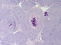
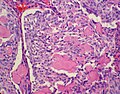
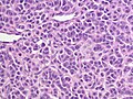
MTC - low mag. (WC)
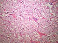
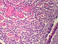
MTC - high mag. (WC)
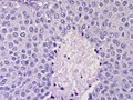
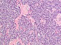
MTC and amyloid - high mag. (WC)
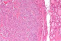
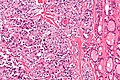
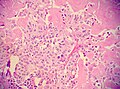
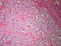
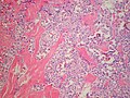
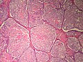
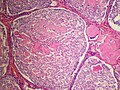
Thyroid - Medullary carcinoma - low power (SKB)
Thyroid - Medullary carcinoma - low power (SKB)
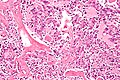
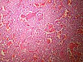
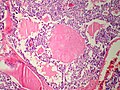
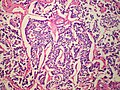
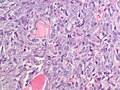
Thyroid - Medullary carcinoma - amyloid (SKB)
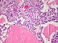
Thyroid - Medullary carcinoma - amyloid - medium power (SKB)
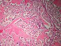
Thyroid - Medullary carcinoma - amyloid - medium power (SKB)
Thyroid - Medullary carcinoma - amyloid - medium power (SKB)
Thyroid - Medullary carcinoma - amyloid - medium power (SKB)
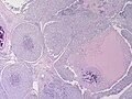
Thyroid - Medullary carcinoma - amyloid - high power (SKB)
Thyroid - Medullary carcinoma - comedonecrosis - low power (SKB)
Thyroid - Medullary carcinoma - comedonecrosis - medium power (SKB)
Thyroid - Medullary carcinoma - comedonecrosis - high power (SKB)
Thyroid - Medullary carcinoma - Spindle cell variant - low power (SKB)
Thyroid - Medullary carcinoma - Spindle cell variant - medium power (SKB)
Thyroid - Medullary carcinoma - Spindle cell variant - low power (SKB)
Thyroid - Medullary carcinoma - Spindle cell variant - high power (SKB)
Thyroid - Medullary carcinoma - Small cell variant - high power (SKB)
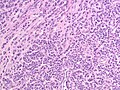
Thyroid - Medullary carcinoma - small cell variant - medium power (SKB)
Thyroid - Medullary carcinoma - small cell variant - high power (SKB)
Thyroid - Medullary carcinoma - medium power (SKB)
Thyroid - Medullary carcinoma - high power (SKB)


.jpg)
.jpg)

Stains
- Congo-red +ve (amyloid present) - mnemonic: CRAP -- congo red amyloid protein.
IHC
Features:[4]
- Calcitonin +ve - it arises from C cells (which produce calcitonin).
- Neuroendocrine markers.
- CEA +ve (often better staining than calcitonin).[5]
- Thyroglobulin usu. -ve.[6]
- TTF-1 +ve
EM
- Neurosecretory granules.
- Feature seen in neuroendocrine tumours.
Images: Neurosecretory granules (ucsf.edu).
Sign out
Lesion, Liver, Core Biopsy:
- METASTATIC MEDULLARY THYROID CARCINOMA, see comment.

Comment:
Stains/IHC confirm the morphologic findings; the tumour stains as follows:
POSITIVE: calcitonin, CEAp, synaptophysin, CD56, TTF-1 (focal, moderate), congo red (confirms the presence of amyloid).
NEGATIVE: thyroglobulin, CDX2.
Micro
The sections show cells of intermediate size without apparent nucleoli, moderate eosinophilic cytoplasm, arranged in nests, focally associated with amorphous acellular cotton candy-like material.
The cotton candy-like material has a light apple-green appearance when polarized.
See also
References
- ↑ 1.0 1.1 Nosé, V. (Apr 2011). "Familial thyroid cancer: a review.". Mod Pathol 24 Suppl 2: S19-33. doi:10.1038/modpathol.2010.147. PMID 21455198.
- ↑ Vainas, I.; Marthopoulos, A.; Chrisoulidou, A.; Raptou, K.; Tziomalos, K.; Pazaitou-Panayiotou, K. (Jul 2013). "Calcitonin stimulation tests for the early diagnosis and follow-up of patients with C cell disease: a descriptive analysis.". Hippokratia 17 (3): 246-51. PMID 24470736.
- ↑ Khurana, R.; Agarwal, A.; Bajpai, VK.; Verma, N.; Sharma, AK.; Gupta, RP.; Madhusudan, KP. (Dec 2004). "Unraveling the amyloid associated with human medullary thyroid carcinoma.". Endocrinology 145 (12): 5465-70. doi:10.1210/en.2004-0780. PMID 15459123.
- ↑ URL: http://pathologyoutlines.com/thyroid.html#medullary. Accessed on: 17 January 2011.
- ↑ SB. 7 January 2010.
- ↑ de Micco, C.; Chapel, F.; Dor, AM.; Garcia, S.; Ruf, J.; Carayon, P.; Henry, JF.; Lebreuil, G. (Mar 1993). "Thyroglobulin in medullary thyroid carcinoma: immunohistochemical study with polyclonal and monoclonal antibodies.". Hum Pathol 24 (3): 256-62. PMID 8454270.