Difference between revisions of "Inflammatory pseudopolyp"
Jump to navigation
Jump to search
| Line 58: | Line 58: | ||
**[[Solitary rectal ulcer]]. | **[[Solitary rectal ulcer]]. | ||
Images: | ===Images=== | ||
<gallery> | |||
Image: Inflammatory polyp -- very low mag.jpg | IP - very low mag. | |||
Image: Inflammatory polyp -- low mag.jpg | IP - low mag. | |||
Image: Inflammatory polyp -- intermed mag.jpg | IP - intermed. mag. | |||
Image: Inflammatory polyp -- high mag.jpg | IP - high mag. | |||
</gallery> | |||
www: | |||
*[http://www.humpath.com/spip.php?article8234&id_document=18554 Pseudopolyp (humpath.com)]. | *[http://www.humpath.com/spip.php?article8234&id_document=18554 Pseudopolyp (humpath.com)]. | ||
*[http://missinglink.ucsf.edu/lm/IDS_106_LowerGI/Lower_GI_histo_small/24-UC-pseudoplp.jpg Pseudopolyp (ucsf.edu)]. | *[http://missinglink.ucsf.edu/lm/IDS_106_LowerGI/Lower_GI_histo_small/24-UC-pseudoplp.jpg Pseudopolyp (ucsf.edu)]. | ||
Revision as of 03:29, 15 December 2013
| Inflammatory pseudopolyp | |
|---|---|
| Diagnosis in short | |
 Inflammatory polyp. H&E stain. | |
|
| |
| LM | polypoid shape, inflammatory cells - esp. neutrophils |
| LM DDx | juvenile polyp, mucosal prolapse, adenomatous polyps |
| Site | colon, rectum, others |
|
| |
| Clinical history | +/-inflammatory bowel disease |
| Prevalence | not common |
| Prognosis | benign |
| Clin. DDx | other types of polyps |
| Inflammatory pseudopolyp | |
|---|---|
| External resources | |
| EHVSC | 9992 |
Inflammatory pseudopolyp is a benign polypoid lesion usually seen in the context of inflammatory bowel disease.
It is also referred to as inflammatory polyp.
General
- Not a true polyp.
- The label inflammatory pseudopolyp = inflammatory bowel disease (IBD).
- If there is no history of IBD... reconsider the diagnosis.
Microscopic
Features:
- Polypoid shape.
- Inflammation - esp. neutrophils - key feature.
Negatives:
- No nuclear atypia.
- May have focal nuclear hyperchromasia and nuclear enlargement.
- No dilated glands.
DDx:
Images
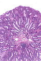
IP - very low mag.
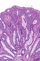
IP - low mag.
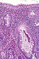
IP - intermed. mag.
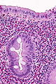
IP - high mag.


www:
Sign out
SIGMOID COLON POLYP, PERI-DIVERTICULAR, BIOPSY:
- INFLAMMATORY PSEUDOPOLYP.
POLYP, DESCENDING COLON, BIOPSY:
- INFLAMED POLYPOID FRAGMENT OF COLORECTAL-TYPE MUCOSA.
-- NEGATIVE FOR DYSPLASIA.
Micro
The sections show a fragment of colorectal mucosa with focal ulceration, acute inflammation and a well-vascularized stroma with plump stromal cells. Occasional stromal cells have nuclear hyperchromasia.
See also
References
- ↑ Aust, DE.; Rüschoff, J. (Jul 2011). "[Polyps of the colorectum: non-neoplastic and non-hamartomatous].". Pathologe 32 (4): 297-302. doi:10.1007/s00292-011-1435-1. PMID 21607734.