Difference between revisions of "Gynecologic cytopathology"
m (→Abnormal cells) |
|||
| (172 intermediate revisions by the same user not shown) | |||
| Line 1: | Line 1: | ||
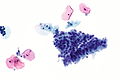
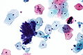
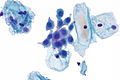
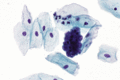
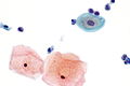
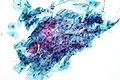
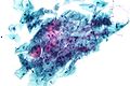
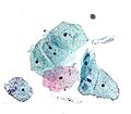
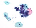
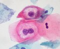
[[Image:Low grade squamous intraepithelial lesion.jpg|thumb|right|250px|A cytology specimen with LSIL. [[Pap stain]]. (WC/Nephron)]] | |||
'''Gynecologic cytopathology''' is a subset of [[cytopathology]]. ''Gynecologic'' usually refers to Pap test specimens, i.e. uterine cervix, vaginal vault; other gynecologic specimens are considered ''non-gynecologic''. | '''Gynecologic cytopathology''' is a subset of [[cytopathology]]. ''Gynecologic'' usually refers to Pap test specimens, i.e. uterine cervix, vaginal vault; other gynecologic specimens are considered ''non-gynecologic''. | ||
This article deals only with cervical cytopathology. An introduction to cytopathology is in the ''[[cytopathology]]'' article. | This article deals only with cervical cytopathology. An introduction to cytopathology is in the ''[[cytopathology]]'' article. | ||
'''Cervical cytology''' redirects to this article. | |||
=Preparation= | |||
The standard for Pap test is the Papanicolaou stain. It is described in the ''[[staining]]'' article and discussed in the context of cytopathology in the ''[[cytopathology]]'' article. | The standard for Pap test is the Papanicolaou stain. It is described in the ''[[staining]]'' article and discussed in the context of cytopathology in the ''[[cytopathology]]'' article. | ||
=Slide marking conventions= | |||
Conventions are important for facilitating communication between various team members. They are discussed in the ''[[cytopathology]]'' article. | Conventions are important for facilitating communication between various team members. They are discussed in the ''[[cytopathology]]'' article. | ||
=Normal cells= | |||
Squamous cell types:<ref>Half-day. 10 November 2008.</ref> | Squamous cell types:<ref>Half-day. 10 November 2008.</ref> | ||
#Intermediate cells: | #Intermediate cells: | ||
| Line 16: | Line 19: | ||
#*Associated with progesterone - (light) blue. | #*Associated with progesterone - (light) blue. | ||
#*This '''is''' the cell of reference in Pap test, i.e. other cells are measured against this cell when assessing a Pap test. | #*This '''is''' the cell of reference in Pap test, i.e. other cells are measured against this cell when assessing a Pap test. | ||
#**Nucleus ~ 7-8 micrometers. | |||
#***Slightly smaller than a [[neutrophil]]. | |||
#Parabasal cells: | #Parabasal cells: | ||
#*Blue-grey. | #*Blue-grey. | ||
#* | #*Associated with atrophy. | ||
#Basal cells: | #Basal cells: | ||
#*Small cells. | #*Small cells. | ||
| Line 29: | Line 34: | ||
Glandular cells:<ref>SM. 14 January 2010.</ref> | Glandular cells:<ref>SM. 14 January 2010.</ref> | ||
*Sheets of cells with regular spacing. | *Sheets of cells with regular spacing. | ||
*Relatively high NC ratio (when compared to intermediate cells). | *Relatively high [[NC ratio]] (when compared to intermediate cells). | ||
*Nucleoli (like most glandular cells). | *Nucleoli (like most glandular cells). | ||
*Nucleus approximately the size of an intermediate cell nucleus. | *Nucleus approximately the size of an intermediate cell nucleus. | ||
===Mix of cells | ===Images=== | ||
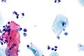
<gallery> | |||
Image: Benign endocervical epithelium -- high mag.jpg | Endocervical epithelium - high mag. (WC) | |||
Image: Benign endocervical epithelium -- very high mag.jpg | Endocervical epithelium - very high mag. (WC) | |||
Image: Benign endocervical epithelium - 2 -- high mag.jpg | Endocervical epithelium - high mag. (WC) | |||
Image: Benign endocervical epithelium - 2 -- very high mag.jpg | Endocervical epithelium - very high mag. (WC) | |||
Image: Benign endocervical epithelium - 3 -- high mag.jpg | Endocervical epithelium - high mag. (WC) | |||
Image: Benign endocervical epithelium - 3 -- very high mag.jpg | Endocervical epithelium - very high mag. (WC) | |||
</gallery> | |||
<gallery> | |||
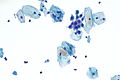
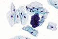
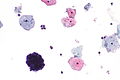
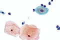
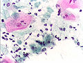
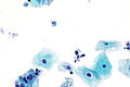
Image:Low-grade_sil_and_endocx.jpg | Endocervical cells and LSIL. (WC) | |||
</gallery> | |||
==Mix of cells== | |||
The mix of cells is dependent on age and hormones:<ref>GR. 4 February 2010.</ref> | The mix of cells is dependent on age and hormones:<ref>GR. 4 February 2010.</ref> | ||
*Progesterone - makes the Pap test blue... more intermediate cells. | *Progesterone - makes the Pap test blue... more intermediate cells. | ||
| Line 40: | Line 58: | ||
*Older patients... more estrogen, glycogen. | *Older patients... more estrogen, glycogen. | ||
== | ==Less common non-malignant cells== | ||
*Clue cells. | |||
*Squamous metaplastic cells. | |||
*Endometrial cells. | |||
*Atrophic cells. | |||
*Tingible body macrophages. | |||
*Navicular cells. | |||
===Clue cells=== | |||
Features: | |||
*Purple squamous cell covered with rod-shaped bacteria. | |||
Notes: | |||
*The cytologic finding of ''[[bacterial vaginosis]]''. | |||
Image: | |||
*[http://www.atsu.edu/faculty/chamberlain/Website/lectures/lecture/image/clue2.jpg Clue cell (atsu.edu)]. | *[http://www.atsu.edu/faculty/chamberlain/Website/lectures/lecture/image/clue2.jpg Clue cell (atsu.edu)]. | ||
*[http:// | |||
===Squamous metaplastic cells=== | |||
Features: | |||
*"Dense" cytoplasm. | |||
*Nucleus ~2X the size of an intermediate cell nucleus. | |||
**Nucleolus (small) - '''important'''. | |||
**Regular/smooth nuclear membrane. | |||
Note: | |||
*Squamous metaplastic cells have a similar appearance to parabasal cells; they cannot be differentiated on morphologic grounds. | |||
*Squamous metaplastic cells have a high NC ratio - they are differentiated from HSIL via nuclear features (dark staining + irregular nuclear contour = HSIL). | |||
**Slight nuc. contour irregularies are accepted, may be darker staining. | |||
====Images==== | |||
<gallery> | |||
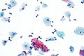
Image: Squamous metaplasia - Pap test -- high mag.jpg | SM - high mag. (WC) | |||
Image: Squamous metaplasia - Pap test -- very high mag.jpg | SM - very high mag. (WC) | |||
Image: Squamous metaplasia - Pap test - alt -- very high mag.jpg | SM - very high mag. (WC) | |||
</gallery> | |||
===Endometrial cells=== | |||
Features:<ref>SM. 14 January 2010.</ref> | |||
*Cluster of cells with a well-defined border that is bilayered, i.e. a clump of (epithelioid) stromal cells surrounded by (flatted) glandular cells. Classically described as a cluster with a ''double contour''; known as ''exodus pattern''.<ref>URL: [http://nih.techriver.net/view.php?patientId=78 http://nih.techriver.net/view.php?patientId=78]. Accessed on: 31 March 2012.</ref> | |||
*Scant cytoplasm. | |||
*Chromatin clumping. | |||
*Raisin-like nuclei - approximately the size of an intermediate cell nucleus. | |||
**Nuclei can be considered normal if nucleus less than 2X the size of an intermediate cell nucleus. | |||
Notes: | |||
*Endometrial cells may appear irregular in the context of an [[intrauterine device]] (IUD); abnormalities in the context of an IUD are often ignored. | |||
**Cytology: cytoplasmic vacuolization, +/-multinucleation. | |||
***May be [[signet ring cell carcinoma|signet ring cell]]-like. | |||
*The presence of endometrial cells on a Pap test on a woman >=40 years old (per Bethesda guidelines) should be noted in the pathology report<ref name=pmid15900572>{{cite journal |author=Thrall MJ, Kjeldahl KS, Savik K, Gulbahce HE, Pambuccian SE |title=Significance of benign endometrial cells in papanicolaou tests from women aged >=40 years |journal=Cancer |volume=105 |issue=4 |pages=207-16 |year=2005 |month=August |pmid=15900572 |doi=10.1002/cncr.21156 |url=}}</ref> - this prompts an endometrial biopsy. | |||
**The practise of reporting ''benign'' endometrial cells in premenopausal women is ''not'' backed by evidence that demonstrates a significant benefit. | |||
====Images==== | |||
<gallery> | |||
Image: Endometrial cells on Pap - 2 -- very high mag.jpg | Endometrial cells - very high mag. | |||
Image: Endometrial cells on Pap - 2 -- very high mag.gif | Endometrial cells - very high mag. | |||
Image: Endometrial cells on Pap - 2a -- very high mag.jpg | Endometrial cells - very high mag. | |||
Image: Endometrial cells on Pap - 2a -- very high mag.gif | Endometrial cells - very high mag. | |||
</gallery> | |||
<gallery> | |||
Image: Endometrial cells on Pap -- high mag.jpg | Endometrial cells - high mag. | |||
Image: Endometrial cells on Pap -- very high mag.jpg | Endometrial cells - very high mag. | |||
Image: Endometrial cells on Pap - alt -- very high mag.jpg | Endometrial cells - very high mag. | |||
</gallery> | |||
www: | |||
*[http://rapids001.techriver.net/nih/patientImages/1826.jpg Endometrial cells - double contour (techriver.net)]. | |||
*[http://nih.techriver.net/view.php?patientId=221 Endometrial cells with "exodus" pattern (techriver.net)]. | |||
===Atrophic cells=== | |||
Features:<ref>DeMay, RM. The Art & Science of Cytopathology: Exfoliative Cytology. 1996. ISBN 0-89189-322-9. PP.116-7.</ref> | |||
*Cells smaller. | |||
*Cytoplasm grey/blue. | |||
*No "dancing"/"sparkling" chromatin. | |||
*+/-"Dirty" background - degenerated cells, inflammatory cells (neutrophils, histiocytes). | |||
**May mimic "dirty" background of tumour, i.e. 'tumour diathesis'. | |||
Notes: | |||
*Usually older women. | |||
*May be a cellular cluster. | |||
DDx: | |||
*[[HSIL]] - chromatin pattern irregular. | |||
===Tingible body macrophages=== | |||
Features: | |||
*Abundant cytoplasm with vacuolization. | |||
*May be seen in the context of chlamydia. | |||
===Navicular cells=== | |||
{{Main|Navicular cell}} | |||
Features: | |||
*Intermediate cells with: | |||
*#Folded edges. | |||
*#Abundant cytoplasmic glycogen - central yellow. | |||
====Images==== | |||
<gallery> | |||
Image: Navicular cell -- very high mag.jpg | NC - very high mag. | |||
Image: Navicular cell - alt -- very high mag.jpg | NC - very high mag. | |||
Image: Navicular cell -- extremely high mag.jpg | NC - extremely high mag. | |||
Image: Navicular cells -- extremely high mag.jpg | NCs - extremely high mag. | |||
</gallery> | |||
==Glycogen halos versus HPV effect== | ==Glycogen halos versus HPV effect== | ||
| Line 96: | Line 181: | ||
|} | |} | ||
=Gynecologic pathology in tables= | |||
==Normal cells== | |||
{| class="wikitable" | {| class="wikitable sortable" | ||
! Cell | ! Cell | ||
! Architecture | ! Architecture | ||
| Line 110: | Line 195: | ||
| Irregular | | Irregular | ||
| '''Blue, abundant''' | | '''Blue, abundant''' | ||
| Small nucleus (~ size of PMN), no [[nucleolus]] | | Small nucleus (~ size of [[PMN]]), no [[nucleolus]] | ||
| - | | - | ||
|- | |- | ||
| Line 125: | Line 210: | ||
| Dense, dark blue | | Dense, dark blue | ||
| 2X IC nucleus, '''nucleolus''', no membrane irreg., no chromatin changes | | 2X IC nucleus, '''nucleolus''', no membrane irreg., no chromatin changes | ||
| DDx: HSIL, basal cell | | DDx: [[HSIL]], basal cell | ||
|- | |- | ||
| Endometrial cell | | Endometrial cell | ||
| '''Well-circumscribed clump/ball of cells with squamoid covering cells''' | | '''Well-circumscribed clump/ball of cells with squamoid covering cells'''; referred to as "exodus" pattern<ref>URL: [http://nih.techriver.net/view.php?patientId=221 http://nih.techriver.net/view.php?patientId=221]. Accessed on: 26 November 2011.</ref> | ||
| Indistinct within cluster | | Indistinct within cluster | ||
| '''Blue, small/very scant''' | | '''Blue, small/very scant''' | ||
| Line 135: | Line 220: | ||
|- | |- | ||
| Glandular (endocervical) cell | | Glandular (endocervical) cell | ||
| '''Sheets of cells with regular spacing, columnar morphology may be apparent, +/- | | '''Sheets of cells with regular spacing, columnar morphology may be apparent, +/-palisading at edge of clump''' | ||
| Often distict | | Often distict | ||
| Blue, '''scant-to-moderate''' | | Blue, '''scant-to-moderate''' | ||
| Line 147: | Line 232: | ||
| Large NC ratio, nuc. membrane irregularities, '''NO chromatin clumping'''<ref>DeMay, RM. The Art & Science of Cytopathology: Exfoliative Cytology. 1996. ISBN 0-89189-322-9. PP.116-7.</ref> | | Large NC ratio, nuc. membrane irregularities, '''NO chromatin clumping'''<ref>DeMay, RM. The Art & Science of Cytopathology: Exfoliative Cytology. 1996. ISBN 0-89189-322-9. PP.116-7.</ref> | ||
| DDx: HSIL | | DDx: HSIL | ||
|- | |||
| [[Radiation changes in cervical cytology|Radiation changes]] | |||
| Single cells/groups | |||
| Well-circumscribed | |||
| vacuolated, usu. abundant | |||
| '''Normal NC ratio''', enlarged nucleus, no nuclear membrane irregularies, +/-multinucleation | |||
| DDx: [[LSIL]], vitamin B12 def. | |||
|} | |} | ||
Note: | |||
*If ''only'' normal cells are present the diagnosis is ''negative for intraepithelial lesion and malignancy'' (NILM). | |||
==Abnormal cells== | |||
{| class="wikitable" | {| class="wikitable" | ||
! Cell | ! Cell | ||
| Line 157: | Line 251: | ||
! DNA | ! DNA | ||
! Other | ! Other | ||
! Image | |||
|- | |- | ||
| Low-grade squamous intraepithelial lesion (LSIL) | | [[Low-grade squamous intraepithelial lesion]] (LSIL) | ||
| Single cells/groups | | Single cells/groups | ||
| Irregular or moderately-circumscribed | | Irregular or moderately-circumscribed | ||
| Line 164: | Line 259: | ||
| Large nucleus (3-4X IC nuc. - see ''Note 1''), perinuclear clearing, nuc. membrane irregularities, chromatin clumping | | Large nucleus (3-4X IC nuc. - see ''Note 1''), perinuclear clearing, nuc. membrane irregularities, chromatin clumping | ||
| DDx: HSIL, reactive changes | | DDx: HSIL, reactive changes | ||
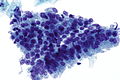
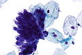
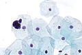
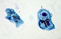
| [[Image:Low-grade squamous intraepithelial lesion - 3 -- very high mag.jpg| thumb|center|150px|LSIL (WC)]] | |||
|- | |- | ||
| High-grade squamous intraepithelial lesion (HSIL) | | [[High-grade squamous intraepithelial lesion]] (HSIL) | ||
| Often single cells, may be groups | | Often single cells, may be groups | ||
| Well-circumscribed | | Well-circumscribed | ||
| Line 171: | Line 267: | ||
| Large nucleus (3-4X IC nuc. - see ''Note 1''), '''nuc. membrane irregularities, clumping of coarse chromatin, dark nuc. staining''', +/- small nucleoli | | Large nucleus (3-4X IC nuc. - see ''Note 1''), '''nuc. membrane irregularities, clumping of coarse chromatin, dark nuc. staining''', +/- small nucleoli | ||
| DDx: squamous metaplasia, atrophy with atypia, superficial endometrial cells | | DDx: squamous metaplasia, atrophy with atypia, superficial endometrial cells | ||
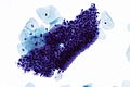
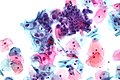
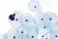
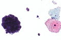
| [[Image:High-grade squamous intraepithelial lesion - 2 -- very high mag.jpg |thumb|center|150px|HSIL (WC)]] | |||
|- | |- | ||
| Atypical squamous cells of | | [[Atypical squamous cells of undetermined significance]] (ASC-US) | ||
| Single cells/groups | | Single cells/groups | ||
| Irregular or moderately-circumscribed | | Irregular or moderately-circumscribed | ||
| Line 178: | Line 275: | ||
| '''Moderately enlarged nucleus''' (~2.5-3.0X IC nuc.), minimal changes in nuclear membrane and chromatin | | '''Moderately enlarged nucleus''' (~2.5-3.0X IC nuc.), minimal changes in nuclear membrane and chromatin | ||
| DDx: LSIL, reactive changes | | DDx: LSIL, reactive changes | ||
| | |||
|- | |- | ||
| Atypical squamous cells cannot exclude HSIL (ASC-H) | | [[Atypical squamous cells, cannot exclude HSIL]] (ASC-H) | ||
| Often single cells, may be groups | | Often single cells, may be groups | ||
| Irregular or moderately-circumscribed | | Irregular or moderately-circumscribed | ||
| Line 185: | Line 283: | ||
| '''Moderately enlarged nucleus''' (~1.5-2.0X IC nuc.), minimal changes in nuclear membrane and chromatin | | '''Moderately enlarged nucleus''' (~1.5-2.0X IC nuc.), minimal changes in nuclear membrane and chromatin | ||
| DDx: HSIL, AIS | | DDx: HSIL, AIS | ||
| | |||
|- | |- | ||
| Atypical glandular | | [[Atypical glandular cells]] (AGC) | ||
| Usu. groups of cells | | Usu. groups of cells | ||
| Usually well-circumscribed (?) | | Usually well-circumscribed (?) | ||
| Line 192: | Line 291: | ||
| Moderately enlarged nucleus (~2X IC nuc.), '''nuc. membrane irregularities, chromatin clumping, dark nuc. staining''', nucleoli | | Moderately enlarged nucleus (~2X IC nuc.), '''nuc. membrane irregularities, chromatin clumping, dark nuc. staining''', nucleoli | ||
| DDx: AIS, HSIL | | DDx: AIS, HSIL | ||
| | |||
|- | |- | ||
| | | [[Adenocarcinoma in situ]] (AIS) | ||
| | | groups; '''rosette formation''' | ||
| Usually well-circumscribed | | Usually well-circumscribed | ||
| Dark blue '''dense''', scant | | Dark blue '''dense''', scant | ||
| Large nucleus (>=2X IC nuc.), '''nuc. membrane irregularities, chromatin clumping, dark nuc. staining''', nucleoli (very common), pseudostratification (as in endocervical AIS) | | Large nucleus (>=2X IC nuc.), '''nuc. membrane irregularities, chromatin clumping, dark nuc. staining''', nucleoli (very common), pseudostratification (as in endocervical AIS) | ||
| DDx: AGC, HSIL | | DDx: AGC, HSIL | ||
| [[Image:Endocervical adenocarcinoma in situ - cyto -- very high mag.jpg|thumb|center|150px|Endocervical AIS (WC)]] | |||
|- | |- | ||
| Features of SCC (see ''Note 2'') | | Features of SCC (see ''Note 2'') | ||
| Line 206: | Line 307: | ||
| Large NC ratio, '''nucleolus''', nuc. membrane irregularities, chromatin clumping | | Large NC ratio, '''nucleolus''', nuc. membrane irregularities, chromatin clumping | ||
| DDx: HSIL | | DDx: HSIL | ||
| | |||
|} | |} | ||
| Line 219: | Line 321: | ||
*By definition, it is not possible to diagnose [[squamous cell carcinoma]] (SCC) on a pap test as one cannot demonstrate stromal invasion. | *By definition, it is not possible to diagnose [[squamous cell carcinoma]] (SCC) on a pap test as one cannot demonstrate stromal invasion. | ||
===HSIL versus LSIL=== | |||
{| class="wikitable" | {| class="wikitable" | ||
! | ! | ||
| Line 253: | Line 355: | ||
| Hypermature (orangeophilic cell present) | | Hypermature (orangeophilic cell present) | ||
|- | |- | ||
| | | Images (example) | ||
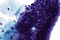
| [ | | [[Image:High-grade_squamous_intraepithelial_lesion.jpg |thumb|center|150px| HSIL (WC)]] [[Image:High-grade squamous intraepithelial lesion - 4 -- very high mag.jpg| thumb|center|150px| HSIL (WC)]] | ||
| [ | | [[Image:Low-grade_squamous_intraepithelial_lesion.jpg |thumb|center|150px| LSIL (WC)]] [[Image:Low-grade_sil_and_endocx.jpg |thumb|center|150px| LSIL & endoCx (WC)]] | ||
|} | |} | ||
| Line 264: | Line 366: | ||
**LSIL cells: classically the size of IC. | **LSIL cells: classically the size of IC. | ||
==Infectious organisms== | |||
{| class="wikitable" | {| class="wikitable sortable" | ||
! Disease | ! Disease | ||
! Organism | ! Organism | ||
| Line 275: | Line 377: | ||
! Image | ! Image | ||
|- | |- | ||
| Trichomoniasis | | [[Trichomoniasis]] | ||
| Trichomonas vaginalis | | Trichomonas vaginalis | ||
| Protozoan | | Protozoan | ||
| ''' | | Pear-shaped '''pale-grey fluffy cytoplasm with well-defined nucleus''', approx. 30 μm. | ||
| Acute inflammation (PMNs) | | Acute inflammation (PMNs), may be seen with Leptothrix (hair-like appearance ~0.5 x 20 μm) | ||
| Sexually transmitted | | Sexually transmitted | ||
| <ref name=Ref_WMSP446>{{Ref WMSP|446}}</ref> | | <ref name=Ref_WMSP446>{{Ref WMSP|446}}</ref> | ||
| [ | | [[Image:Trichomonas_pap_test.jpg |thumb|150px|center|T. vaginalis - Pap stain (WC)]] [[Image:Trichomonas - Pap - 3 -- very high mag.jpg |thumb|150px|center|Trichomonas - Pap stain (WC)]] | ||
|- | |- | ||
| Candidiasis | | [[Gynecologic_cytopathology#Candida|Candidiasis]] | ||
| Candida albicans | | Candida albicans | ||
| Fungi | | [[Fungi]] | ||
| '''Branching hyphae ~= 1/2 the dia. of IC nucleus, red''' | | '''Branching hyphae ~= 1/2 the dia. of IC nucleus, red''' | ||
| PMNs | | PMNs | ||
| ? | | ? | ||
| ? | | ? | ||
| [ | | [[Image:Candida_pap_1.jpg|thumb|150px|center| Candida on Pap test (WC)]] | ||
|- | |- | ||
| Herpes | | [[Gynecologic_cytopathology#Herpes simplex virus|Herpes]] | ||
| [[Herpes simplex virus]] (HSV 1 - less commonly, HSV 2 - more commonly) | | [[Herpes simplex virus]] (HSV 1 - less commonly, HSV 2 - more commonly) | ||
| Virus | | [[Virus]] | ||
| Large ground glass nuclei then multinucleation with moulding & inclusions with clear halo | | Large ground glass nuclei then multinucleation with moulding & inclusions with clear halo | ||
| ? | | ? | ||
| Sexually transmitted | | Sexually transmitted | ||
| ? | | ? | ||
| [ | | [[Image:Herpes_simplex_virus_pap_test.jpg |thumb|center|150px| HSV (WC)]] [[Image:Herpes_simplex_virus_pap_test_2.jpg |thumb|center|150px| HSV (WC)]] | ||
|- | |- | ||
| Actinomycetes | | [[Gynecologic_cytopathology#Actinomycetes|Actinomycetes]] | ||
| Actinomycetes | | Actinomycetes | ||
| Gram-positive bacteria | | Gram-positive bacteria | ||
| '''Clusters of cocci in chains - hyphae-like appearance''' | | '''Clusters of cocci in chains - hyphae-like appearance''' | ||
| | | low power: pom-pom ''or'' fuzzy ball-like appearance | ||
| Should prompt removal of IUD, if present. | | Should prompt removal of [[IUD]], if present. | ||
| <ref name=Ref_WMSP446>{{Ref WMSP|446}}</ref> | | <ref name=Ref_WMSP446>{{Ref WMSP|446}}</ref> | ||
| [http://www. | | [http://www.gfmer.ch/selected_images_v2/detail_list.php?cat1=4&cat2=23&cat3=552&cat4=6&stype=n Actinomycetes (gfmer.ch)], [http://o.quizlet.com/Swjzk-aC7Ah8aYQmYzQ8Kg_m.jpg Actinomycetes (quizlet.com)] | ||
|- | |- | ||
| Bacterial vaginosis (see ''Note 1'') | | [[Bacterial vaginosis]] (see ''Note 1'') | ||
| Gardnerella vaginalis | | Gardnerella vaginalis | ||
| Gram-variable rod | | Gram-variable rod | ||
| Line 318: | Line 420: | ||
| Fishy smell | | Fishy smell | ||
| ? | | ? | ||
| [ | | [[File:Vaginose-G15.jpg|thumb|center|150px|Bacterial vaginosis (WC)]] [http://www.atsu.edu/faculty/chamberlain/Website/lectures/lecture/image/clue2.jpg Clue cell (atsu.edu)] | ||
|} | |} | ||
| Line 324: | Line 426: | ||
*Usually not reported. | *Usually not reported. | ||
=Adequacy of specimens= | |||
There is a generally accepted standard for cervical (liquid-based) cytology specimens:<ref>UHN PCY50001.08 P.10.</ref> | There is a generally accepted standard for cervical (liquid-based) cytology specimens:<ref>UHN PCY50001.08 P.10.</ref> | ||
*>5000 squamous cells/slide, if no abnormality is present. | *>5000 squamous cells/slide, if no abnormality is present. | ||
| Line 333: | Line 435: | ||
Note: | Note: | ||
*The standard for conventional pap smears is: 8000-12000 squamous cells | *The standard for conventional pap smears is: 8000-12000 (well-visualized) squamous cells.<ref name=pmid12645338>{{Cite journal | last1 = Sheffield | first1 = MV. | last2 = Simsir | first2 = A. | last3 = Talley | first3 = L. | last4 = Roberson | first4 = AJ. | last5 = Elgert | first5 = PA. | last6 = Chhieng | first6 = DC. | title = Interobserver variability in assessing adequacy of the squamous component in conventional cervicovaginal smears. | journal = Am J Clin Pathol | volume = 119 | issue = 3 | pages = 367-73 | month = Mar | year = 2003 | doi = | PMID = 12645338 }}</ref> | ||
===Transformation zone (TZ)=== | ===Transformation zone (TZ)=== | ||
The presence of the TZ should be commented on:<ref>GR. 4 February 2010.</ref> | The presence of the TZ should be commented on:<ref>GR. 4 February 2010.</ref> | ||
| Line 343: | Line 446: | ||
*Young nulliparous. | *Young nulliparous. | ||
=Specific entities - infectious= | |||
==Candida== | ==Candida== | ||
{{Main|Candidiasis}} | |||
===General=== | |||
*Common. | |||
*May be asymptomatic. | |||
*Usually ''Candida albicans''. | |||
===Cytology=== | |||
Features: | Features: | ||
*Typically in clusters - lead to darkened clusters of squamous cells (at low power). | *Typically in clusters - lead to darkened clusters of squamous cells (at low power). | ||
| Line 352: | Line 463: | ||
*Presence should be noted in the pathology report. | *Presence should be noted in the pathology report. | ||
Images: | ====Images==== | ||
<gallery> | |||
Image:Candida_pap_1.jpg | Candida on Pap test - example 1. (WC) | |||
Image:Candida_pap_2.jpg | Candida on Pap test - example 2. (WC) | |||
</gallery> | |||
<gallery> | |||
Image: Candida - Pap test -- high mag.jpg | Candida - high mag. (WC) | |||
Image: Candida - Pap test -- very high mag.jpg | Candida - very high mag. (WC) | |||
</gallery> | |||
=====www===== | |||
*[http://www.flickr.com/photos/moorepix4u2c/1425271033/in/set-72157602113534479/ Candida on Pap test (flickr.com)]. | *[http://www.flickr.com/photos/moorepix4u2c/1425271033/in/set-72157602113534479/ Candida on Pap test (flickr.com)]. | ||
== | ==Trichomoniasis== | ||
===General=== | |||
*Caused by ''Trichomonas vaginalis'' - a protozoa. | |||
*Sexually transmitted. | *Sexually transmitted. | ||
*Common. | |||
*Occasionally found in [[urine cytology]] specimens.<ref>{{cite journal |authors=Doxtader EE, Elsheikh TM |title=Diagnosis of trichomoniasis in men by urine cytology |journal=Cancer Cytopathol |volume=125 |issue=1 |pages=55–59 |date=January 2017 |pmid=27636204 |doi=10.1002/cncy.21778 |url=}}</ref> | |||
===Cytopathology=== | ===Cytopathology=== | ||
Features: | Features: | ||
*Low power: grey blob with a nucleus | *Low power: grey blob with a nucleus, may be pear-shaped: | ||
**Size: approximately 30 micrometres.<ref name=Ref_WMSP446>{{Ref WMSP|446}}</ref> | **Size: approximately 30 micrometres.<ref name=Ref_WMSP446>{{Ref WMSP|446}}</ref> | ||
**Shape: usually oval, may have teardrop-shaped. | **Shape: usually oval, may have teardrop-shaped. | ||
| Line 376: | Line 498: | ||
Notes: | Notes: | ||
*Trichomonas is tricky - it is easy to miss if one is not suspicious, in the context of inflammation. | *Trichomonas is tricky - it is easy to miss if one is not suspicious, in the context of inflammation. | ||
*May vaguely resemble a neutrophil: | *May vaguely resemble a [[neutrophil]]: | ||
**Flagellum useful to differentiate. | **Flagellum useful to differentiate. | ||
**Neutrophil has multiple lobulations of the nucleus. | **Neutrophil has multiple lobulations of the nucleus. | ||
*May be seen in association of Leptothrix. | |||
**Appearance: long, hair-like. | |||
**Size: ~0.5 x 20 micrometres. | |||
Images | ====Images==== | ||
<gallery> | |||
Image:Trichomonas_pap_test.jpg | T. vaginalis - Pap stain. (WC) | |||
*[http:// | Image:Pap_test_trichomonas.JPG | T. vaginalis - Pap stain. (WC) | ||
*[http:// | Image:Trichomonas_vaginalis_01.jpg | Trichomonas vaginalis - Giemsa stain. (WC) | ||
</gallery> | |||
<gallery> | |||
Image: Trichomonas - Pap - 2 -- high mag.jpg | Trichomonas - high mag. | |||
Image: Trichomonas - Pap - 2 -- very high mag.jpg | Trichomonas - very high mag. | |||
</gallery> | |||
<gallery> | |||
Image: Trichomonas - Pap - 3 -- high mag.jpg | Trichomonas - high mag. | |||
Image: Trichomonas - Pap - 3 -- very high mag.jpg | Trichomonas - very high mag. | |||
</gallery> | |||
=====www===== | |||
*[http://nih.techriver.net/view.php?patientId=305 Trichomonas and Leptothrix (nih.techriver.net)]. | |||
*[http://nih.techriver.net/view.php?patientId=325 Trichomonas and Leptothrix (nih.techriver.net)]. | |||
==Herpes simplex virus== | ==Herpes simplex virus== | ||
{{Main|Herpes simplex virus}} | |||
===General=== | |||
*May be ''HSV1'' or ''HSV2''. | |||
**Classically HSV2 based on epidemiology and location. | |||
===Cytology=== | |||
Features:<ref name=Ref_WMSP446>{{Ref WMSP|446}}</ref> | Features:<ref name=Ref_WMSP446>{{Ref WMSP|446}}</ref> | ||
#Early: Large "ground-glass" nuclei - nuclei with hazy & uniformly dull appearance. | #Early: Large "ground-glass" nuclei - nuclei with hazy & uniformly dull appearance. | ||
#Late: multi-nucleation with moulding of nuclei and nuclear inclusions surrounded by a clear halo. | #Late: multi-nucleation with moulding of nuclei and nuclear inclusions surrounded by a clear halo. | ||
DDx: | |||
*[[Reactive endocervical cells]] - may be multinucleated. | |||
====Image==== | |||
<gallery> | |||
Image:Herpes simplex virus pap test.jpg | HSV on pap test. (WC) | |||
</gallery> | |||
==Actinomycetes== | ==Actinomycetes== | ||
| Line 406: | Line 557: | ||
*''Mycete'' = fungus.<ref>URL: [http://en.wiktionary.org/wiki/-mycete#English http://en.wiktionary.org/wiki/-mycete#English]. Accessed on: 14 September 2011.</ref> | *''Mycete'' = fungus.<ref>URL: [http://en.wiktionary.org/wiki/-mycete#English http://en.wiktionary.org/wiki/-mycete#English]. Accessed on: 14 September 2011.</ref> | ||
== | DDx - sulfur granule:<ref name=asc_cockle>URL: [http://www.cytology-asc.com/cec/normal/index.htm#cockle http://www.cytology-asc.com/cec/normal/index.htm#cockle]. Accessed on: 10 April 2012.</ref> | ||
*Hematoidin (cockleburr) crystal - radiating crystal, refractile, classically golden-brown. | |||
==Bacterial vaginosis== | |||
===General=== | |||
*Benign. | |||
*Very common. | |||
*Classically associated with ''Gardnerella vaginalis''.<ref name=pmid3493202>{{cite journal |author=Scott TG, Smyth CJ, Keane CT |title=In vitro adhesiveness and biotype of Gardnerella vaginalis strains in relation to the occurrence of clue cells in vaginal discharges |journal=Genitourinary medicine |volume=63 |issue=1 |pages=47–53 |year=1987 |month=February |pmid=3493202 |pmc=1194007 |doi= |url=}}</ref><ref name=pmid22082330>{{Cite journal | last1 = Polatti | first1 = F. | title = Bacterial vaginosis, Atopobium vaginae and nifuratel. | journal = Curr Clin Pharmacol | volume = 7 | issue = 1 | pages = 36-40 | month = Feb | year = 2012 | doi = | PMID = 22082330 }}</ref> | |||
Clinical: | |||
*Fishy odor. | |||
Treatment: | |||
*Antibiotics (metronidazole or clindamycin).<ref name=pmid22082330/> | |||
===Cytopathology=== | |||
Features: | |||
*Purple squamous cell covered with rod-shaped micro-organisms. | |||
Image: | |||
*[http://www.atsu.edu/faculty/chamberlain/Website/lectures/lecture/image/clue2.jpg Clue cell (atsu.edu)]. | |||
===Stains=== | |||
*Gram stain +ve/-ve. | |||
**''Gardnerella vaginalis'' is a gram variable rod.<ref name=pmid6399409>{{cite journal |author=Taylor-Robinson D |title=The bacteriology of Gardnerella vaginalis |journal=Scand J Urol Nephrol Suppl |volume=86 |issue= |pages=41–55 |year=1984 |pmid=6399409 |doi= |url=}}</ref> | |||
===Sign out=== | |||
*Usually not reported. | |||
=Squamous intraepithelial lesions= | |||
*Abbreviated ''SIL''. | |||
General: | General: | ||
*The nucleus makes it SIL. | *The nucleus makes it SIL. | ||
| Line 413: | Line 595: | ||
Management (in short): | Management (in short): | ||
*LSIL = repeat Pap test in 6 months. | *LSIL = repeat Pap test in 6 months. | ||
*HSIL = | *HSIL = referral for coloposcopy. | ||
==Low-grade squamous intraepithelial lesion | ==Low-grade squamous intraepithelial lesion== | ||
*Abbreviated '''LSIL'''. | |||
{{Main|Low-grade squamous intraepithelial lesion}} | |||
===General=== | |||
*Usually regress, i.e. will disappear on their own. | *Usually regress, i.e. will disappear on their own. | ||
*Low inter-rater concordance.<ref name=pmid22007754>{{Cite journal | last1 = Bigras | first1 = G. | last2 = Wilson | first2 = J. | last3 = Russell | first3 = L. | last4 = Johnson | first4 = G. | last5 = Morel | first5 = D. | last6 = Saddik | first6 = M. | title = Interobserver concordance in the assessment of features used for the diagnosis of cervical atypical squamous cells and squamous intraepithelial lesions (ASC-US, ASC-H, LSIL and HSIL). | journal = Cytopathology | volume = 24 | issue = 1 | pages = 44-51 | month = Feb | year = 2013 | doi = 10.1111/j.1365-2303.2011.00930.x | PMID = 22007754 }}</ref> | |||
===Cytopathology=== | ===Cytopathology=== | ||
Features: | Features: | ||
#Nuclei 3x size of intermediate cell - '''key feature'''. † | |||
#Irregular nuclear border. | |||
#+/-Perinuclear 'cavity' (clearing). | |||
#*The best perinuclear halos have a sharp punched-out edge. | |||
#Chromatin clumping/irregular & granular. | |||
Images: | Note: | ||
*[http://www.flickr.com/photos/moorepix4u2c/1440144102/in/set-72157602113534479/ LSIL | * † Nucleus diameter ~21-24 μm. | ||
* In the context of exams: 2 of criteria 1-3 is enough to call LSIL.<ref>Chan, S. 26 April 2012.</ref> | |||
====Images==== | |||
<gallery> | |||
Image:Low-Grade_SIL_with_HPV_Effect.jpg | LSIL with HPV effect. (WC) | |||
Image:Low_grade_squamous_intraepithelial_lesion.jpg | LSIL. (WC) | |||
Image:ThinPrep_Pap_smear_HPV.jpeg | LSIL. (WC) | |||
</gallery> | |||
www: | |||
*[http://www.flickr.com/photos/moorepix4u2c/1440144102/in/set-72157602113534479/ Possible LSIL (flickr.com)]. | |||
===Sign out=== | |||
<pre> | |||
Low grade squamous intraepithelial lesion (LSIL). | |||
</pre> | |||
==High-grade intraepithelial lesion | ====Cannot exclude HSIL==== | ||
*Often progress to cervical cancer. | <pre> | ||
At least low grade squamous intraepithelial lesion; CANNOT EXCLUDE high-grade squamous intraepithelial lesion. | |||
</pre> | |||
==High-grade squamous intraepithelial lesion== | |||
*Abbreviated '''HSIL'''. | |||
{{Main|Squamous intraepithelial lesion of the uterine cervix}} | |||
===General=== | |||
*Often progress to [[cervical cancer]]. | |||
===Cytopathology=== | ===Cytopathology=== | ||
Features: | Features: | ||
*Often single cells. | *Often single cells, may be in clusters. | ||
*Blue cells - nucleus and cytoplasm. | *Blue cells - nucleus and cytoplasm. | ||
*Increased NC ratio - '''key feature'''. | *Increased NC ratio - '''key feature'''. | ||
*Irregular nuclear border. | **Irregular nuclear border. | ||
*Chromatin clumping. | **Chromatin clumping. | ||
Note: | |||
*Nucleoli uncommon - should prompt consideration of [[squamous carcinoma]]. | |||
DDx: | |||
*[[LSIL]]. | |||
*[[ASC-H]]. | |||
*[[Squamous carcinoma]]. | |||
Image | ====Images==== | ||
<gallery> | |||
Image:High-Grade_SIL.jpg | HSIL. (WC) | |||
Image:High-grade_squamous_intraepithelial_lesion.jpg | HSIL. (WC/Nephron) | |||
</gallery> | |||
===Squamous cell carcinoma | ===Squamous cell carcinoma=== | ||
*Abbreviated ''SCC''. | |||
{{Main|Squamous cell carcinoma}} | {{Main|Squamous cell carcinoma}} | ||
*Some believe that one can diagnosis SCC on a pap test. | *Some believe that one can diagnosis SCC on a pap test. | ||
| Line 450: | Line 670: | ||
Features suggestive of invasion: | Features suggestive of invasion: | ||
*Nucleoli. | *Loose clumps of ovoid-to-spindled cells with: | ||
* | **+/-Orange/red cytoplasm (orangeophilic cytoplasm). | ||
*Necrotic debris. | **Nucleoli - '''key feature'''. | ||
* | **Coarse chromatin. | ||
**Nuclear hyperchromasia. | |||
*Necrotic debris - often obscures cell borders: | |||
**Anucleate, fragmented cells - cytoplasm-like material. | |||
**Neutrophils. | |||
Note: | |||
*Nucleoli DDx: | |||
**[[Reactive squamous epithelium of the uterine cervix|Reactive changes]]. | |||
**Glandular lesions ([[adenocarcinoma in situ]], atypical glandular cells). | |||
Image: | Image: | ||
*[http://commons.wikimedia.org/wiki/File:Squamous_cell_carcinoma_in_the_cervix,_pap_stain.jpg HSIL with features suggestive of invasion (WC)] | *[http://commons.wikimedia.org/wiki/File:Squamous_cell_carcinoma_in_the_cervix,_pap_stain.jpg HSIL with features suggestive of invasion (WC)] | ||
== | =Glandular lesions= | ||
*Abbreviated '' | ==Adenocarcinoma in situ== | ||
*Abbreviated ''AIS''. | |||
''Adenocarcinoma in situ'' on Pap test is classically divided into: | |||
Adenocarcinoma on Pap test is classically divided into: | |||
*Endocervical. | *Endocervical. | ||
*Uterine. | *Uterine. | ||
*Extra-uterine. | *Extra-uterine. | ||
Adenocarcinoma vs. squamous carcinoma: | |||
*"Feathering" - seen in adenocarcinoma<ref>URL: [http://www.cytology-asc.com/cec/endocx/ http://www.cytology-asc.com/cec/endocx/]. Accessed on: 13 September 2011.</ref> more commonly on smears.<ref name=pmid18335553>{{Cite journal | last1 = Belsley | first1 = NA. | last2 = Tambouret | first2 = RH. | last3 = Misdraji | first3 = J. | last4 = Muzikansky | first4 = A. | last5 = Russell | first5 = DK. | last6 = Wilbur | first6 = DC. | title = Cytologic features of endocervical glandular lesions: comparison of SurePath, ThinPrep, and conventional smear specimen preparations. | journal = Diagn Cytopathol | volume = 36 | issue = 4 | pages = 232-7 | month = Apr | year = 2008 | doi = 10.1002/dc.20782 | PMID = 18335553 | URL=http://onlinelibrary.wiley.com/doi/10.1002/dc.20782/pdf }}</ref> | *Adenocarcinoma: | ||
*"Birdtails" - seen on liquid preparations. | **Mucin vacuole. | ||
**Eccentric nucleus. | |||
*Endocervical adenocarcinoma in situ: | |||
**"Feathering" - seen in adenocarcinoma<ref>URL: [http://www.cytology-asc.com/cec/endocx/ http://www.cytology-asc.com/cec/endocx/]. Accessed on: 13 September 2011.</ref> more commonly on smears.<ref name=pmid18335553>{{Cite journal | last1 = Belsley | first1 = NA. | last2 = Tambouret | first2 = RH. | last3 = Misdraji | first3 = J. | last4 = Muzikansky | first4 = A. | last5 = Russell | first5 = DK. | last6 = Wilbur | first6 = DC. | title = Cytologic features of endocervical glandular lesions: comparison of SurePath, ThinPrep, and conventional smear specimen preparations. | journal = Diagn Cytopathol | volume = 36 | issue = 4 | pages = 232-7 | month = Apr | year = 2008 | doi = 10.1002/dc.20782 | PMID = 18335553 | URL=http://onlinelibrary.wiley.com/doi/10.1002/dc.20782/pdf }}</ref> | |||
**"Birdtails" - seen on liquid preparations. | |||
*Squamous carcinoma: | |||
**Orangeophilic cytoplasm. | |||
**Central nucleus. | |||
Images: | ===Images=== | ||
www: | |||
*[http://www.cytology-asc.com/cec/endocx/endo4.html Feathering in adenocarcinoma (cytology-asc.com)]. | *[http://www.cytology-asc.com/cec/endocx/endo4.html Feathering in adenocarcinoma (cytology-asc.com)]. | ||
*[http://www.edupathonline.com/apps/blog/show/3692069-endocervical-ais AIS (edupathonline.com)]. | *[http://www.edupathonline.com/apps/blog/show/3692069-endocervical-ais AIS (edupathonline.com)]. | ||
== | ==Endocervical adenocarcinoma in situ== | ||
{{Main|Endocervical adenocarcinoma in situ}} | |||
*[[AKA]] ''adenocarcinoma in situ of the endocervix''. | |||
===General=== | |||
*Associated with [[HPV]]. | *Associated with [[HPV]]. | ||
**May be seen in conjunction with a SIL. | |||
*Management - like AGC and other types of AIS: coloscopy +/- endometrial biopsy. | |||
===Cytopathology=== | ===Cytopathology=== | ||
Features: | Features: | ||
*Cluster of small cells. | *Cluster of small cells with: | ||
** | **Moderate nuclear enlargement. | ||
*Nucleoli - '''key feature''' | **Coarse chromatin. | ||
**Nucleoli - prominent - '''key feature'''. | |||
**+/-Mitoses. | |||
*"Feathering" - picket fence-like arrangement of the cells at the edge of the cell cluster. | |||
*Apoptotic/necrotic cells. | |||
Negatives: | Negatives: | ||
| Line 495: | Line 734: | ||
**Cilia on cells is a feature of benignancy and should sway the pathologist away from adenocarcinoma. | **Cilia on cells is a feature of benignancy and should sway the pathologist away from adenocarcinoma. | ||
Image: | DDx: | ||
*[http:// | *[[AGC]] - no prominent nucleoli, not 3-dimensional. | ||
*[[Endometrial adenocarcinoma in situ]]. | |||
*Metastatic [[colorectal adenocarcinoma]]. | |||
*Lower uterine segment epithelium<ref name=Ref_GP167>{{Ref GP|167}}</ref> - esp. [[proliferative phase endometrium]] - mitoses rare, NC ratio normal. | |||
===Image=== | |||
<gallery> | |||
Image: Endocervical adenocarcinoma in situ - cyto -- high mag.jpg | ECAIS - high mag. (WC) | |||
Image: Endocervical adenocarcinoma in situ - cyto -- very high mag.jpg | ECAIS - very high mag. (WC) | |||
Image: Endocervical adenocarcinoma in situ - cyto -- very high mag.gif | ECAIS - very high mag. (WC) | |||
Image: Endocervical adenocarcinoma in situ - cyto - alt -- high mag.jpg | ECAIS - high mag. (WC) | |||
Image: Endocervical adenocarcinoma in situ - cyto - alt -- very high mag.jpg | ECAIS - very high mag. (WC) | |||
</gallery> | |||
<gallery> | |||
Image:Cervical_AIS,_ThinPrep.jpg | Endocervical AIS. (WC/euthman) | |||
</gallery> | |||
==Endometrial adenocarcinoma in situ== | |||
*[[AKA]] ''adenocarcinoma in situ of the endometrium''. | |||
===General=== | |||
*Management - like AGC and other types of AIS: coloscopy + endometrial biopsy. | |||
===Cytopathology=== | |||
Features: | |||
*Single cells or cluster of small cells with: | |||
**Moderate nuclear enlargement ~2x intermediate cell nucleus. | |||
**Nuclear hyperchromasia. | |||
**Coarse chromatin. | |||
**Nucleoli - prominent - '''key feature'''. | |||
**+/-Mitoses. | |||
**+/-Intracytoplasmic neutrophils. | |||
*Apoptotic/necrotic cells. | |||
*+/-[[Psammoma bodies]]. | |||
**Suggestive of [[serous carcinoma of the endometrium]]. | |||
DDx: | |||
*[[AGC]] - no prominent nucleoli, not 3-dimensional. | |||
*[[Endocervical adenocarcinoma in situ]]. | |||
=Waffle categories= | |||
*Like all [[waffle diagnosis|waffle diagnoses]], these should be used sparingly. | |||
==Atypical squamous cells of undetermined significance== | |||
*Abbreviated ''ASC-US'' or ''ASCUS''. | |||
===General=== | |||
*This is a [[waffle diagnosis|waffle category]] that should be used sparingly. | |||
**The ASCUS/LSIL rate is used as a [[quality]] measure<ref name=pmid10757336>{{Cite journal | last1 = Duggan | first1 = MA. | title = Cytologic and histologic diagnosis and significance of controversial squamous lesions of the uterine cervix. | journal = Mod Pathol | volume = 13 | issue = 3 | pages = 252-60 | month = Mar | year = 2000 | doi = 10.1038/modpathol.3880046 | PMID = 10757336 | URL = http://www.nature.com/modpathol/journal/v13/n3/full/3880046a.html }}</ref> - the specific ratios are dependent on how they specimens are processed.<ref>URL: [http://www.cap.org/apps/docs/proficiency_testing/CYP07600.pdf http://www.cap.org/apps/docs/proficiency_testing/CYP07600.pdf]. Accessed on: 2 May 2012.</ref> | |||
*Diagnosis may be an indication for HPV testing. | |||
===Cytology=== | |||
Features: | |||
*Squamous differentiation: | |||
**Central nucleus. | |||
**Dense/solid-appearing cytoplasm. | |||
*Nuclear size >2.5X IC nucleus, but <3X IC nucleus. | |||
*+/-Orange/red cytoplasmic (orangeophilic cytoplasm).<ref name=pmid16299739>{{Cite journal | last1 = Owens | first1 = CL. | last2 = Ali | first2 = SZ. | title = Atypical squamous cells in exfoliative urinary cytology: clinicopathologic correlates. | journal = Diagn Cytopathol | volume = 33 | issue = 6 | pages = 394-8 | month = Dec | year = 2005 | doi = 10.1002/dc.20344 | PMID = 16299739 }}</ref> | |||
Note: | |||
*One should '''not''' see [[nucleoli]]. | |||
**Nucleoli are seen in [[reactive changes]] and [[squamous cell carcinoma of the uterine cervix]]. | |||
*The IC nucleus is ~ 8 μm.<ref>URL: [http://www.curran.pwp.blueyonder.co.uk/cytology.htm http://www.curran.pwp.blueyonder.co.uk/cytology.htm]. Accessed on: 5 November 2012.</ref> | |||
DDx: | |||
*[[NILM]]. | |||
*[[LSIL]]. | |||
===Images=== | |||
<gallery> | |||
Image: Atypical squamous cell of undetermined significance - 1 -- high mag.jpg | ASCUS - high mag. (WC) | |||
Image: Atypical squamous cell of undetermined significance - 1 -- very high mag.jpg | ASCUS - very high mag. (WC) | |||
Image: Atypical squamous cell of undetermined significance - 1a -- high mag.jpg | ASCUS - high mag. (WC) | |||
Image: Atypical squamous cell of undetermined significance - 1a -- very high mag.jpg | ASCUS - very high mag. (WC) | |||
Image: Atypical squamous cell of undetermined significance - 1b -- very high mag.jpg | ASCUS - very high mag. (WC) | |||
</gallery> | |||
===Sign out=== | |||
<pre> | |||
Atypical squamous cells of undetermined significance (ASC-US). | |||
</pre> | |||
==Atypical squamous cells, cannot exclude high-grade squamous intraepithelial lesion== | |||
*Abbreviated ''ASC-H''. | |||
===General=== | |||
*This is a [[waffle diagnosis|waffle category]] that should be used very rarely. | |||
*Higher HPV positivity vs. ASC-US.<ref name=pmid16136595>{{Cite journal | last1 = Srodon | first1 = M. | last2 = Parry Dilworth | first2 = H. | last3 = Ronnett | first3 = BM. | title = Atypical squamous cells, cannot exclude high-grade squamous intraepithelial lesion: diagnostic performance, human papillomavirus testing, and follow-up results. | journal = Cancer | volume = 108 | issue = 1 | pages = 32-8 | month = Feb | year = 2006 | doi = 10.1002/cncr.21388 | PMID = 16136595 }}</ref> | |||
*Management - like HSIL: colposcopy. | |||
===Cytology=== | |||
Features: | |||
*Atypia that falls short of diagnosing [[HSIL]]: | |||
**Increased NC ratio. | |||
*Architecture: cell clusters or rare single cells. | |||
DDx:<ref name=pmid16686950>{{Cite journal | last1 = Chivukula | first1 = M. | last2 = Shidham | first2 = VB. | title = ASC-H in Pap test--definitive categorization of cytomorphological spectrum. | journal = Cytojournal | volume = 3 | issue = | pages = 14 | month = | year = 2006 | doi = 10.1186/1742-6413-3-14 | PMID = 16686950 | PMC = 1524979 | URL = http://www.ncbi.nlm.nih.gov/pmc/articles/pmid/16686950/?tool=pubmed }}</ref> | |||
*[[HSIL]]. | |||
*[[NILM]] - atrophy, parabasal cells. | |||
*[[LSIL]]. | |||
==Atypical glandular cells== | |||
*Abbreviated ''AGC''. | |||
*Previously ''atypical glandular cells of undetermined significance'', abbreviated ''AGUS''. | |||
===General=== | |||
*[[Waffle diagnosis]]. | |||
*Clinical management, like AIS: coloscopy +/- endometrial biopsy. | |||
May represent either: | |||
*Endocervical cells, i.e. atypical endocervical cells (AEC). | |||
*Endometrial cells, i.e. atypical endometrial cell (AEM). | |||
===Microscopic=== | |||
Features: | |||
*Atypical glandular cells: | |||
**Cell cluster with cells with a diameter <= 2x intermediate cell nucleus. | |||
**Some features of nuclear atypia, e.g. irregular nuclear membrane, granular chromatin, nuclear hyperchromasia, nuclear enlargement. | |||
DDx: | |||
*Adenocarcinoma in situ. | |||
=Uncommon stuff= | |||
==Follicular cervicitis== | |||
===General=== | |||
*Uncommon. (???) | |||
*Finding may be associated with ''[[Chlamydia trachomatis]]''.<ref name=pmid6893939>{{Cite journal | last1 = Hare | first1 = MJ. | last2 = Toone | first2 = E. | last3 = Taylor-Robinson | first3 = D. | last4 = Evans | first4 = RT. | last5 = Furr | first5 = PM. | last6 = Cooper | first6 = P. | last7 = Oates | first7 = JK. | title = Follicular cervicitis--colposcopic appearances and association with Chlamydia trachomatis. | journal = Br J Obstet Gynaecol | volume = 88 | issue = 2 | pages = 174-80 | month = Feb | year = 1981 | doi = | PMID = 6893939 }}</ref> | |||
===Cytology=== | |||
Features:<ref name=pmid12485172>{{Cite journal | last1 = Halford | first1 = JA. | title = Cytological features of chronic follicular cervicitis in liquid-based specimens: a potential diagnostic pitfall. | journal = Cytopathology | volume = 13 | issue = 6 | pages = 364-70 | month = Dec | year = 2002 | doi = | PMID = 12485172 }}</ref> | |||
*Discohesive clusters of small (lymphoid) cells with interspersed: | |||
**Tingible-body macrophages. | |||
**[[Plasma cells]]. | |||
DDx: | |||
*[[AGC]] - nuclei larger, more cohesive | |||
Image: | |||
*[http://nih.techriver.net/view.php?patientId=146 Follicular cervicitis (nih.techriver.net)]. | |||
==Hematoidin crystal== | |||
*[[AKA]] ''hematoidin cockleburr''. | |||
*[[AKA]] ''cockleburr''. | |||
===General=== | |||
*Rare. | |||
*Benign. | |||
*Associated with hemorrhage in pregnancy.<ref name=pmid8465632>{{Cite journal | last1 = Minassian | first1 = H. | last2 = Schinella | first2 = R. | last3 = Reilly | first3 = JC. | title = Crystalline bodies in cervical smears. Clinicocytologic correlation. | journal = Acta Cytol | volume = 37 | issue = 2 | pages = 149-52 | month = | year = | doi = | PMID = 8465632 }}</ref> | |||
Note: | |||
*Overlap with ''crystalline bodies''. (???) | |||
**''Crystalline bodies'' associated with pregnancy, and OCP use.<ref name=pmid8465632/> | |||
===Cytology=== | |||
Features:<ref name=asc_cockle>URL: [http://www.cytology-asc.com/cec/normal/index.htm#cockle http://www.cytology-asc.com/cec/normal/index.htm#cockle]. Accessed on: 10 April 2012.</ref><ref name=pmid3866455>{{Cite journal | last1 = Zaharopoulos | first1 = P. | last2 = Wong | first2 = JY. | last3 = Keagy | first3 = N. | title = Hematoidin crystals in cervicovaginal smears. Report of two cases. | journal = Acta Cytol | volume = 29 | issue = 6 | pages = 1029-34 | month = | year = | doi = | PMID = 3866455 }}</ref> | |||
*Radiating crystal. | |||
*Refractile. | |||
*Classically golden-brown. | |||
*+/-Surrounded by macrophages. | |||
DDx: | |||
*Sulfur granule of ''[[Actinomycetes]]''. | |||
Images: | |||
*[http://www.cytology-asc.com/cec/normal/images/cockleburrs%20%281%29.jpg Cockleburr crystal (cytology-asc.com)].<ref name=asc_cockle>URL: [http://www.cytology-asc.com/cec/normal/index.htm#cockle http://www.cytology-asc.com/cec/normal/index.htm#cockle]. Accessed on: 10 April 2012.</ref> | |||
*[http://www.cytology-asc.com/cec/normal/images/cockleburrs%20%282%29.jpg Cockleburr crystal (cytology-asc.com)]. | |||
*[http://www.cytology-asc.com/cec/normal/images/cockleburrs%20%283%29.jpg Cockleburr crystal (cytology-asc.com)]. | |||
==Carpet beetle larval parts== | |||
===General=== | |||
*Uncommon distinctive contaminant.<ref name=pmid3859134>{{Cite journal | last1 = Bechtold | first1 = E. | last2 = Staunton | first2 = CE. | last3 = Katz | first3 = SS. | title = Carpet beetle larval parts in cervical cytology specimens. | journal = Acta Cytol | volume = 29 | issue = 3 | pages = 345-52 | month = | year = | doi = | PMID = 3859134 }}</ref> | |||
*Fragment of a beetle. | |||
*Benign. | |||
===Cytology=== | |||
Features: | |||
*Slender long structure - fern-like. | |||
Note: | |||
*One may have a complete insect.<ref>URL: [http://www.cytology-asc.com/cec/normal/index.htm#dustmite http://www.cytology-asc.com/cec/normal/index.htm#dustmite]. Accessed on: 10 April 2012.</ref> | |||
Image: | |||
*[http://www.archivesofpathology.org/na101/home/literatum/publisher/pinnacle/journals/content/arpa/2005/15432165-129.6/1543-2165%282005%29129%5B809%3Aufics%5D2.0.co%3B2/production/images/large/i1543-2165-129-6-809-f01.jpeg Carpet beetle (archivesofpathology.org)]. | |||
==Radiation changes in cervical cytology== | |||
{{Main|Radiation changes}} | |||
===General=== | |||
*Radiation is used to treat cervical cancer. | |||
===Cytology=== | |||
Features:<ref name=pmid2887465>{{Cite journal | last1 = Gupta | first1 = S. | last2 = Mukherjee | first2 = K. | last3 = Gupta | first3 = YN. | last4 = Kumar | first4 = M. | title = Sequential radiation changes in cytology of vaginal smears in carcinoma of cervix uteri during radiotherapy. | journal = Int J Gynaecol Obstet | volume = 25 | issue = 4 | pages = 303-8 | month = Aug | year = 1987 | doi = | PMID = 2887465 }}</ref> | |||
*Architecture: single cells/groups. | |||
*Cell borders: well-circumscribed. | |||
*Cytoplasm: vacuolated, usually abundant. | |||
*Nucleus: | |||
**Enlarged nucleus - but '''normal NC ratio'''. | |||
**No nuclear membrane irregularies. | |||
**Chromatin: "smudgy". | |||
**+/-Multinucleation. | |||
DDx: | |||
*[[LSIL]]. | |||
*Vitamin B12 deficiency. | |||
Images: | |||
*[http://screening.iarc.fr/atlascyto_detail.php?flag=0&lang=1&Id=cyt17095&cat=E2f1 Radiation changes (iarc.fr)]. | |||
*[http://screening.iarc.fr/atlascyto_detail.php?flag=0&lang=1&Id=cyt17099&cat=E2f1 Radiation changes (iarc.fr)]. | |||
==Cornflaking artifact== | |||
===General=== | |||
*Processing artifact - due to air under the cover slip.<ref name=asc_cornflake>URL: [http://www.cytology-asc.com/cec/normal/index.htm#cornflake http://www.cytology-asc.com/cec/normal/index.htm#cornflake]. Accessed on: 10 April 2012.</ref> | |||
===Cytology=== | |||
Features:<ref name=asc_cornflake>URL: [http://www.cytology-asc.com/cec/normal/index.htm#cornflake http://www.cytology-asc.com/cec/normal/index.htm#cornflake]. Accessed on: 10 April 2012.</ref> | |||
*Central brown discolourization in squamous cells. | |||
Images: | |||
*[http://www.cytology-asc.com/cec/normal/index_cornflakes.jpg Corn-flaking artifact (cytology-asc.com)].<ref name=asc_cornflake>URL: [http://www.cytology-asc.com/cec/normal/index.htm#cornflake http://www.cytology-asc.com/cec/normal/index.htm#cornflake]. Accessed on: 10 April 2012.</ref> | |||
*[http://www.cytology-asc.com/cec/normal/index_Cornflaking%20x20.jpg Corn-flaking artifact (cytology-asc.com)]. | |||
==Endocervical repair== | |||
===General=== | |||
*Benign. | |||
===Cytology=== | |||
Features: | |||
*Cluster of (2-dimensional) glandular cells with: | |||
**Streaming (school of fish-appearance). | |||
**Prominent nucleoli. | |||
**[[Neutrophil]]s. | |||
Image: | |||
*[http://nih.techriver.net/view.php?patientId=316 Reactive endocervical cells (nih.techriver.net)]. | |||
=Historical= | |||
==Maturation index== | |||
*Abbreviated ''MI''. | |||
===General=== | |||
*Based on vaginal wall scrape. | |||
===Definition=== | |||
<math>MI = P : I : S </math>. | |||
Where: | |||
*P = number of parabasal cells / 300 squamous cells * 100 %. | |||
*I = number of intermediate cells / 300 squamous cells * 100 %. | |||
*S = number of superficial cells / 300 squamous cells * 100 %. | |||
===Interpretation=== | |||
Common patterns: | |||
*Superficial predominant, no parabasal = high estrogen effect. | |||
*Parabasal predominant, no superficial = atrophy. | |||
Examples: | |||
* ''70 : 30 : 0'' is an atrophic pattern. | |||
* ''0 : 30 : 70'' is a high estrogen pattern. | |||
Note: | |||
*Significant inflammation distorts the result. | |||
=See also= | |||
*[[Cytopathology]]. | *[[Cytopathology]]. | ||
*[[Basics]]. | *[[Basics]]. | ||
*[[Cervix]]. | *[[Cervix]]. | ||
*[[Uterus]]. | *[[Uterus]]. | ||
*[[Gynecologic pathology]]. | |||
=References= | |||
{{reflist|2}} | {{reflist|2}} | ||
=External links= | |||
*[http://www.cytology-asc.com/cec/normal/index.htm Collection of usual benign findings on pap tests (cytology-asc.com)]. | *[http://www.cytology-asc.com/cec/normal/index.htm Collection of usual benign findings on pap tests (cytology-asc.com)]. | ||
*[http://nih.techriver.net/ Bethesda system atlas (techriver.net)]. | *[http://nih.techriver.net/ Bethesda system atlas (techriver.net)]. | ||
*[http://www.i2k.com/~suzanne/normalpap.htm Normal pap test - drawing (i2k.com)]. | |||
[[Category:Cytopathology]] | [[Category:Cytopathology]] | ||
Latest revision as of 20:38, 2 June 2022

Gynecologic cytopathology is a subset of cytopathology. Gynecologic usually refers to Pap test specimens, i.e. uterine cervix, vaginal vault; other gynecologic specimens are considered non-gynecologic.
This article deals only with cervical cytopathology. An introduction to cytopathology is in the cytopathology article.
Cervical cytology redirects to this article.
Preparation
The standard for Pap test is the Papanicolaou stain. It is described in the staining article and discussed in the context of cytopathology in the cytopathology article.
Slide marking conventions
Conventions are important for facilitating communication between various team members. They are discussed in the cytopathology article.
Normal cells
Squamous cell types:[1]
- Intermediate cells:
- In clusters or single.
- 30-50 micrometres in diameter.
- Associated with progesterone - (light) blue.
- This is the cell of reference in Pap test, i.e. other cells are measured against this cell when assessing a Pap test.
- Nucleus ~ 7-8 micrometers.
- Slightly smaller than a neutrophil.
- Nucleus ~ 7-8 micrometers.
- Parabasal cells:
- Blue-grey.
- Associated with atrophy.
- Basal cells:
- Small cells.
- Rarely seen.
- Superficial cell:[2]
- Nucleus smaller than for intermediate cell.
- Cytoplasm red.
- Orange staining superficial cells are hypermature - suggests (abnormal) keratinization.
Glandular cells:[3]
- Sheets of cells with regular spacing.
- Relatively high NC ratio (when compared to intermediate cells).
- Nucleoli (like most glandular cells).
- Nucleus approximately the size of an intermediate cell nucleus.
Images
Endocervical epithelium - high mag. (WC)
Endocervical epithelium - very high mag. (WC)
Endocervical epithelium - high mag. (WC)
Endocervical epithelium - very high mag. (WC)
Endocervical epithelium - high mag. (WC)
Endocervical epithelium - very high mag. (WC)


Endocervical cells and LSIL. (WC)

Mix of cells
The mix of cells is dependent on age and hormones:[4]
- Progesterone - makes the Pap test blue... more intermediate cells.
- Yonger patients have a mix of cells.
- Menopausal patients... more parabasal cells.
- Older patients... more estrogen, glycogen.
Less common non-malignant cells
- Clue cells.
- Squamous metaplastic cells.
- Endometrial cells.
- Atrophic cells.
- Tingible body macrophages.
- Navicular cells.
Clue cells
Features:
- Purple squamous cell covered with rod-shaped bacteria.
Notes:
- The cytologic finding of bacterial vaginosis.
Image:
Squamous metaplastic cells
Features:
- "Dense" cytoplasm.
- Nucleus ~2X the size of an intermediate cell nucleus.
- Nucleolus (small) - important.
- Regular/smooth nuclear membrane.
Note:
- Squamous metaplastic cells have a similar appearance to parabasal cells; they cannot be differentiated on morphologic grounds.
- Squamous metaplastic cells have a high NC ratio - they are differentiated from HSIL via nuclear features (dark staining + irregular nuclear contour = HSIL).
- Slight nuc. contour irregularies are accepted, may be darker staining.
Images
SM - high mag. (WC)

SM - very high mag. (WC)
SM - very high mag. (WC)


Endometrial cells
Features:[5]
- Cluster of cells with a well-defined border that is bilayered, i.e. a clump of (epithelioid) stromal cells surrounded by (flatted) glandular cells. Classically described as a cluster with a double contour; known as exodus pattern.[6]
- Scant cytoplasm.
- Chromatin clumping.
- Raisin-like nuclei - approximately the size of an intermediate cell nucleus.
- Nuclei can be considered normal if nucleus less than 2X the size of an intermediate cell nucleus.
Notes:
- Endometrial cells may appear irregular in the context of an intrauterine device (IUD); abnormalities in the context of an IUD are often ignored.
- Cytology: cytoplasmic vacuolization, +/-multinucleation.
- May be signet ring cell-like.
- Cytology: cytoplasmic vacuolization, +/-multinucleation.
- The presence of endometrial cells on a Pap test on a woman >=40 years old (per Bethesda guidelines) should be noted in the pathology report[7] - this prompts an endometrial biopsy.
- The practise of reporting benign endometrial cells in premenopausal women is not backed by evidence that demonstrates a significant benefit.
Images
Endometrial cells - very high mag.
Endometrial cells - very high mag.
Endometrial cells - very high mag.
Endometrial cells - very high mag.


Endometrial cells - high mag.
Endometrial cells - very high mag.
Endometrial cells - very high mag.


www:
- Endometrial cells - double contour (techriver.net).
- Endometrial cells with "exodus" pattern (techriver.net).
Atrophic cells
Features:[8]
- Cells smaller.
- Cytoplasm grey/blue.
- No "dancing"/"sparkling" chromatin.
- +/-"Dirty" background - degenerated cells, inflammatory cells (neutrophils, histiocytes).
- May mimic "dirty" background of tumour, i.e. 'tumour diathesis'.
Notes:
- Usually older women.
- May be a cellular cluster.
DDx:
- HSIL - chromatin pattern irregular.
Tingible body macrophages
Features:
- Abundant cytoplasm with vacuolization.
- May be seen in the context of chlamydia.
Features:
- Intermediate cells with:
- Folded edges.
- Abundant cytoplasmic glycogen - central yellow.
Images
NC - very high mag.
NC - very high mag.
NC - extremely high mag.
NCs - extremely high mag.


Glycogen halos versus HPV effect
| HPV effect (koilocyte) | Glycogen halo | |
|---|---|---|
| Discolouration of halo | Clear | Yellow |
| Nuclear changes | Associated with nuc. changes | Normal nuclei |
| Cell-to-cell variability | No - all clear | Yes - some yellow some clear |
Gynecologic pathology in tables
Normal cells
| Cell | Architecture | Cell borders | Cytoplasm | DNA | DDx |
|---|---|---|---|---|---|
| Intermediate cell (IC) | Single cells | Irregular | Blue, abundant | Small nucleus (~ size of PMN), no nucleolus | - |
| Superficial cell (SC) | Single cells | Irregular | Red, abundant | Small nucleus, 1/2 size of IC nucleus, no nucleolus | - |
| Squamous metaplastic cell | Single cells/clumps of cells | Smooth/oviod shape | Dense, dark blue | 2X IC nucleus, nucleolus, no membrane irreg., no chromatin changes | DDx: HSIL, basal cell |
| Endometrial cell | Well-circumscribed clump/ball of cells with squamoid covering cells; referred to as "exodus" pattern[9] | Indistinct within cluster | Blue, small/very scant | Small, dark, nuclear moulding, degenerative changes (chromatin clumping) | DDx: HSIL, basal cell. |
| Glandular (endocervical) cell | Sheets of cells with regular spacing, columnar morphology may be apparent, +/-palisading at edge of clump | Often distict | Blue, scant-to-moderate | Nucleus ~ size of an IC nucleus, no membrane irreg., no chromatin changes | DDx: endometrial cell |
| Atrophy | Single cells/groups | Well-circumscribed | Grey/blue dense, may be scant | Large NC ratio, nuc. membrane irregularities, NO chromatin clumping[10] | DDx: HSIL |
| Radiation changes | Single cells/groups | Well-circumscribed | vacuolated, usu. abundant | Normal NC ratio, enlarged nucleus, no nuclear membrane irregularies, +/-multinucleation | DDx: LSIL, vitamin B12 def. |
Note:
- If only normal cells are present the diagnosis is negative for intraepithelial lesion and malignancy (NILM).
Abnormal cells
| Cell | Architecture | Cell borders | Cytoplasm | DNA | Other | Image |
|---|---|---|---|---|---|---|
| Low-grade squamous intraepithelial lesion (LSIL) | Single cells/groups | Irregular or moderately-circumscribed | Blue, abundant - NC ratio ~ 1:3 | Large nucleus (3-4X IC nuc. - see Note 1), perinuclear clearing, nuc. membrane irregularities, chromatin clumping | DDx: HSIL, reactive changes | |
| High-grade squamous intraepithelial lesion (HSIL) | Often single cells, may be groups | Well-circumscribed | Dark blue, scant - NC ratio ~ 1:2 | Large nucleus (3-4X IC nuc. - see Note 1), nuc. membrane irregularities, clumping of coarse chromatin, dark nuc. staining, +/- small nucleoli | DDx: squamous metaplasia, atrophy with atypia, superficial endometrial cells | |
| Atypical squamous cells of undetermined significance (ASC-US) | Single cells/groups | Irregular or moderately-circumscribed | Blue, abundant cytoplasm | Moderately enlarged nucleus (~2.5-3.0X IC nuc.), minimal changes in nuclear membrane and chromatin | DDx: LSIL, reactive changes | |
| Atypical squamous cells, cannot exclude HSIL (ASC-H) | Often single cells, may be groups | Irregular or moderately-circumscribed | Blue, moderate-to-scant cytoplasm | Moderately enlarged nucleus (~1.5-2.0X IC nuc.), minimal changes in nuclear membrane and chromatin | DDx: HSIL, AIS | |
| Atypical glandular cells (AGC) | Usu. groups of cells | Usually well-circumscribed (?) | Dark blue dense, scant | Moderately enlarged nucleus (~2X IC nuc.), nuc. membrane irregularities, chromatin clumping, dark nuc. staining, nucleoli | DDx: AIS, HSIL | |
| Adenocarcinoma in situ (AIS) | groups; rosette formation | Usually well-circumscribed | Dark blue dense, scant | Large nucleus (>=2X IC nuc.), nuc. membrane irregularities, chromatin clumping, dark nuc. staining, nucleoli (very common), pseudostratification (as in endocervical AIS) | DDx: AGC, HSIL | |
| Features of SCC (see Note 2) | Large clusters of cells with irreg. edge and "streaming", +/-blood, necrotic debris | Poorly seen | Dark blue dense, scant | Large NC ratio, nucleolus, nuc. membrane irregularities, chromatin clumping | DDx: HSIL |


Note 1:
- LSIL/HSIL nucleus - at least 3X IC nucleus.
- ASCUS nucleus - at least 2.5X IC nucleus.
- 3X is not an absolute requirement to call SIL, i.e. SIL may be called with a smaller nucleus in circumstances where other nuclear features are at the extremus of malignant.
- ASCUS nucleus - at least 2.5X IC nucleus.
- Large nuclear size, membrane irregularities, "clumpy" chromatin and dark nuc. staining - are the key features.
- Perinuclear clearing is quite subjective.
- The best perinuclear halos have a sharp punched-out edge.
- Perinuclear clearing is quite subjective.
Note 2:
- By definition, it is not possible to diagnose squamous cell carcinoma (SCC) on a pap test as one cannot demonstrate stromal invasion.
HSIL versus LSIL
| HSIL | LSIL | |
|---|---|---|
| NC ratio[11] - see Note 1 | ~1:2 | ~1:3 |
| Nuclear membrane irregularities | Marked - distinct notches | Moderate |
| Chromatin granularity | Coarse, clumped, +/-nucleolus (red) | Coarse, no nucleolus |
| Cytoplasmic staining | Dark | Light |
| Perinuclear clearing | Usually absent | Often present |
| Binucleation | Uncommon | May be present |
| Maturity of squamous cell | Normal maturity | Hypermature (orangeophilic cell present) |
| Images (example) |


Note 1:
- The single most useful feature is NC ratio but it is not definitive; NC ratio should be evaluated in the context of nuclear irregularities (nuclear membrane smoothness, chromatin pattern, presence of nucleolus).[11]
- It may be easier to think in terms of cell size - approximate values are:
- HSIL cells: < 1/2 size of IC.
- LSIL cells: classically the size of IC.
Infectious organisms
| Disease | Organism | Group | Dx features | Associated features | Clinical | Reference | Image |
|---|---|---|---|---|---|---|---|
| Trichomoniasis | Trichomonas vaginalis | Protozoan | Pear-shaped pale-grey fluffy cytoplasm with well-defined nucleus, approx. 30 μm. | Acute inflammation (PMNs), may be seen with Leptothrix (hair-like appearance ~0.5 x 20 μm) | Sexually transmitted | [12] | |
| Candidiasis | Candida albicans | Fungi | Branching hyphae ~= 1/2 the dia. of IC nucleus, red | PMNs | ? | ? | |
| Herpes | Herpes simplex virus (HSV 1 - less commonly, HSV 2 - more commonly) | Virus | Large ground glass nuclei then multinucleation with moulding & inclusions with clear halo | ? | Sexually transmitted | ? | |
| Actinomycetes | Actinomycetes | Gram-positive bacteria | Clusters of cocci in chains - hyphae-like appearance | low power: pom-pom or fuzzy ball-like appearance | Should prompt removal of IUD, if present. | [12] | Actinomycetes (gfmer.ch), Actinomycetes (quizlet.com) |
| Bacterial vaginosis (see Note 1) | Gardnerella vaginalis | Gram-variable rod | "Clue cell": bacterial clusters attached to a purple squamous cell | ? Assoc. | Fishy smell | ? | Clue cell (atsu.edu) |


Note 1:
- Usually not reported.
Adequacy of specimens
There is a generally accepted standard for cervical (liquid-based) cytology specimens:[13]
- >5000 squamous cells/slide, if no abnormality is present.
- If abnormal cells are present, any number of cells is acceptable.
- This works-out to approx. 4 cells/HPF.
- Where: HPF = area seen at 400X with an eye piece diameter is ~22 mm.
- 10 HPFs are counted and a table is used to see whether the sample is adequate.
- This works-out to approx. 4 cells/HPF.
- If abnormal cells are present, any number of cells is acceptable.
Note:
- The standard for conventional pap smears is: 8000-12000 (well-visualized) squamous cells.[14]
Transformation zone (TZ)
The presence of the TZ should be commented on:[15]
- An adequate TZ is 10 cells - endocervical cells or squamous metaplastic cells (per Bethesda).
Difficulties in obtaining a TZ may arise in the following populations:
- Pregnant (endocervical canal not sampled).
- Menopausal.
- Young nulliparous.
Specific entities - infectious
Candida
General
- Common.
- May be asymptomatic.
- Usually Candida albicans.
Cytology
Features:
- Typically in clusters - lead to darkened clusters of squamous cells (at low power).
- May appear to "shish kabob" the cell; may appear to puncture the cell membrane (as they overlie it).
- Red staining hyphae; width of hyphae ~= 1/2 the diameter of an intermediate cell nucleus; branches.
Notes:
- Presence should be noted in the pathology report.
Images
Candida on Pap test - example 1. (WC)
Candida on Pap test - example 2. (WC)

Candida - high mag. (WC)
Candida - very high mag. (WC)


www
Trichomoniasis
General
- Caused by Trichomonas vaginalis - a protozoa.
- Sexually transmitted.
- Common.
- Occasionally found in urine cytology specimens.[16]
Cytopathology
Features:
- Low power: grey blob with a nucleus, may be pear-shaped:
- Size: approximately 30 micrometres.[12]
- Shape: usually oval, may have teardrop-shaped.
- Flagellum - hair-thin locomotive stucture, usu. barely visible at 200X - diagnostic feature.
Cytopathological associations:
- Acute inflammation (neutrophils), often marked - key feature at low power.
- Reactive squamous cells with:
- Nucleoli,
- Perinuclear halos, and
- Moth-eaten cytoplasm; cytoplasm that has multiple vacuoles with star-like spaces.
Notes:
- Trichomonas is tricky - it is easy to miss if one is not suspicious, in the context of inflammation.
- May vaguely resemble a neutrophil:
- Flagellum useful to differentiate.
- Neutrophil has multiple lobulations of the nucleus.
- May be seen in association of Leptothrix.
- Appearance: long, hair-like.
- Size: ~0.5 x 20 micrometres.
Images
T. vaginalis - Pap stain. (WC)
T. vaginalis - Pap stain. (WC)
Trichomonas vaginalis - Giemsa stain. (WC)


Trichomonas - high mag.
Trichomonas - very high mag.


Trichomonas - high mag.
Trichomonas - very high mag.

www
Herpes simplex virus
General
- May be HSV1 or HSV2.
- Classically HSV2 based on epidemiology and location.
Cytology
Features:[12]
- Early: Large "ground-glass" nuclei - nuclei with hazy & uniformly dull appearance.
- Late: multi-nucleation with moulding of nuclei and nuclear inclusions surrounded by a clear halo.
DDx:
- Reactive endocervical cells - may be multinucleated.
Image
HSV on pap test. (WC)
Actinomycetes
General
- Presence should prompt removal of intrauterine device (IUD), if present.[12]
- Gram-positive bacteria.
- Microorganism part of the large Actinobacteria group.
Cytopathology
Features:[12]
- Clusters of filamentous bacteria.
- Hyphae-like appearance/"filamentous".
Notes:
- Mycete = fungus.[17]
DDx - sulfur granule:[18]
- Hematoidin (cockleburr) crystal - radiating crystal, refractile, classically golden-brown.
Bacterial vaginosis
General
Clinical:
- Fishy odor.
Treatment:
- Antibiotics (metronidazole or clindamycin).[20]
Cytopathology
Features:
- Purple squamous cell covered with rod-shaped micro-organisms.
Image:
Stains
- Gram stain +ve/-ve.
- Gardnerella vaginalis is a gram variable rod.[21]
Sign out
- Usually not reported.
Squamous intraepithelial lesions
- Abbreviated SIL.
General:
- The nucleus makes it SIL.
- The cytoplasm determines the grade (LSIL vs. HSIL).
Management (in short):
- LSIL = repeat Pap test in 6 months.
- HSIL = referral for coloposcopy.
Low-grade squamous intraepithelial lesion
- Abbreviated LSIL.
General
- Usually regress, i.e. will disappear on their own.
- Low inter-rater concordance.[22]
Cytopathology
Features:
- Nuclei 3x size of intermediate cell - key feature. †
- Irregular nuclear border.
- +/-Perinuclear 'cavity' (clearing).
- The best perinuclear halos have a sharp punched-out edge.
- Chromatin clumping/irregular & granular.
Note:
- † Nucleus diameter ~21-24 μm.
- In the context of exams: 2 of criteria 1-3 is enough to call LSIL.[23]
Images
LSIL with HPV effect. (WC)
LSIL. (WC)
LSIL. (WC)


www:
Sign out
Low grade squamous intraepithelial lesion (LSIL).
Cannot exclude HSIL
At least low grade squamous intraepithelial lesion; CANNOT EXCLUDE high-grade squamous intraepithelial lesion.
High-grade squamous intraepithelial lesion
- Abbreviated HSIL.
General
- Often progress to cervical cancer.
Cytopathology
Features:
- Often single cells, may be in clusters.
- Blue cells - nucleus and cytoplasm.
- Increased NC ratio - key feature.
- Irregular nuclear border.
- Chromatin clumping.
Note:
- Nucleoli uncommon - should prompt consideration of squamous carcinoma.
DDx:
Images

HSIL. (WC)
HSIL. (WC/Nephron)

Squamous cell carcinoma
- Abbreviated SCC.
- Some believe that one can diagnosis SCC on a pap test.
- This is nonsense, as SCC implies invasion which cannot be seen on a pap test.
Features suggestive of invasion:
- Loose clumps of ovoid-to-spindled cells with:
- +/-Orange/red cytoplasm (orangeophilic cytoplasm).
- Nucleoli - key feature.
- Coarse chromatin.
- Nuclear hyperchromasia.
- Necrotic debris - often obscures cell borders:
- Anucleate, fragmented cells - cytoplasm-like material.
- Neutrophils.
Note:
- Nucleoli DDx:
- Reactive changes.
- Glandular lesions (adenocarcinoma in situ, atypical glandular cells).
Image:
Glandular lesions
Adenocarcinoma in situ
- Abbreviated AIS.
Adenocarcinoma in situ on Pap test is classically divided into:
- Endocervical.
- Uterine.
- Extra-uterine.
Adenocarcinoma vs. squamous carcinoma:
- Adenocarcinoma:
- Mucin vacuole.
- Eccentric nucleus.
- Endocervical adenocarcinoma in situ:
- Squamous carcinoma:
- Orangeophilic cytoplasm.
- Central nucleus.
Images
www:
Endocervical adenocarcinoma in situ
- AKA adenocarcinoma in situ of the endocervix.
General
- Associated with HPV.
- May be seen in conjunction with a SIL.
- Management - like AGC and other types of AIS: coloscopy +/- endometrial biopsy.
Cytopathology
Features:
- Cluster of small cells with:
- Moderate nuclear enlargement.
- Coarse chromatin.
- Nucleoli - prominent - key feature.
- +/-Mitoses.
- "Feathering" - picket fence-like arrangement of the cells at the edge of the cell cluster.
- Apoptotic/necrotic cells.
Negatives:
- Lack cilia.
- Cilia on cells is a feature of benignancy and should sway the pathologist away from adenocarcinoma.
DDx:
- AGC - no prominent nucleoli, not 3-dimensional.
- Endometrial adenocarcinoma in situ.
- Metastatic colorectal adenocarcinoma.
- Lower uterine segment epithelium[26] - esp. proliferative phase endometrium - mitoses rare, NC ratio normal.
Image
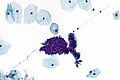
ECAIS - high mag. (WC)
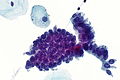
ECAIS - very high mag. (WC)
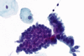
ECAIS - very high mag. (WC)
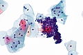
ECAIS - high mag. (WC)
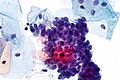
ECAIS - very high mag. (WC)


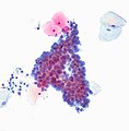
Endocervical AIS. (WC/euthman)

Endometrial adenocarcinoma in situ
- AKA adenocarcinoma in situ of the endometrium.
General
- Management - like AGC and other types of AIS: coloscopy + endometrial biopsy.
Cytopathology
Features:
- Single cells or cluster of small cells with:
- Moderate nuclear enlargement ~2x intermediate cell nucleus.
- Nuclear hyperchromasia.
- Coarse chromatin.
- Nucleoli - prominent - key feature.
- +/-Mitoses.
- +/-Intracytoplasmic neutrophils.
- Apoptotic/necrotic cells.
- +/-Psammoma bodies.
- Suggestive of serous carcinoma of the endometrium.
DDx:
- AGC - no prominent nucleoli, not 3-dimensional.
- Endocervical adenocarcinoma in situ.
Waffle categories
- Like all waffle diagnoses, these should be used sparingly.
Atypical squamous cells of undetermined significance
- Abbreviated ASC-US or ASCUS.
General
- This is a waffle category that should be used sparingly.
- Diagnosis may be an indication for HPV testing.
Cytology
Features:
- Squamous differentiation:
- Central nucleus.
- Dense/solid-appearing cytoplasm.
- Nuclear size >2.5X IC nucleus, but <3X IC nucleus.
- +/-Orange/red cytoplasmic (orangeophilic cytoplasm).[29]
Note:
- One should not see nucleoli.
- Nucleoli are seen in reactive changes and squamous cell carcinoma of the uterine cervix.
- The IC nucleus is ~ 8 μm.[30]
DDx:
Images
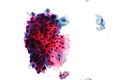
ASCUS - high mag. (WC)
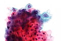
ASCUS - very high mag. (WC)
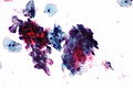
ASCUS - high mag. (WC)
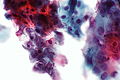
ASCUS - very high mag. (WC)
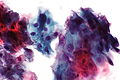
ASCUS - very high mag. (WC)


Sign out
Atypical squamous cells of undetermined significance (ASC-US).
Atypical squamous cells, cannot exclude high-grade squamous intraepithelial lesion
- Abbreviated ASC-H.
General
- This is a waffle category that should be used very rarely.
- Higher HPV positivity vs. ASC-US.[31]
- Management - like HSIL: colposcopy.
Cytology
Features:
- Atypia that falls short of diagnosing HSIL:
- Increased NC ratio.
- Architecture: cell clusters or rare single cells.
DDx:[32]
Atypical glandular cells
- Abbreviated AGC.
- Previously atypical glandular cells of undetermined significance, abbreviated AGUS.
General
- Waffle diagnosis.
- Clinical management, like AIS: coloscopy +/- endometrial biopsy.
May represent either:
- Endocervical cells, i.e. atypical endocervical cells (AEC).
- Endometrial cells, i.e. atypical endometrial cell (AEM).
Microscopic
Features:
- Atypical glandular cells:
- Cell cluster with cells with a diameter <= 2x intermediate cell nucleus.
- Some features of nuclear atypia, e.g. irregular nuclear membrane, granular chromatin, nuclear hyperchromasia, nuclear enlargement.
DDx:
- Adenocarcinoma in situ.
Uncommon stuff
Follicular cervicitis
General
- Uncommon. (???)
- Finding may be associated with Chlamydia trachomatis.[33]
Cytology
Features:[34]
- Discohesive clusters of small (lymphoid) cells with interspersed:
- Tingible-body macrophages.
- Plasma cells.
DDx:
- AGC - nuclei larger, more cohesive
Image:
Hematoidin crystal
General
- Rare.
- Benign.
- Associated with hemorrhage in pregnancy.[35]
Note:
- Overlap with crystalline bodies. (???)
- Crystalline bodies associated with pregnancy, and OCP use.[35]
Cytology
- Radiating crystal.
- Refractile.
- Classically golden-brown.
- +/-Surrounded by macrophages.
DDx:
- Sulfur granule of Actinomycetes.
Images:
- Cockleburr crystal (cytology-asc.com).[18]
- Cockleburr crystal (cytology-asc.com).
- Cockleburr crystal (cytology-asc.com).
Carpet beetle larval parts
General
- Uncommon distinctive contaminant.[37]
- Fragment of a beetle.
- Benign.
Cytology
Features:
- Slender long structure - fern-like.
Note:
- One may have a complete insect.[38]
Image:
Radiation changes in cervical cytology
General
- Radiation is used to treat cervical cancer.
Cytology
Features:[39]
- Architecture: single cells/groups.
- Cell borders: well-circumscribed.
- Cytoplasm: vacuolated, usually abundant.
- Nucleus:
- Enlarged nucleus - but normal NC ratio.
- No nuclear membrane irregularies.
- Chromatin: "smudgy".
- +/-Multinucleation.
DDx:
- LSIL.
- Vitamin B12 deficiency.
Images:
Cornflaking artifact
General
- Processing artifact - due to air under the cover slip.[40]
Cytology
Features:[40]
- Central brown discolourization in squamous cells.
Images:
Endocervical repair
General
- Benign.
Cytology
Features:
- Cluster of (2-dimensional) glandular cells with:
- Streaming (school of fish-appearance).
- Prominent nucleoli.
- Neutrophils.
Image:
Historical
Maturation index
- Abbreviated MI.
General
- Based on vaginal wall scrape.
Definition
.

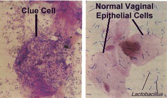
Where:
- P = number of parabasal cells / 300 squamous cells * 100 %.
- I = number of intermediate cells / 300 squamous cells * 100 %.
- S = number of superficial cells / 300 squamous cells * 100 %.
Interpretation
Common patterns:
- Superficial predominant, no parabasal = high estrogen effect.
- Parabasal predominant, no superficial = atrophy.
Examples:
- 70 : 30 : 0 is an atrophic pattern.
- 0 : 30 : 70 is a high estrogen pattern.
Note:
- Significant inflammation distorts the result.
See also
References
- ↑ Half-day. 10 November 2008.
- ↑ SM. 14 January 2010.
- ↑ SM. 14 January 2010.
- ↑ GR. 4 February 2010.
- ↑ SM. 14 January 2010.
- ↑ URL: http://nih.techriver.net/view.php?patientId=78. Accessed on: 31 March 2012.
- ↑ Thrall MJ, Kjeldahl KS, Savik K, Gulbahce HE, Pambuccian SE (August 2005). "Significance of benign endometrial cells in papanicolaou tests from women aged >=40 years". Cancer 105 (4): 207-16. doi:10.1002/cncr.21156. PMID 15900572.
- ↑ DeMay, RM. The Art & Science of Cytopathology: Exfoliative Cytology. 1996. ISBN 0-89189-322-9. PP.116-7.
- ↑ URL: http://nih.techriver.net/view.php?patientId=221. Accessed on: 26 November 2011.
- ↑ DeMay, RM. The Art & Science of Cytopathology: Exfoliative Cytology. 1996. ISBN 0-89189-322-9. PP.116-7.
- ↑ 11.0 11.1 Slater, DN.; Rice, S.; Stewart, R.; Melling, SE.; Hewer, EM.; Smith, JH. (Aug 2005). "Proposed Sheffield quantitative criteria in cervical cytology to assist the diagnosis and grading of squamous intra-epithelial lesions, as some Bethesda system definitions require amendment.". Cytopathology 16 (4): 168-78. doi:10.1111/j.1365-2303.2005.00264.x. PMID 16048503. http://www3.interscience.wiley.com/journal/118661591/abstract?CRETRY=1&SRETRY=0.
- ↑ 12.0 12.1 12.2 12.3 12.4 12.5 Humphrey, Peter A; Dehner, Louis P; Pfeifer, John D (2008). The Washington Manual of Surgical Pathology (1st ed.). Lippincott Williams & Wilkins. pp. 446. ISBN 978-0781765275.
- ↑ UHN PCY50001.08 P.10.
- ↑ Sheffield, MV.; Simsir, A.; Talley, L.; Roberson, AJ.; Elgert, PA.; Chhieng, DC. (Mar 2003). "Interobserver variability in assessing adequacy of the squamous component in conventional cervicovaginal smears.". Am J Clin Pathol 119 (3): 367-73. PMID 12645338.
- ↑ GR. 4 February 2010.
- ↑ Doxtader EE, Elsheikh TM (January 2017). "Diagnosis of trichomoniasis in men by urine cytology". Cancer Cytopathol 125 (1): 55–59. doi:10.1002/cncy.21778. PMID 27636204.
- ↑ URL: http://en.wiktionary.org/wiki/-mycete#English. Accessed on: 14 September 2011.
- ↑ 18.0 18.1 18.2 URL: http://www.cytology-asc.com/cec/normal/index.htm#cockle. Accessed on: 10 April 2012.
- ↑ Scott TG, Smyth CJ, Keane CT (February 1987). "In vitro adhesiveness and biotype of Gardnerella vaginalis strains in relation to the occurrence of clue cells in vaginal discharges". Genitourinary medicine 63 (1): 47–53. PMC 1194007. PMID 3493202. https://www.ncbi.nlm.nih.gov/pmc/articles/PMC1194007/.
- ↑ 20.0 20.1 Polatti, F. (Feb 2012). "Bacterial vaginosis, Atopobium vaginae and nifuratel.". Curr Clin Pharmacol 7 (1): 36-40. PMID 22082330.
- ↑ Taylor-Robinson D (1984). "The bacteriology of Gardnerella vaginalis". Scand J Urol Nephrol Suppl 86: 41–55. PMID 6399409.
- ↑ Bigras, G.; Wilson, J.; Russell, L.; Johnson, G.; Morel, D.; Saddik, M. (Feb 2013). "Interobserver concordance in the assessment of features used for the diagnosis of cervical atypical squamous cells and squamous intraepithelial lesions (ASC-US, ASC-H, LSIL and HSIL).". Cytopathology 24 (1): 44-51. doi:10.1111/j.1365-2303.2011.00930.x. PMID 22007754.
- ↑ Chan, S. 26 April 2012.
- ↑ URL: http://www.cytology-asc.com/cec/endocx/. Accessed on: 13 September 2011.
- ↑ Belsley, NA.; Tambouret, RH.; Misdraji, J.; Muzikansky, A.; Russell, DK.; Wilbur, DC. (Apr 2008). "Cytologic features of endocervical glandular lesions: comparison of SurePath, ThinPrep, and conventional smear specimen preparations.". Diagn Cytopathol 36 (4): 232-7. doi:10.1002/dc.20782. PMID 18335553.
- ↑ Nucci, Marisa R.; Oliva, Esther (2009). Gynecologic Pathology: A Volume in Foundations in Diagnostic Pathology Series (1st ed.). Churchill Livingstone. pp. 167. ISBN 978-0443069208.
- ↑ Duggan, MA. (Mar 2000). "Cytologic and histologic diagnosis and significance of controversial squamous lesions of the uterine cervix.". Mod Pathol 13 (3): 252-60. doi:10.1038/modpathol.3880046. PMID 10757336.
- ↑ URL: http://www.cap.org/apps/docs/proficiency_testing/CYP07600.pdf. Accessed on: 2 May 2012.
- ↑ Owens, CL.; Ali, SZ. (Dec 2005). "Atypical squamous cells in exfoliative urinary cytology: clinicopathologic correlates.". Diagn Cytopathol 33 (6): 394-8. doi:10.1002/dc.20344. PMID 16299739.
- ↑ URL: http://www.curran.pwp.blueyonder.co.uk/cytology.htm. Accessed on: 5 November 2012.
- ↑ Srodon, M.; Parry Dilworth, H.; Ronnett, BM. (Feb 2006). "Atypical squamous cells, cannot exclude high-grade squamous intraepithelial lesion: diagnostic performance, human papillomavirus testing, and follow-up results.". Cancer 108 (1): 32-8. doi:10.1002/cncr.21388. PMID 16136595.
- ↑ Chivukula, M.; Shidham, VB. (2006). "ASC-H in Pap test--definitive categorization of cytomorphological spectrum.". Cytojournal 3: 14. doi:10.1186/1742-6413-3-14. PMC 1524979. PMID 16686950. https://www.ncbi.nlm.nih.gov/pmc/articles/PMC1524979/.
- ↑ Hare, MJ.; Toone, E.; Taylor-Robinson, D.; Evans, RT.; Furr, PM.; Cooper, P.; Oates, JK. (Feb 1981). "Follicular cervicitis--colposcopic appearances and association with Chlamydia trachomatis.". Br J Obstet Gynaecol 88 (2): 174-80. PMID 6893939.
- ↑ Halford, JA. (Dec 2002). "Cytological features of chronic follicular cervicitis in liquid-based specimens: a potential diagnostic pitfall.". Cytopathology 13 (6): 364-70. PMID 12485172.
- ↑ 35.0 35.1 Minassian, H.; Schinella, R.; Reilly, JC.. "Crystalline bodies in cervical smears. Clinicocytologic correlation.". Acta Cytol 37 (2): 149-52. PMID 8465632.
- ↑ Zaharopoulos, P.; Wong, JY.; Keagy, N.. "Hematoidin crystals in cervicovaginal smears. Report of two cases.". Acta Cytol 29 (6): 1029-34. PMID 3866455.
- ↑ Bechtold, E.; Staunton, CE.; Katz, SS.. "Carpet beetle larval parts in cervical cytology specimens.". Acta Cytol 29 (3): 345-52. PMID 3859134.
- ↑ URL: http://www.cytology-asc.com/cec/normal/index.htm#dustmite. Accessed on: 10 April 2012.
- ↑ Gupta, S.; Mukherjee, K.; Gupta, YN.; Kumar, M. (Aug 1987). "Sequential radiation changes in cytology of vaginal smears in carcinoma of cervix uteri during radiotherapy.". Int J Gynaecol Obstet 25 (4): 303-8. PMID 2887465.
- ↑ 40.0 40.1 40.2 URL: http://www.cytology-asc.com/cec/normal/index.htm#cornflake. Accessed on: 10 April 2012.