|
|
| (22 intermediate revisions by 2 users not shown) |
| Line 1: |
Line 1: |
| This article deals with '''dermatologic neoplasms'''. It includes '''dermatologic cancer''', which can be deadly. Collectively, dermatologic cancers are the most common form of cancer. | | This article deals with '''dermatologic neoplasms''', also known as '''skin tumours'''. It includes '''dermatologic cancer''', which can be deadly. Collectively, dermatologic cancers are the most common form of cancer. |
|
| |
|
| An introduction to dermatopathy is found in the ''[[dermatopathology]]'' article. Non-malignant disease is covered in the ''[[non-malignant skin disease]]'' article. | | An introduction to dermatopathy is found in the ''[[dermatopathology]]'' article. Non-malignant disease is covered in the ''[[non-malignant skin disease]]'' article. |
| Line 8: |
Line 8: |
|
| |
|
| ==Squamous cell carcinoma of the skin== | | ==Squamous cell carcinoma of the skin== |
| {{Main|Squamous carcinoma}}
| |
| *Abbreviated ''skin SCC'', ''SCC of the skin'', and ''SCC of skin''. | | *Abbreviated ''skin SCC'', ''SCC of the skin'', and ''SCC of skin''. |
| ===General===
| | {{Main|Squamous cell carcinoma of the skin}} |
| Precursor:<ref name=Ref_PBoD8_1180>{{Ref PBoD8|1180}}</ref>
| |
| *[[Actinic keratosis]] (solar keratosis).
| |
| **Clinical: yellow-brown scaly, patches, sandpaper sensation.
| |
| | |
| Risk factors:<ref name=Ref_PBoD8_1180>{{Ref PBoD8|1180}}</ref>
| |
| *Sun exposure.
| |
| *Immune suppression (e.g. organ transplant recipients).
| |
| | |
| Notes:
| |
| *[[Keratoacanthoma]].
| |
| **Some don't believe this entity exists.
| |
| ***These people sign this entity as ''low grade squamous cell carcinoma, keratoacanthoma type''.<ref>RS. 17 May 2010.</ref>
| |
| | |
| ===Microscopic===
| |
| *See ''[[squamous cell carcinoma]]''.
| |
| | |
| High risk features - for SCC of the skin:<ref>URL: [http://www.cap.org/apps/docs/committees/cancer/cancer_protocols/2011/SkinSquamousCell_11protocol.pdf http://www.cap.org/apps/docs/committees/cancer/cancer_protocols/2011/SkinSquamousCell_11protocol.pdf]. Accessed on: 29 March 2012.</ref>
| |
| *Primary site is ear ''or'' lip.†
| |
| *Clark level IV/V = reticular dermis or deeper.
| |
| *>=2 mm thickness -- measured from ''granular layer'' (stratum granulosum) ''or'' ulcer base to deepest aspect.
| |
| *[[Lymphovascular invasion]].
| |
| *Perineural invasion.
| |
| *Poorly differentiated.
| |
| | |
| Note:
| |
| * † The words used are "hair-bearing lip" - but there is considerable confusion about this as the AJCC manual contradicts itself.<ref name=pmid21151529>{{Cite journal | last1 = Buethe | first1 = D. | last2 = Warner | first2 = C. | last3 = Miedler | first3 = J. | last4 = Cockerell | first4 = CJ. | title = Focus Issue on Squamous Cell Carcinoma: Practical Concerns Regarding the 7th Edition AJCC Staging Guidelines. | journal = J Skin Cancer | volume = 2011 | issue = | pages = 156391 | month = | year = 2011 | doi = 10.1155/2011/156391 | PMID = 21151529 | PMC = 2990020 | URL = http://www.hindawi.com/journals/jsc/2011/156391/ }}</ref>
| |
| | |
| DDx:
| |
| *[[Inverted follicular keratosis]].
| |
| *[[Bowen disease]].
| |
| *[[Malignant melanoma]].
| |
| *[[Paget disease of the breast]].
| |
| *[[Eccrine carcinoma]]
| |
| | |
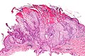
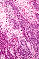
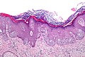
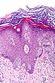
| ====Bowen disease====
| |
| '''Bowen disease''' is ''[[squamous cell carcinoma]] in situ'' of the skin.
| |
| *Its histomorphologic appearance may be similar to [[Paget disease of the breast]]/[[Extramammary Paget disease]], Toker cell hyperplasia and [[melanoma]].
| |
| **[[IHC]] is used to separate the entities definitively.
| |
| | |
| Histologic DDx of Bowen disease:
| |
| *Benign Toker cell hyperplasia.
| |
| *[[Malignant melanoma]].
| |
| *[[Paget disease of the breast]].
| |
| *[[Eccrine carcinoma]].
| |
| | |
| =====Images=====
| |
| <gallery>
| |
| Image:Bowen_disease_%281%29.jpg | Bowen disease - 1. (WC)
| |
| Image:Bowen_disease_%282%29.jpg | Bowen disease - 2. (WC)
| |
| Image:Bowen_disease_%283%29.jpg | Bowen disease - 3. (WC)
| |
| </gallery>
| |
| ===IHC===
| |
| Bowen's disease panel:
| |
| *CK5/6 +ve.<ref>RS. May 2010.</ref>
| |
| **Usu. -ve in [[Paget disease of the breast]]/[[Extramammary Paget disease]].
| |
| *S100 -ve, HMB-45 -ve.
| |
| **Both typically +ve in melanoma.
| |
| *CEA -ve<ref name=emed_pagets>URL: [http://emedicine.medscape.com/article/1101235-workup#a0721 http://emedicine.medscape.com/article/1101235-workup#a0721]. Accessed on: 2 September 2011.</ref> (+ve in [[Paget disease of the breast]]/[[Extramammary Paget disease]], -ve in Toker cells).
| |
| *CK7 -ve.
| |
| **Toker cells CK7 +ve.<ref name=pmid19601945>{{Cite journal | last1 = Nofech-Mozes | first1 = S. | last2 = Hanna | first2 = W. | title = Toker cells revisited. | journal = Breast J | volume = 15 | issue = 4 | pages = 394-8 | month = | year = | doi = 10.1111/j.1524-4741.2009.00743.x | PMID = 19601945 }}</ref>
| |
| | |
| ===Sign-out===
| |
| ====Invasive SCC====
| |
| <pre>
| |
| SKIN, SITE, BIOPSY:
| |
| - MODERATELY-DIFFERENTIATED INVASIVE SQUAMOUS CELL CARCINOMA, SEE COMMENT.
| |
| - NEGATIVE FOR LYMPHOVASCULAR INVASION.
| |
| - NEGATIVE FOR PERINEURAL INVASION.
| |
| | |
| COMMENT:
| |
| The nearest margin (lateral margin) is 1 mm. The tumour is 9 mm in maximal dimension.
| |
| </pre>
| |
| | |
| <pre>
| |
| SKIN LESION, SITE, EXCISION:
| |
| - INVASIVE SQUAMOUS CELL CARCINOMA, MODERATELY-DIFFERENTIATED.
| |
| -- TUMOUR GREATEST DIMENSION: ___ CM.
| |
| -- TUMOUR THICKNESS: ___ MM.
| |
| -- LATERAL MARGINS: NEGATIVE FOR IN SITU CARCINOMA AND INVASIVE CARCINOMA.
| |
| -- DEEP MARGIN: NEGATIVE FOR INVASIVE CARCINOMA.
| |
| -- NEAREST MARGIN: 1 MM, LATERAL MARGIN.
| |
| -- NEGATIVE FOR LYMPHOVASCULAR INVASION.
| |
| -- NEGATIVE FOR PERINEURAL INVASION.
| |
| - EXTENSIVE SOLAR ELASTOSIS.
| |
| </pre>
| |
| | |
| <pre>
| |
| SKIN, SITE, BIOPSY:
| |
| - INVASIVE SQUAMOUS CELL CARCINOMA, SEE TUMOUR SUMMARY.
| |
| | |
| TUMOUR SUMMARY:
| |
| Histologic type: squamous cell carcinoma, type not otherwise specified.
| |
| Histologic grade: moderately differentiated.
| |
| Greatest dimension: ___ cm.
| |
| Tumour thickness: ___ mm.
| |
| Peripheral margin: negative for invasive carcinoma and in situ carcinoma.
| |
| Deep margin (invasive component): negative for invasive carcinoma.
| |
| Closest margin: deep margin, ___ mm.
| |
| Lymphovascular invasion: not identified.
| |
| Perineural invasion: not identified.
| |
| </pre>
| |
| | |
| ====Bowen's disease====
| |
| <pre>
| |
| SKIN LESION, RIGHT EAR, BIOPSY:
| |
| - SQUAMOUS CELL CARCINOMA IN SITU (BOWEN'S DISEASE), INCOMPLETELY EXCISED.
| |
| | |
| COMMENT:
| |
| Complete excision of the lesion is recommended.
| |
| </pre>
| |
|
| |
|
| ==Melanoma== | | ==Melanoma== |
| {{Main|Malignant melanoma}} | | {{Main|Malignant melanoma}} |
| ===General===
| |
| *Known as the great mimicker in pathology; it may look like many things. | | *Known as the great mimicker in pathology; it may look like many things. |
|
| |
| ===Microscopic===
| |
| Features:
| |
| *Classic appearance of melanoma:
| |
| **Loosely cohesive; mix of small nests of cells, single cells.
| |
| **Usu. mixed of spindle and ovoid cell morphology.
| |
| **+/-Occasional large binucleated cells.
| |
| **+/-Cytoplasm: brown pigment (melanin).
| |
| **+/-Prominent (large) red nucleoli (like in ''serous carcinoma'' of the ovary).
| |
| **Often marked nuclear pleomorphism - variation in cell size, shape & staining (like in ''serous carcinoma'' of the ovary).
| |
| **[[Nuclear pseudoinclusions]] (like in ''papillary thyroid carcinoma'').
| |
|
| |
|
| =Less common malignant= | | =Less common malignant= |
| ==Dermatofibrosarcoma protuberans== | | ==Dermatofibrosarcoma protuberans== |
| *Abbreviated ''DFSP''. | | *Abbreviated ''DFSP''. |
| ===General===
| | {{Main|Dermatofibrosarcoma protuberans}} |
| *Dermal location.
| |
| *Destroys adnexal structures.
| |
| *Occasionally transforms to a (more aggressive) [[adult fibrosarcoma|fibrosarcoma]].<ref name=pmid21128251>{{Cite journal | last1 = Stacchiotti | first1 = S. | last2 = Pedeutour | first2 = F. | last3 = Negri | first3 = T. | last4 = Conca | first4 = E. | last5 = Marrari | first5 = A. | last6 = Palassini | first6 = E. | last7 = Collini | first7 = P. | last8 = Keslair | first8 = F. | last9 = Morosi | first9 = C. | title = Dermatofibrosarcoma protuberans-derived fibrosarcoma: clinical history, biological profile and sensitivity to imatinib. | journal = Int J Cancer | volume = 129 | issue = 7 | pages = 1761-72 | month = Oct | year = 2011 | doi = 10.1002/ijc.25826 | PMID = 21128251 }}</ref>
| |
| | |
| Treatment:<ref name=Ref_PBoD8_1183>{{Ref PBoD8|1183}}</ref>
| |
| *Wide excision.
| |
| *May include [[imatinib]] (Gleevec).
| |
| | |
| ===Gross===
| |
| Features:<ref name=Ref_PCPBoD8_600>{{Ref PCPBoD8|600}}</ref>
| |
| *Firm plaque, often bosselated, usually on the trunk.
| |
| *+/-Ulceration.
| |
| | |
| Images:
| |
| *[http://dermatlas.med.jhmi.edu/derm/display.cfm?ImageID=-375107780 Protuberant DFSP (dermatlas.med.jhmi.edu)].
| |
| *[http://dermatlas.med.jhmi.edu/derm/indexDisplay.cfm?ImageID=-1421097348 Huge DFSP on back (dermatlas.med.jhmi.edu)].
| |
| *[http://dermatlas.med.jhmi.edu/derm/indexDisplay.cfm?ImageID=-109598044 Protuberant DFSP - gross and histology (dermatlas.med.jhmi.edu)].
| |
| | |
| ===Microscopic===
| |
| Features:<ref name=Ref_PBoD8_1183>{{Ref PBoD8|1183}}</ref>
| |
| *Dermal spindle cell lesion with storiform pattern.
| |
| **Spokes of the wheel-pattern.
| |
| *Contains adipose tissue within the tumour -- '''key feature'''.
| |
| **Described as "honeycomb pattern" and "Swiss cheese pattern".
| |
| | |
| Notes:
| |
| *Adnexal structure within tumour are preserved -- this is unusual for a malignant tumour -- '''important'''.
| |
| | |
| | |
| DDx:
| |
| *[[Dermatofibroma]] - main DDx - has entrapment of collagen bundles at the edge of the lesion.
| |
| *[[Dermatomyofibroma]].<ref name=Ref_Derm504>{{Ref Derm|504}}</ref>
| |
| *[[Nodular fasciitis]].
| |
| | |
| DDx of storiform pattern:
| |
| *DFSP.
| |
| *Dermatofibroma.
| |
| *[[Solitary fibrous tumour]].
| |
| *[[Undifferentiated pleomorphic sarcoma]].
| |
| | |
| ====Images====
| |
| <gallery>
| |
| Image:SkinTumors-P9280838.JPG | DFSP with fat entrapped. (WC)
| |
| Image:SkinTumors-P9270829.JPG | DFSP - high mag. (WC)
| |
| Image:Storiform_pattern_-_intermed_mag.jpg | DFSP - storiform pattern - intermed. mag. (WC/Nephron)
| |
| Image:Storiform_pattern_-_very_high_mag.jpg | DFSP - storiform pattern - very high mag. (WC/Nephron)
| |
| </gallery>
| |
| www:
| |
| *[http://webpathology.com/image.asp?case=317&n=1 DFSP (webpathology.com)].
| |
| | |
| ===IHC===
| |
| Panel:<ref>AP. May 2009.</ref>
| |
| *CD34 +ve.
| |
| **Usually negative in dermatofibroma.<ref name=pmid7694515>{{cite journal |author=Abenoza P, Lillemoe T |title=CD34 and factor XIIIa in the differential diagnosis of dermatofibroma and dermatofibrosarcoma protuberans |journal=Am J Dermatopathol |volume=15 |issue=5 |pages=429–34 |year=1993 |month=October |pmid=7694515 |doi= |url=}}</ref><ref name=pmid9129699>{{cite journal |author=Goldblum JR, Tuthill RJ |title=CD34 and factor-XIIIa immunoreactivity in dermatofibrosarcoma protuberans and dermatofibroma |journal=Am J Dermatopathol |volume=19 |issue=2 |pages=147–53 |year=1997 |month=April |pmid=9129699 |doi= |url=}}</ref>
| |
| *Factor XIIIa -ve.
| |
| **Usually positive in dermatofibroma.<ref name=pmid7694515>{{cite journal |author=Abenoza P, Lillemoe T |title=CD34 and factor XIIIa in the differential diagnosis of dermatofibroma and dermatofibrosarcoma protuberans |journal=Am J Dermatopathol |volume=15 |issue=5 |pages=429–34 |year=1993 |month=October |pmid=7694515 |doi= |url=}}</ref><ref name=pmid9129699>{{cite journal |author=Goldblum JR, Tuthill RJ |title=CD34 and factor-XIIIa immunoreactivity in dermatofibrosarcoma protuberans and dermatofibroma |journal=Am J Dermatopathol |volume=19 |issue=2 |pages=147–53 |year=1997 |month=April |pmid=9129699 |doi= |url=}}</ref>
| |
| *S100 -ve (screen for melanoma).
| |
| *Caldesmin -ve (screen for muscle differentiation).
| |
| *Beta-catenin. (???)
| |
| *MIB1 (proliferation marker).
| |
| **Should not be confused with ''MIB-1'' a gene that regulates [[apoptosis]].
| |
| | |
| ===Molecular===
| |
| A characteristic [[translocation]] is seen:<ref>{{Ref PBoD8|1249}}</ref>
| |
| t(17;22)(q22;q15) COLA1/PDGFB.
| |
|
| |
|
| ==Cutaneous B-cell lymphoma== | | ==Cutaneous B-cell lymphoma== |
| Line 224: |
Line 36: |
| ==Cutaneous T-cell lymphoma== | | ==Cutaneous T-cell lymphoma== |
| *Abbreviated CTCL. | | *Abbreviated CTCL. |
| | | {{Main|Cutaneous T-cell lymphoma}} |
| ===General===
| |
| *''Mycosis fungoides'' - is a subtype (???).
| |
| *CTCL is more common than cutaneous B-cell lymphoma (CBCL).<ref>URL: [http://emedicine.medscape.com/article/1099540-overview http://emedicine.medscape.com/article/1099540-overview]. Accessed on: 24 August 2010.</ref><ref>URL: [http://emedicine.medscape.com/article/1098342-overview http://emedicine.medscape.com/article/1098342-overview]. Accessed on: 24 August 2010.</ref>
| |
| | |
| Stages - like [[Kaposi sarcoma]]:
| |
| *Patch.
| |
| *Plaque.
| |
| *Nodular.
| |
| | |
| ===Microscopic===
| |
| *Atypical lymphocytes:
| |
| **Have folded "cerebriform" nuclei; ''Sezary-Lutzner cells''.<ref name=Ref_Klatt385>{{Ref Klatt|385}}</ref>
| |
| *Grouping:
| |
| **Nests in the epidermis - known as "Pautrier microabscesses".
| |
| **Single lymphocytes in epidermis - without accompanying edema.
| |
| **Short linear arrays of lymphocytes along the basal layer of the epidermis; "epidermotropism".<ref name=Ref_Klatt385>{{Ref Klatt|385}}</ref>
| |
| | |
| DDx:
| |
| *[[Lymphomatoid papulosis]].
| |
| | |
| ====Images====
| |
| <gallery>
| |
| Image:Cutaneous_T-cell_lymphoma_-_very_high_mag.jpg | CTCL - very high mag. (WC/Nephron)
| |
| Image:Cutaneous_T-cell_lymphoma_-_intermed_mag.jpg | CTCL - intermed. mag. (WC/Nephron)
| |
| </gallery>
| |
| www:
| |
| *[http://www.jci.org/articles/view/24826/figure/2 CTCL (jci.org)].
| |
| *[http://www.mdconsult.com/das/book/body/199872830-2/0/1709/I4-u1.0-B978-0-443-06694-8..50117-2--f2.fig CTCL (mdconsult.com)].
| |
| | |
| ===IHC===
| |
| Key stain:
| |
| *CD4 +ve.<ref>{{Ref PBoD8|1185}}</ref>
| |
| | |
| Other stains:
| |
| *CD3 +ve.
| |
| *CD8 -ve.
| |
| *CD20 -ve (to r/o significant B cell population).
| |
| *CD30 -ve.
| |
| *CD5 +ve.
| |
| *CD7 -ve (often lost first in T cell lymphomas).
| |
| *Ki-67 high.
| |
| *CD56 -ve.
| |
|
| |
|
| ==Merkel cell carcinoma== | | ==Merkel cell carcinoma== |
| Line 294: |
Line 64: |
|
| |
|
| ==Trichilemmal carcinoma== | | ==Trichilemmal carcinoma== |
| ===General===
| | {{Main|Trichilemmal carcinoma}} |
| *Super rare.
| |
| *Not well-described.
| |
| | |
| ===Microscopic===
| |
| Features:<ref>{{Ref Derm|399-400}}</ref>
| |
| *Clear (glycogen-rich) cytoplasm in center of lesion.
| |
| *Peripheral palisading at edge of lesion - root sheath differentiation (hair follicle).
| |
| *Contiguous with hair follicle ''or'' assoc. with [[trichilemmoma]].
| |
| | |
| DDx:
| |
| *[[Squamous cell carcinoma]], clear cell variant.
| |
| *[[Basal cell carcinoma]], clear cell variant.
| |
| *[[Trichilemmoma]].
| |
|
| |
|
| ==Lymphomatoid papulosis== | | ==Lymphomatoid papulosis== |
| Line 352: |
Line 109: |
| ==Atypical fibroxanthoma== | | ==Atypical fibroxanthoma== |
| *Abbreviated ''AFX''. | | *Abbreviated ''AFX''. |
| ===General===
| | {{Main|Atypical fibroxanthoma}} |
| *Typically head & neck region.<ref>URL: [http://emedicine.medscape.com/article/1056204-overview http://emedicine.medscape.com/article/1056204-overview]. Accessed on 2 September 2011.</ref>
| |
| *Thought to be related to [[pleomorphic undifferentiated sarcoma]];<ref name=pmid21664889>{{Cite journal | last1 = Withers | first1 = AH. | last2 = Brougham | first2 = ND. | last3 = Barber | first3 = RM. | last4 = Tan | first4 = ST. | title = Atypical fibroxanthoma and malignant fibrous histiocytoma. | journal = J Plast Reconstr Aesthet Surg | volume = | issue = | pages = | month = Jun | year = 2011 | doi = 10.1016/j.bjps.2011.05.004 | PMID = 21664889 }}</ref><ref name=pmid23319144>{{Cite journal | last1 = Tchernev | first1 = G. | last2 = Tronnier | first2 = M. | last3 = Ananiev | first3 = J. | last4 = Taneva | first4 = T. | last5 = Patterson | first5 = JW. | last6 = Gulubova | first6 = M. | last7 = Trafeli | first7 = JP. | last8 = Gegova | first8 = A. | last9 = Harrell | first9 = M. | title = Atypical fibroxanthoma-a diagnosis of exclusion! | journal = Wien Med Wochenschr | volume = 163 | issue = 15-16 | pages = 380-386 | month = Aug | year = 2013 | doi = 10.1007/s10354-012-0173-1 | PMID = 23319144 }}</ref> some say it is the same thing.<ref name=danny>Ghazarian, Danny; 16 September 2011.</ref>
| |
| *Usually benign.
| |
| **May metastasize - case report-type of occurrence.<ref>{{Cite journal | last1 = New | first1 = D. | last2 = Bahrami | first2 = S. | last3 = Malone | first3 = J. | last4 = Callen | first4 = JP. | title = Atypical fibroxanthoma with regional lymph node metastasis: report of a case and review of the literature. | journal = Arch Dermatol | volume = 146 | issue = 12 | pages = 1399-404 | month = Dec | year = 2010 | doi = 10.1001/archdermatol.2010.206 | PMID = 20713774 | URL = http://archderm.jamanetwork.com/article.aspx?articleid=422416 }}</ref>
| |
| | |
| Clinical:
| |
| *Rapid growth.
| |
| *Elderly.
| |
| *Good prognosis.<ref name=pmid20526171>{{Cite journal | last1 = Beer | first1 = TW. | last2 = Drury | first2 = P. | last3 = Heenan | first3 = PJ. | title = Atypical fibroxanthoma: a histological and immunohistochemical review of 171 cases. | journal = Am J Dermatopathol | volume = 32 | issue = 6 | pages = 533-40 | month = Aug | year = 2010 | doi = 10.1097/DAD.0b013e3181c80b97 | PMID = 20526171 }}</ref>
| |
| | |
| ===Microscopic===
| |
| Features:<ref name=Ref_Derm521>{{Ref Derm|521}}</ref>
| |
| *Dermal lesion - '''key point'''.
| |
| *Marked nuclear atypia.
| |
| *Mitoses.
| |
| *Mulitnucleated cells.
| |
| *Foamy cytoplasm - '''key feature'''.
| |
| | |
| DDx:
| |
| *[[Melanoma]].
| |
| *[[Pleomorphic undifferentiated sarcoma]] (MFH) - deeper than the dermis.
| |
| *[[Leiomyosarcoma]].
| |
| *Sarcomatoid [[squamous carcinoma]].
| |
| | |
| Notes:
| |
| *No Grenz zone. (???)
| |
| | |
| Image:
| |
| *[http://dermatology.cdlib.org/141/case_reports/afx/1.jpg AFX (cdlib.org)].<ref name=pmid18319023>{{Cite journal | last1 = Vandergriff | first1 = TW. | last2 = Reed | first2 = JA. | last3 = Orengo | first3 = IF. | title = An unusual presentation of atypical fibroxanthoma. | journal = Dermatol Online J | volume = 14 | issue = 1 | pages = 6 | month = | year = 2008 | doi = | PMID = 18319023 }}</ref>
| |
| | |
| ===IHC===
| |
| Features:<ref name=Ref_Derm521>{{Ref Derm|521}}</ref>
| |
| *S100 -ve (done to r/o melanoma).
| |
| *34betaE12 -ve.
| |
| *p63 -ve (done to exclude SCC)
| |
| **Scant staining not considered +ve.
| |
| *Desmin -ve (done to r/o leiomyosarcoma).
| |
| | |
| ===Sign out===
| |
| ====Incompletely excised====
| |
| <pre>
| |
| SKIN LESION, MID BACK, SHAVE BIOPSY:
| |
| - ATYPICAL SPINDLE CELL NEOPLASM, SEE MICRO AND COMMENT.
| |
| | |
| COMMENT:
| |
| The diagnosis of atypical fibroxanthoma (AFX) is favoured. The main differential
| |
| diagnosis is pleomorphic undifferentiated sarcoma.
| |
| | |
| The extent of the lesion cannot be determined, as it is present at the deep margin.
| |
| | |
| This lesion should be re-excised, as it could represent an aggressive malignancy.
| |
| </pre>
| |
|
| |
|
| =Benign= | | =Benign= |
| Line 411: |
Line 116: |
| *Benign sweat duct tumour. | | *Benign sweat duct tumour. |
| *Eccrine differentiation. | | *Eccrine differentiation. |
| *Usually close to lower eyelid.<ref>{{Ref PBoD8|1177}}</ref> | | *Usually close to lower [[eyelid]].<ref>{{Ref PBoD8|1177}}</ref> |
|
| |
|
| ===Microscopic=== | | ===Microscopic=== |
| Line 444: |
Line 149: |
|
| |
|
| Images: | | Images: |
| *[http://archive.ispub.com/journal/the-internet-journal-of-dermatology/volume-7-number-1/cutaneous-mixed-tumor.article-g01.fs.jpg Chondroid syringoma - low mag. (ispub.com)].<ref name=ispub_mts>URL: [http://www.ispub.com/journal/the_internet_journal_of_dermatology/volume_7_number_2_23/article/cutaneous_mixed_tumor.html http://www.ispub.com/journal/the_internet_journal_of_dermatology/volume_7_number_2_23/article/cutaneous_mixed_tumor.html]. Access on: 21 September 2011.</ref> | | *[https://www.dermnetnz.org/topics/apocrine-mixed-tumour-pathology Chondroid syringoma (DermnetNZ)]. |
| *[http://archive.ispub.com/journal/the-internet-journal-of-dermatology/volume-7-number-1/cutaneous-mixed-tumor.article-g02.fs.jpg Chondroid syringoma - high mag. (ispub.com)].<ref name=ispub_mts>URL: [http://www.ispub.com/journal/the_internet_journal_of_dermatology/volume_7_number_2_23/article/cutaneous_mixed_tumor.html http://www.ispub.com/journal/the_internet_journal_of_dermatology/volume_7_number_2_23/article/cutaneous_mixed_tumor.html]. Access on: 21 September 2011.</ref>
| |
|
| |
|
| ==Dermal cylindroma== | | ==Dermal cylindroma== |
| ===General===
| | {{Main|Dermal cylindroma}} |
| *Benign skin lesion.
| |
| **Occasionally malignant.<ref name=pmid16882695/>
| |
| *Should not be confused with ''cylindroma'' ([[adenoid cystic carcinoma]]).
| |
| *May be related to ''[[eccrine spiradenoma]]''.<ref name=pmid6302142>{{Cite journal | last1 = Gerber | first1 = JE. | last2 = Descalzi | first2 = ME. | title = Eccrine spiradenoma and dermal cylindroma. | journal = J Cutan Pathol | volume = 10 | issue = 1 | pages = 73-8 | month = Feb | year = 1983 | doi = | PMID = 6302142 }}</ref><ref name=pmid8936072>{{Cite journal | last1 = Lee | first1 = MW. | last2 = Kelly | first2 = JW. | title = Dermal cylindroma and eccrine spiradenoma. | journal = Australas J Dermatol | volume = 37 | issue = 1 | pages = 48-9 | month = Feb | year = 1996 | doi = | PMID = 8936072 }}</ref>
| |
| | |
| May be familial:<ref name=pmid16882695/>
| |
| *Familial cylindromatosis (autosomal dominant).
| |
| *Brook–Spiegler syndrome.
| |
| | |
| ===Gross===
| |
| *Classically scalp - usually head and neck or face.
| |
| | |
| ===Microscopic===
| |
| Features:<ref name=pmid16882695/>
| |
| *Nests of cells that fit together like a jigsaw puzzle - the borders of the nests are opposed and undulate.
| |
| *#Basaloid cells with scant cytoplasm and dark nuclei palisade around the edge of the nests.
| |
| *#Larger cells with moderate eosinophilic cytoplasm and lighter staining nuclei are at the centre of the nests.
| |
| *Cells nests surrounded by a band of hyaline (i.e. glassy, eosinophilic, acellular) material ~ 2X thickness of a basilar cell - '''key feature'''.
| |
| **This is basement membrane.
| |
|
| |
| DDx:
| |
| *[[Eccrine spiradenoma]].
| |
| *[[Basal cell carcinoma]] - has [[myxoid stroma]].
| |
| | |
| ====Images====
| |
| <gallery>
| |
| Image:Dermal_cylindroma_intermed_mag.jpg | Dermal cylindroma. (WC/Nephron)
| |
| Image:Dermal_cylindroma_intermed_mag_deep.jpg | Dermal cylindroma - high mag. (WC/Nephron)
| |
| </gallery>
| |
| www:
| |
| *[http://jcp.bmj.com/content/60/2/145/F7.large.jpg Dermal cylindroma (bmj.com)].<ref name=pmid16882695>{{Cite journal | last1 = Obaidat | first1 = NA. | last2 = Alsaad | first2 = KO. | last3 = Ghazarian | first3 = D. | title = Skin adnexal neoplasms--part 2: an approach to tumours of cutaneous sweat glands. | journal = J Clin Pathol | volume = 60 | issue = 2 | pages = 145-59 | month = Feb | year = 2007 | doi = 10.1136/jcp.2006.041608 | PMID = 16882695 | PMC = 1860616 | URL = http://www.ncbi.nlm.nih.gov/pmc/articles/PMC1860616/?tool=pubmed }}</ref>
| |
| | |
| ===Stains===
| |
| *PAS +ve (basement membrane).<ref name=pmid16882695/>
| |
|
| |
|
| ==Keratoacanthoma== | | ==Keratoacanthoma== |
| Line 510: |
Line 180: |
| ==Trichilemmoma== | | ==Trichilemmoma== |
| *May be spelled ''tricholemmoma''. | | *May be spelled ''tricholemmoma''. |
| ===General===
| | {{Main|Trichilemmoma}} |
| *Benign neoplasm with features of the pilosebaceous follicular epithelium.<ref>URL: [http://emedicine.medscape.com/article/1059940-overview http://emedicine.medscape.com/article/1059940-overview]. Accessed on: 2 September 2011.</ref>
| |
| *Associated with ''nevus sebaceous''.<ref name=pmid16503928>{{Cite journal | last1 = Baykal | first1 = C. | last2 = Buyukbabani | first2 = N. | last3 = Yazganoglu | first3 = KD. | last4 = Saglik | first4 = E. | title = [Tumors associated with nevus sebaceous]. | journal = J Dtsch Dermatol Ges | volume = 4 | issue = 1 | pages = 28-31 | month = Jan | year = 2006 | doi = 10.1111/j.1610-0387.2006.05855.x | PMID = 16503928 }}</ref>
| |
| *Muliple trichilemmomas associated with [[Cowden syndrome]].<ref name=Ref_Derm386>{{Ref Derm|386}}</ref>
| |
| | |
| ===Microscopic===
| |
| Features:<ref name=Ref_Derm386>{{Ref Derm|386}}</ref>
| |
| *Superficial dermal lesion contiguous with the epidermis:
| |
| **Core of lesion:
| |
| ***Cuboidal cells with round nuclei, eosinophilic-clear cytoplasm.
| |
| **Periphery of lesion:
| |
| ***Surrounded by hyaline band.
| |
| ***Peripheral palisading.
| |
|
| |
|
| DDx:
| | ==Poroma== |
| *[[Trichilemmal carcinoma]].
| | {{Main|Poroma}} |
| *[[Basal cell carcinoma]].
| |
| *[[Inverted follicular keratosis]].
| |
| | |
| Images:
| |
| *[http://ccr.cancer.gov/staff/images/9033_12822_Lee_1520.jpg Trichilemmoma - low mag. (cancer.gov)].<ref name=lee>URL: [http://ccr.cancer.gov/staff/gallery.asp?profileid=12822 http://ccr.cancer.gov/staff/gallery.asp?profileid=12822]. Accessed on: 2 September 2011.</ref>
| |
| *[http://ccr.cancer.gov/staff/images/9033_12822_Lee_1521.jpg Trichilemmoma - high mag. (cancer.gov)].<ref name=lee/>
| |
| *[http://dermimages.med.jhmi.edu/images/trichilemmoma_1_060109.jpg Trichilemmoma (jhmi.edu)].<ref>URL: [http://dermatlas.med.jhmi.edu/derm/indexDisplay.cfm?ImageID=667496720 http://dermatlas.med.jhmi.edu/derm/indexDisplay.cfm?ImageID=667496720]. Accessed on: 2 September 2011.</ref>
| |
| *[http://www.flickr.com/photos/40981620@N04/3812019838/in/pool-1185084@N23/ Trichilemmoma - low mag. (flickr.com/Irlam)].
| |
| *[http://www.flickr.com/photos/40981620@N04/3812019930/in/pool-1185084@N23/ Trichilemmoma - intermed. mag. (flickr.com/Irlam)].
| |
| *[http://www.flickr.com/photos/40981620@N04/3811204517/in/pool-1185084@N23/ Trichilemmoma - high mag. (flickr.com/Irlam)].
| |
| | |
| ==Eccrine poroma==
| |
| ===General=== | |
| *Benign tumour arising from the distal sweat duct.
| |
| *Erythematous - gross.
| |
| | |
| ===Microscopic===
| |
| Features:<ref>URL: [http://www.pathconsultddx.com/pathCon/diagnosis?pii=S1559-8675(06)70190-5 http://www.pathconsultddx.com/pathCon/diagnosis?pii=S1559-8675(06)70190-5]. Accessed on: 2 July 2010.</ref>
| |
| *Broad sheets of basaloid cells - attached to the epidermis - containing ductal structures - '''key feature'''.
| |
| *Biphasic stroma:
| |
| *#Edematous stroma.
| |
| *#Sclerotic stroma.
| |
| *Moderate nuclear pleomorphism.
| |
| *+/-Occasional mitoses.
| |
| | |
| Notes:
| |
| *Area above gland appears crusted.
| |
| | |
| DDx:
| |
| *[[Trichilemmoma]].
| |
| *[[Nodular hidradenoma]].
| |
| | |
| Images:
| |
| *[http://www.flickr.com/photos/40981620@N04/3808316834/in/photostream/ Eccrine poroma - low mag. (flickr.com)]
| |
| *[http://www.flickr.com/photos/40981620@N04/3807502071/in/photostream Eccrine poroma - intermed. mag. (flickr.com)].
| |
|
| |
|
| ==Nodular hidradenoma== | | ==Nodular hidradenoma== |
| *[[AKA]] ''eccrine acrospiroma''.<ref name=pmid18319032>{{Cite journal | last1 = Punia | first1 = RP. | last2 = Garg | first2 = S. | last3 = Bal | first3 = A. | last4 = Mohan | first4 = H. | title = Pigmented nodular hidradenoma masquerading as nodular malignant melanoma. | journal = Dermatol Online J | volume = 14 | issue = 1 | pages = 15 | month = | year = 2008 | doi = | PMID = 18319032 |URL = http://dermatology.cdlib.org/141/case_presentations/hidradenoma/punia.html }}</ref> | | *[[AKA]] ''eccrine acrospiroma''.<ref name=pmid18319032>{{Cite journal | last1 = Punia | first1 = RP. | last2 = Garg | first2 = S. | last3 = Bal | first3 = A. | last4 = Mohan | first4 = H. | title = Pigmented nodular hidradenoma masquerading as nodular malignant melanoma. | journal = Dermatol Online J | volume = 14 | issue = 1 | pages = 15 | month = | year = 2008 | doi = | PMID = 18319032 |URL = http://dermatology.cdlib.org/141/case_presentations/hidradenoma/punia.html }}</ref> |
| ===General===
| | {{Main|Nodular hidradenoma}} |
| *Benign adnexal tumour.<ref name=pmid9537017>{{Cite journal | last1 = Stratigos | first1 = AJ. | last2 = Olbricht | first2 = S. | last3 = Kwan | first3 = TH. | last4 = Bowers | first4 = KE. | title = Nodular hidradenoma. A report of three cases and review of the literature. | journal = Dermatol Surg | volume = 24 | issue = 3 | pages = 387-91 | month = Mar | year = 1998 | doi = | PMID = 9537017 }}</ref>
| |
| | |
| Typical locations:<ref name=pmid18319032/>
| |
| *Scalp.
| |
| *Face.
| |
| *Trunk, anterior.
| |
| | |
| ===Microscopic===
| |
| Features:<ref name=pmid9537017/>
| |
| *Well-circumscribed dermal lesions with:
| |
| **Back-to-back nests with a whorled appearance.
| |
| **Spaces between cells.
| |
| **Nuclei ovoid and centrally placed in the cell.
| |
| ***No nucleolus.
| |
| **Cystic spaces with degenerated cells.
| |
| | |
| DDx:
| |
| *[[Eccrine poroma]].
| |
| | |
| ====Images====
| |
| <gallery>
| |
| Image:Nodular_hidradenoma_-_low_mag.jpg | Nodular hidradenoma - low mag. (WC/Nephron)
| |
| Image:Nodular_hidradenoma_-_intermed_mag.jpg | Nodular hidradenoma - intermed. mag. (WC/Nephron)
| |
| Image:Nodular_hidradenoma_-_very_high_mag.jpg | Nodular hidradenoma - very high mag. (WC/Nephron)
| |
| </gallery>
| |
| ===IHC===
| |
| Features:<ref name=pmid9537017/>
| |
| *CAM5.2 +ve.
| |
| *AE1/AE3 +ve.
| |
| *EMA +ve.
| |
| *S100 -ve.
| |
| *Desmin -ve.
| |
|
| |
|
| ==Trichoblastoma== | | ==Trichoblastoma== |
| *[[AKA]] ''trichoepithelioma''.
| | {{Main|Trichoblastoma}} |
| **''Trichoepithelioma'' is considered a superficial version of trichoblastoma; WHO lumps the two entities together.<ref name=Ref_Derm383>{{Ref Derm|383}}</ref>
| |
| ===General===
| |
| *Benign.
| |
| **Maligant counterpart of trichoepithelioma: [[trichilemmal carcinoma]].
| |
| *May be familial:
| |
| **Multiple familial trichoepithelioma.<ref name=pmid15289313>{{Cite journal | last1 = Salhi | first1 = A. | last2 = Bornholdt | first2 = D. | last3 = Oeffner | first3 = F. | last4 = Malik | first4 = S. | last5 = Heid | first5 = E. | last6 = Happle | first6 = R. | last7 = Grzeschik | first7 = KH. | title = Multiple familial trichoepithelioma caused by mutations in the cylindromatosis tumor suppressor gene. | journal = Cancer Res | volume = 64 | issue = 15 | pages = 5113-7 | month = Aug | year = 2004 | doi = 10.1158/0008-5472.CAN-04-0307 | PMID = 15289313 }}</ref>
| |
| **Brooke-Spiegler syndrome.
| |
| | |
| ===Microscopic===
| |
| Features:<ref>URL: [http://emedicine.medscape.com/article/1060049-workup#a0723 http://emedicine.medscape.com/article/1060049-workup#a0723]. Accessed on: 31 August 2011.</ref>
| |
| *Well-circumscribed cell nest in the superficial dermis.
| |
| *Surrounding by a fibrous stroma.
| |
| *Basaloid cells with [[peripheral palisading]].
| |
| *+/-Surround keratin-filled cysts.
| |
| *Fibroblasts-like cell aggregate, similar to a follicular papillae (papillary-mesenchymal body).
| |
| | |
| Notes:
| |
| *Very rarely an artefactual cleft - as in [[basal cell carcinoma]].
| |
| | |
| Variants:
| |
| *Desmoplastic trichoblastoma.
| |
| | |
| DDx:
| |
| *[[Basal cell carcinoma]] - usu. mitoses, [[myxoid stroma]] and '''no''' papillary-mesenchymal bodies.
| |
| *[[Dermal cylindroma]] - has hyaline stroma.
| |
| *[[Trichofolliculoma]].
| |
| *[[Sebaceous carcinoma]], well-differentiated - has some cells with clear vacuolated cytoplasm.
| |
| | |
| ====Images====
| |
| <gallery>
| |
| Image:Trichoepithelioma_-_low_mag.jpg | Trichoepithelioma - low mag. (WC/Nephron)
| |
| Image:Trichoepithelioma_-_high_mag.jpg | Trichoepithelioma - high mag. (WC/Nephron)
| |
| </gallery>
| |
| www:
| |
| *[http://img.medscape.com/pi/emed/ckb/dermatology/1048885-1055824-1060049-1348744.jpg Papillary-mesenchymal body (medscape.com)].<ref>URL: [http://emedicine.medscape.com/article/1060049-workup#a0723 http://emedicine.medscape.com/article/1060049-workup#a0723 Papillary-mesenchymal body (emedicine.medscape.com)]. Accessed on: 22 August 2012.</ref>
| |
| *[http://skinpathologyatlas.com/tumors/hair/images/trichofollic-20x-pal.jpg Papillary-mesenchymal body (skinpathologyatlas.com)].<ref>URL: [http://skinpathologyatlas.com/tumors/hair/trichofollic.htm http://skinpathologyatlas.com/tumors/hair/trichofollic.htm]. Accessed on: 22 August 2012.</ref>
| |
| *[http://www.dermnetnz.org/common/image.php?path=/pathology/img/t/trichoepitheliomafigure3.jpg Trichoepithelioma (dermnetnz.org)].
| |
| | |
| ===Sign out===
| |
| <pre>
| |
| SKIN LESION, NOSE, BIOPSY:
| |
| - TRICHOBLASTOMA, COMPLETELY EXCISED.
| |
| </pre>
| |
| | |
| ====Micro====
| |
| The sections show well-circumscribed dermal nests of basaloid cells with peripheral palisading surrounded by a dense fibrous stroma. There is no artefactual clefting between the stroma and basaloid cell nests. Mitotic activity is minimal. Smaller hyperchromatic spindled-to-epithelioid cells in clusters (papillary-mesenchymal bodies) are found within the basaloid cells nests.
| |
| | |
| The epidermis show maturation to the surface and does not have basal atypia.
| |
| | |
| The lesion is completely excised in the plane of section.
| |
|
| |
|
| ==Trichofolliculoma== | | ==Trichofolliculoma== |
| | {{Main|Trichofolliculoma}} |
|
| |
|
| ===General===
| |
| *Benign.
| |
|
| |
| ===Microscopic===
| |
| Features:<ref name=Ref_Derm382>{{Ref Derm|382}}</ref>
| |
| *Irregular hair follicle (basilar nest of cells with an acellular hair shaft) with:
| |
| **Smaller satellites (follicles) consisting of well-circumscribed basilar cells.
| |
|
| |
| Note:
| |
| *Lack artificial clefting between the (basilar) nests and stroma (seen in [[BCC]]).
| |
| *Surrounding stroma does not have a basophilic tingle (seen in [[BCC]]).
| |
|
| |
| DDx:
| |
| *[[Trichoblastoma]].
| |
| *[[Basal cell carcinoma]].
| |
|
| |
| ====Images====
| |
| www:
| |
| *[http://www.dermatopathonline.com/trichofolliculoma2.html Trichofolliculoma - several images (dermatopathonline.com)].
| |
| <gallery>
| |
| Image:SkinTumors-P6190340.JPG | Trichofolliculoma. (WC)
| |
| </gallery>
| |
| ==Apocrine carcinoma of the skin== | | ==Apocrine carcinoma of the skin== |
| ===General=== | | ===General=== |
| Line 699: |
Line 219: |
|
| |
|
| ===IHC=== | | ===IHC=== |
| *GCDFP-15 (gross cystic disease fluid protein-15) +ve.<ref name=pmid7678545/> | | *[[GCDFP-15]] (gross cystic disease fluid protein-15) +ve.<ref name=pmid7678545/> |
|
| |
|
| ==Dermatomyofibroma== | | ==Dermatomyofibroma== |
| Line 743: |
Line 263: |
|
| |
|
| Note: | | Note: |
| *The ''digital papillary adenoma'' is considered malignant; the AFIP says these are best classified as ''adenocarcinomas'', i.e. ''digital papillary adenocarcinoma''.<ref name=pmid10843279>{{Cite journal | last1 = Duke | first1 = WH. | last2 = Sherrod | first2 = TT. | last3 = Lupton | first3 = GP. | title = Aggressive digital papillary adenocarcinoma (aggressive digital papillary adenoma and adenocarcinoma revisited). | journal = Am J Surg Pathol | volume = 24 | issue = 6 | pages = 775-84 | month = Jun | year = 2000 | doi = | PMID = 10843279 }}</ref> | | *The ''digital papillary adenoma'' is considered malignant; the AFIP says these are best classified as ''adenocarcinomas'', i.e. ''[[digital papillary adenocarcinoma]]''.<ref name=pmid10843279>{{Cite journal | last1 = Duke | first1 = WH. | last2 = Sherrod | first2 = TT. | last3 = Lupton | first3 = GP. | title = Aggressive digital papillary adenocarcinoma (aggressive digital papillary adenoma and adenocarcinoma revisited). | journal = Am J Surg Pathol | volume = 24 | issue = 6 | pages = 775-84 | month = Jun | year = 2000 | doi = | PMID = 10843279 }}</ref> |
|
| |
|
| ===Microscopic=== | | ===Microscopic=== |
| Line 756: |
Line 276: |
|
| |
|
| DDx: | | DDx: |
| *Digital papillary adenocarcinoma - location important. | | *[[Digital papillary adenocarcinoma]] - location important. |
| *[[Tubular apocrine adenoma]] (tubulopapillary hidradenoma<ref name=pmid1566975>{{Cite journal | last1 = Fox | first1 = SB. | last2 = Cotton | first2 = DW. | title = Tubular apocrine adenoma and papillary eccrine adenoma. Entities or unity? | journal = Am J Dermatopathol | volume = 14 | issue = 2 | pages = 149-54 | month = Apr | year = 1992 | doi = | PMID = 1566975 }}</ref>) - a related tumour.<ref name=pmid8238787>{{Cite journal | last1 = Ishiko | first1 = A. | last2 = Shimizu | first2 = H. | last3 = Inamoto | first3 = N. | last4 = Nakmura | first4 = K. | title = Is tubular apocrine adenoma a distinct clinical entity? | journal = Am J Dermatopathol | volume = 15 | issue = 5 | pages = 482-7 | month = Oct | year = 1993 | doi = | PMID = 8238787 }}</ref> | | *[[Tubular apocrine adenoma]] (tubulopapillary hidradenoma<ref name=pmid1566975>{{Cite journal | last1 = Fox | first1 = SB. | last2 = Cotton | first2 = DW. | title = Tubular apocrine adenoma and papillary eccrine adenoma. Entities or unity? | journal = Am J Dermatopathol | volume = 14 | issue = 2 | pages = 149-54 | month = Apr | year = 1992 | doi = | PMID = 1566975 }}</ref>) - a related tumour.<ref name=pmid8238787>{{Cite journal | last1 = Ishiko | first1 = A. | last2 = Shimizu | first2 = H. | last3 = Inamoto | first3 = N. | last4 = Nakmura | first4 = K. | title = Is tubular apocrine adenoma a distinct clinical entity? | journal = Am J Dermatopathol | volume = 15 | issue = 5 | pages = 482-7 | month = Oct | year = 1993 | doi = | PMID = 8238787 }}</ref> |
|
| |
|
| Line 772: |
Line 292: |
| *Vimentin +ve. | | *Vimentin +ve. |
| *CEA +ve. | | *CEA +ve. |
| *EMA +ve. | | *[[EMA]] +ve. |
| *S-100 +ve. | | *S-100 +ve. |
|
| |
|


