Course:Introduction to Neuropathology
Course Neuropathology is a online collection of images and descriptions of specimens used for teaching medical students and residents.
This page is divided into following courses:
- Basic neuropathology - preclinical medical education
- Molecular neuropathology - ideal for bachelor of molecular medicine or oncology
- Advanced neuropathology - clinical medical education
Basic neuropathology
Day one
Meningioma
Picture 1

Picture 2
Picture 3
Picture 4
Picture 5


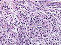
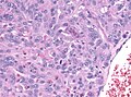
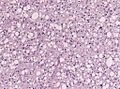
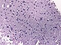
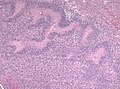
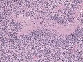
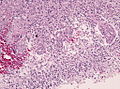
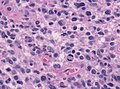
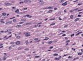
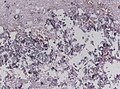
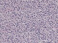
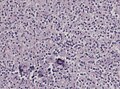
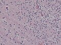
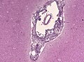
This H&E stain displays parts of a moderately cellular tumor growing with ovoid elongated nuclei (Pictures 1+2). There are no clear-cut cell borders discernible in light microscopy. This interveawing is called a syncytium. WHO Grading of the tumour is dependent of the mitotic activity (Picture 3) or histological hallmarks such as prominent nucleoli, high nuclear to cytoplasmic ratio, CNS infiltation etc.. Focal nuclear clearing (Nuclear pseudoinclusions, Picture 4+5) are typical cut phenomenon. The round calcified inclusions (Psammoma bodies) are characteristic for a meningioma.
- See also: Virtual slide (Meningioma I, HE, usz.ch)
Other language: German
|
|---|
|
Das H&E-Präparat zeigt Anteile eines mäßig zelldichten in Zügen und Wirbeln orientierten Tumors mit ovoid elongierten Tumorzellkernen (Bilder 1+2). Eindeutige Zellgrenzen lassen sich lichtmikroskopisch nicht definieren, was als pseudosynzytialer Aspekt bezeichnet wird. Die Wertung des Tumors nach WHO ist abhängig von der mitotischen Aktivität (Bild 3), oder durch histologische Kriterien wie z.B. prominente Nukleolen, Kern-Plasma-Relation, ZNS-Infiltration etc.. Lochkernzellen (Bild 4+5) sind hingegen ein typisches Anschnittphänomen. Auch rundliche Verkalkungen (Bild 5), sogenannte Psammomkörper sind charakteristisch für Meningeome. |
Astrocytoma
Picture 1
Picture 2
Picture 3


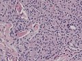
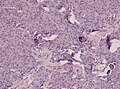
This specimen contains fragments of a diffusely growing tumor with only slight inclreased cell density and focally microcysts withing the neuropil background (Picture 1). Although many of the astrocytic tumour cells look quite similiar there is increased pleomorphism, mostly larger and a more dense chromatin. There are no mitoses seen in this tumour (Picture 2). The tumour borders are not clear, there is just a decrease of cell density a the tumor infiltration zone. In this area one is not always sure which cells are still neoplastic and which cells are normal or reactive astrocytes of the normal brain (Picture 3).
Other language: German
|
|---|
|
Das Präparat besteht aus Fragmenten eines diffus wachsenden, gering zelldichten Tumors, welcher fokal mikrozystische Auflockerungen aufweist (Bild 1). Trotz hoher Ähnlichkeit der astrozytären Tumorzellen finden sich fokal vermehrte Pleomorphie der Kerne, diese meist etwas größer und chromatindichter. Mitosen finden sich in diesem Tumor nicht. (Bild 2). Die Tumorgrenzen sind unscharf, man beobachtet lediglich eine diffus abnehmende Zelldichte im Bereich der Infiltrationszone, in der man nicht genau sagen kann, welches noch Tumorzellen sind und welche bereits normale bzw. reaktiv veränderte Astrozyten darstellen (Bild 3). |
Glioblastoma
Picture 1
Picture 2
Picture 3
Picture 4
Picture 5


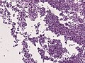
Already at low magnification extraordinary pleomorphism is evident in the cellular tumour (Picture 1). Major hallmark are extensive line-shaped necroses. Tumour cells bordering these necrotic centers are arranged in a pseudopalisading fashion (Picture 2). In addition the glioblastoma harbors neoagniogenesis with vascular proliferations of enlarged endothelial in several layers (Picture 3). The tumour cells are quite pleomorphic, somitimes round, sometimes polygonal. Plenty of mitoses are seen (Picture 4). The tumour cells show slender, elongated cytoplasmic processes, in part still resting on a neuropil-like (fibrillary) background that resembles normal CNS tissue (Picture 5).
Other language: German
|
|---|
|
Schon bei niedriger Vergrößerung erkennt man einen ausserordentlich pleomophen, zelldichten Tumor (Bild 1), dessen herausragendes Merkmal ausgeprägte strichförmige Nekroseareale sind. Die angrenzenden Tumorzellen sind entlang dieser Nekrosebezirke in Pseudopalisadenstellung aufgereiht (Bild 2). Zusätzlich finden sich innerhalb des Tumors Zeichen einer Gefäßneubildung, bei der die Gefäßwände deutlich verbreiter sind und Endothelzellen in mehreren Reihen aufweisen (Bild 3). Die Tumorzellen sind recht pleomorph , teils rundlich teils polygonal bei heterogenem Kernchromatin. Es finden sich reichlich Kernteilungsfiguren (Mitosen, Bild 4). Die Tumorzellen zeigen schmale, jedoch längere Zytoplasmaausläufer, die nur noch abschnittsweise auf einem typischen Neuropil-artigen (fibrillären) Untergrund zu liegen kommen, wie man ihn noch vom normalen ZNS kennt (Bild 5). |
Oligodendroglioma
Picture 1
Picture 2
Picture 3
Picture 4


The sample consists mainly of tumour particles that show extensive calcifications which are already visible at low magnification (Picture 1). The tumour cells show perinuclear halos - a fixation artefact seen typically in oligodendrogliomas (Picture 2). This "fried-egg" appearance at higher cell density results in the so called "honeycomb" appearance. Oligodendrogliomas usually have monomprhic round nuclei with scant chromatin. Between the tumour cells delicate capillaries are present. In contrast to astrocytomas the tumor border is more pronounced, the infiltrative pattern is less evident (Picture 4). In absence of necrosis and almost no mitotic activity this tumour corresponds to WHO Grade II.
Other language: German
|
|---|
|
Das Präparat besteht hauptsächlich aus Tumorpartikeln, welche an mehreren Stellen deutliche Verkalkungen aufweisen. Diese lassen sich schon bei niedriger Vergrößerung beobachten (Bild 1). Die Tumorzellen zeigen ein für Oligodendrogliome typisches Präparationsartefakt bei der durch die Paraffineinbettung ein optisch leeres Zytoplasma entsteht (Bild 2). Dieser sogenannte "Spiegeleieffekt" lässt bei hoher Zelldichte auch an ein Honigwabenmuster denken. Oligodendrogliome besitzen in der Regel relativ monomorphe rundliche Kerne mit mäßigem Chromatingehalt. Zwischen den Tumorzellen verlaufen sehr feine Kapillaren. Im Gegensatz zu Astrozytomen nimmt im Randbereich die Zelldichte abrupter ab, sodaß der Tumor ein weniger diffuses Wachstum aufzeigt (Bild 4). Bei Fehlen von Nekrosen und nahezu nicht vorhandenen Mitosefiguren entspricht der Tumor einem WHO Grad II. |
Day two
Purulent meningitis
Picture 1
Picture 2
Picture 3


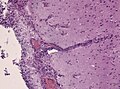
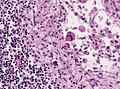
This autopsy specimen from cerebral hemisphere shows diffuse clouding of the arachnoidea and pia mater by a dense cellular infiltrate as seen in this low magnification (Picture 1). In higher magnification the infiltrate consists mainly of neutrophil granulocytes (Picture 2). Because the granulocytes are also seen along capillaries in the Virchow-Robin space, we consider this a meningoencephalitis (Picture 3).
Other language: German
|
|---|
|
Der Gewebeschnitt zeigt Anteile des cerebralen Cortex, dessen Subrarachnoidalraum durch ein zellreiches Infiltrat verlegt ist, welches schon bei niedriger Vergrösserung gut zu erkennen ist (Bild 1). Dieses stellt sich in höherer Vergrößerung als überwiegend aus segmentkernigen neutrophilen Granulozyten bestehend dar (Bild 2). Da die Granulozyten auch entlang der Kapillaren in dem sogenannten Virchow-Robinschen Raum eindringen, spricht man genauer von einer Meningoenzephalitis (Bild 3). |
Tuberculous meningitis
Picture 1
Picture 2
Picture 3
Picture 4


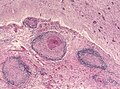
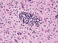
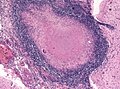
This autopsy specimen was sampled in coronar section from the optic chiasm and displays numerous granulomas (Picture 1). In the brain parenchyma mutlifocal smaller infiltrates consisting of lymphocytes and monocytes. The adjacent astrocytes show reactive changes with broadly swollen, pink cytoplasm ("gemistocytes", Picture 2). The granulomas are surronded by lymphocytes and in between so called epitheloid cells with chromatin-dense, elongated, sole-like nuclei and occassionally Langhans giant cells (Picture 3). The center of the granuloma is often necrotic, such "caeseating necroses" are typical for tuberculous meningitis (Picture 4).
Other language: German
|
|---|
|
Der Gewebeschnitt in der Frontalebene auf Höhe des Chiasmas, zeigt basal zahlreiche Granulome (Bild 1). Im ZNS-Parenchym finden sich multifokal kleinere rundzellige Infiltrate aus überwiegend lympho-monozytären Zellen, die angrenzenden Astrozyten sind reaktiv verändert mit pinkfarbenem, geschwollenem Zytoplasma ("Gemistozyten" Bild 2). Die Granulome besitzen aussen einen Saum aus Lymphozyten an den sich nach innen sogenannte Epitheloidzellen mit länglichen, chromatindichten, Schuhsohlenartig geformten Kernen, Makrophagen sowie mehrkernige Riesenzellen vom Langhans-Typ (Bild 3) anschliessen. Das Zentrum der Granulome ist oft nekrotisch verändert, solche sogenannten "verkäsende Nekrosen" sind typisch für eine Tuberkulöse Meningitis (Bild 4) |
Molecular neuropathology
Coming soon.
Adavanced neuropathology
Coming soon.