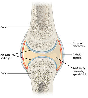
Joints
Joints are important for locomotion. This article collects tidbits about their pathology.
A general differential diagnosis for joints is dealt with in the cartilage article.
Septic arthritis
- Septic joint redirects here.
General
- Uncommon.
- May be treated in a one-stage or two-stage procedure.[1]
Microscopic
Features:
- Microorganisms - key feature.
- Inflammatory cells, esp. neutrophils. ‡
- +/-Osteomyelitis.
Note:
- ‡ There are several criteria for the number of neurophils in the context of prosthetic joints - see prosthetic joint infection.
Sign out
RIGHT FEMORAL HEAD AND SURROUNDING TISSUE, REMOVAL:
- OSTEOMYELITIS, ACUTE.
- OSTEONECROSIS, FOCAL.
- DEGENERATIVE JOINT DISEASE.
- SOFT TISSUE WITH FOCAL NECROSIS AND COCCI MICROORGANISMS, CORRELATION
  WITH MICROBIOLOGY SUGGESTED.
- NEGATIVE FOR MALIGNANCY.
Prosthetic joint infection
General
- Bits of tissue come for frozen section to r/o infection.
- The interface membrane (not the pseudocapsule) should be sampled to obtain a high sensitivity.[2]
Microscopic
Features:
- Neutrophils - key feature.
Notes:
- Various criteria for the number of neutrophils exist (see below).[3]
- Finding of plasma cells and lymphocytes is not contributory for the diagnosis of infection.[3]
- Granulomatous inflammation that isn't of a foreign-body type should get the usual work-up.[5]
Feldman criteria
Athanasou criterion
Morawietz criteria
- 23 neutrophils/10 HPF, where the field diameter = 0.625 mm.[4]
Notes:
- The 0.625 mm field diameter is uncommon; it corresponds to a 25 mm eye piece with a 40x objective (25 mm/40 = 0.625 mm).
- The PMNs/area is: 23 PMNs / (pi/4*0.625^2) = 7.49 PMNs/mm*mm
- If one uses a 22 mm eye piece microscope and the 40x objective, the field diameter is 0.55 mm; thus, one would need to count PMNs in 12.91 HPFs ( 3.0680 mm*mm / 0.2376 mm*mm = 12.91 ) to get the same sample area.
A comparison of the criteria
A study by Bori et al.[3] compared the criteria of Feldman and Athanasou:
| Measure | Feldman | Athanasou |
|---|---|---|
| Sensitivity | 29% | 71% |
| Specificity | 100% | 64% |
Sign out
Tissue from Left Hip, Revision Arthroplasty:
- Connective tissue with focally increased neutrophils (at least
  11 neutrophils/HPF).
- Negative for microorganisms with routine stains.
- NEGATIVE for malignancy.

Comment:
The findings raise the possibility of a joint infection; correlation with
cultures is required.

1 HPF = 0.2376 mm*mm.
Block letters
HEMATOMA, RIGHT HIP, IRRIGATION AFTER HIP REPLACEMENT/TISSUE REMOVAL:
- FIBROADIPOSE TISSUE WITH POSTSURGICAL CHANGES INCLUDING:
-- FOREIGN-BODY TYPE MULTINUCLEATED GIANT CELLS AND FOREIGN MATERIAL.
-- GRANULATION TISSUE.
-- FAT NECROSIS (FOCAL).
- FIBRIN, BLOOD.
- NEUTROPHILS IN BLOOD VESSEL WALLS/PERIVASCULAR (FOCAL), SEE COMMENT.
- NEGATIVE FOR MICROORGANISMS WITH ROUTINE STAIN.

COMMENT:
Up to 17 neutrophils per high power field (0.55 mm field diameter) are seen.
FEMUR, LEFT, BIOPSY:
- BONE MARROW WITH A PATCHY MILD FOCAL PROMINENCE OF PLASMA CELLS AND NEUTROPHILS.
- FIBROUS TISSUE WITH HEMOSIDERIN, ISOLATED NEUTROPHILS AND RARE
MACROPHAGES.
- BONE FRAGMENTS.
- NEGATIVE FOR MICROABSCESS FORMATION.
- NEGATIVE FOR MICROORGANISMS WITH ROUTINE STAINS.
BONE AND SOFT TISSUE, LEFT HIP, EXCISION:
- FIBROADIPOSE TISSUE, SKELETAL MUSCLE AND FRAGMENTS OF BONE WITH SURGICAL CHANGES.
- NEGATIVE FOR MICROABSCESS FORMATION.
- NEGATIVE FOR MICROORGANISMS WITH ROUTINE STAINS.
Juxta-articular myxoma
- Abbreviated JAM.
General
- Classically present as a mass or with pain.[6]
- May recur.
- Described as (microscopically) indistinguishable from intramuscular myxoma.[7]
Gross
Features:
- Close to large joints - classically around the knee.
- >85% of cases around the knee in one large series.[6]
Microscopic
Features:[6]
- Myxomatous stroma.
- +/-Cystic changes.
- Large nuclei with hyperchromasia and nuclear scalloping.[8]
DDx:
- Ganglion cyst.
- Intramuscular myxoma - surrounded by muscle.
Images:
Morton neuroma
Main article: Morton neuroma
Ganglion cyst
Main article: Ganglion cyst
Rheumatoid arthritis
Main article: Rheumatoid arthritis
Acute synovitis
Main article: Acute synovitis
See also
References
- ↑ Chen CE, Wang JW, Juhn RJ (October 2008). "Total hip arthroplasty for primary septic arthritis of the hip in adults". Int Orthop 32 (5): 573–80. doi:10.1007/s00264-007-0366-1. PMC 2551720. PMID 17483946. https://www.ncbi.nlm.nih.gov/pmc/articles/PMC2551720/.
- ↑ 2.0 2.1 Bori, G.; Muñoz-Mahamud, E.; Garcia, S.; Mallofre, C.; Gallart, X.; Bosch, J.; Garcia, E.; Riba, J. et al. (Apr 2011). "Interface membrane is the best sample for histological study to diagnose prosthetic joint infection.". Mod Pathol 24 (4): 579-84. doi:10.1038/modpathol.2010.219. PMID 21131917.
- ↑ 3.0 3.1 3.2 3.3 3.4 Bori, G.; Soriano, A.; García, S.; Mallofré, C.; Riba, J.; Mensa, J. (Jun 2007). "Usefulness of histological analysis for predicting the presence of microorganisms at the time of reimplantation after hip resection arthroplasty for the treatment of infection.". J Bone Joint Surg Am 89 (6): 1232-7. doi:10.2106/JBJS.F.00741. PMID 17545426.
- ↑ 4.0 4.1 Morawietz, L.; Tiddens, O.; Mueller, M.; Tohtz, S.; Gansukh, T.; Schroeder, JH.; Perka, C.; Krenn, V. (Jun 2009). "Twenty-three neutrophil granulocytes in 10 high-power fields is the best histopathological threshold to differentiate between aseptic and septic endoprosthesis loosening.". Histopathology 54 (7): 847-53. doi:10.1111/j.1365-2559.2009.03313.x. PMID 19635104.
- ↑ URL: http://path.upmc.edu/cases/case174.html. Accessed on: 8 January 2012.
- ↑ 6.0 6.1 6.2 Meis, JM.; Enzinger, FM. (Jun 1992). "Juxta-articular myxoma: a clinical and pathologic study of 65 cases.". Hum Pathol 23 (6): 639-46. PMID 1592386.
- ↑ Humphrey, Peter A; Dehner, Louis P; Pfeifer, John D (2008). The Washington Manual of Surgical Pathology (1st ed.). Lippincott Williams & Wilkins. pp. 624. ISBN 978-0781765275.
- ↑ URL: http://path.upmc.edu/cases/case38/micro.html. Accessed on: 2 January 2011.
