Difference between revisions of "GATA3"
Jump to navigation
Jump to search
m (vauthors) |
|||
| (9 intermediate revisions by the same user not shown) | |||
| Line 6: | Line 6: | ||
| Abbrev = | | Abbrev = | ||
| Synonyms = | | Synonyms = | ||
| Similar = [[thrombomodulin]] | | Similar = [[thrombomodulin]], mammaglobin | ||
| Clones = | | Clones = | ||
| Use = bladder versus prostate, bladder versus SCC | | Use = bladder versus prostate, bladder versus SCC, breast versus other, parathyroid versus thyroid | ||
| Subspecial = [[Genitourinary pathology]], [[Breast pathology]] | | Subspecial = [[Genitourinary pathology]], [[Breast pathology]] | ||
| Pattern = nuclear | | Pattern = nuclear | ||
| Line 22: | Line 22: | ||
**[[Lobular breast carcinoma]].<ref name=pmid24061521>{{Cite journal | last1 = Ellis | first1 = CL. | last2 = Chang | first2 = AG. | last3 = Cimino-Mathews | first3 = A. | last4 = Argani | first4 = P. | last5 = Youssef | first5 = RF. | last6 = Kapur | first6 = P. | last7 = Montgomery | first7 = EA. | last8 = Epstein | first8 = JI. | title = GATA-3 immunohistochemistry in the differential diagnosis of adenocarcinoma of the urinary bladder. | journal = Am J Surg Pathol | volume = 37 | issue = 11 | pages = 1756-60 | month = Nov | year = 2013 | doi = 10.1097/PAS.0b013e31829cdba7 | PMID = 24061521 }}</ref> | **[[Lobular breast carcinoma]].<ref name=pmid24061521>{{Cite journal | last1 = Ellis | first1 = CL. | last2 = Chang | first2 = AG. | last3 = Cimino-Mathews | first3 = A. | last4 = Argani | first4 = P. | last5 = Youssef | first5 = RF. | last6 = Kapur | first6 = P. | last7 = Montgomery | first7 = EA. | last8 = Epstein | first8 = JI. | title = GATA-3 immunohistochemistry in the differential diagnosis of adenocarcinoma of the urinary bladder. | journal = Am J Surg Pathol | volume = 37 | issue = 11 | pages = 1756-60 | month = Nov | year = 2013 | doi = 10.1097/PAS.0b013e31829cdba7 | PMID = 24061521 }}</ref> | ||
*[[Chromophobe renal cell carcinoma]] ~50% of cases.<ref name=pmid24145643/> | *[[Chromophobe renal cell carcinoma]] ~50% of cases.<ref name=pmid24145643/> | ||
*[[Trophoblastic tumours]] +ve | *[[Trophoblastic tumours]] +ve<ref>{{Cite journal | last1 = Mirkovic | first1 = J. | last2 = Elias | first2 = K. | last3 = Drapkin | first3 = R. | last4 = Barletta | first4 = JA. | last5 = Quade | first5 = B. | last6 = Hirsch | first6 = MS. | title = GATA3 expression in gestational trophoblastic tissues and tumours. | journal = Histopathology | volume = 67 | issue = 5 | pages = 636-44 | month = Nov | year = 2015 | doi = 10.1111/his.12681 | PMID = 25753145 }}</ref> including [[choriocarcinoma]].<ref name=pmid26772394 >{{cite journal |authors=Osman H, Cheng L, Ulbright TM, Idrees MT |title=The utility of CDX2, GATA3, and DOG1 in the diagnosis of testicular neoplasms: an immunohistochemical study of 109 cases |journal=Hum Pathol |volume=48 |issue= |pages=18–24 |date=February 2016 |pmid=26772394 |doi=10.1016/j.humpath.2015.09.028 |url=}}</ref> | ||
*Parathyroid - [[parathyroid hyperplasia]], [[parathyroid adenoma]], [[parathyroid carcinoma]].<ref name=pmid25046229>{{Cite journal | last1 = Ordóñez | first1 = NG. | title = Value of GATA3 immunostaining in the diagnosis of parathyroid tumors. | journal = Appl Immunohistochem Mol Morphol | volume = 22 | issue = 10 | pages = 756-61 | month = | year = | doi = 10.1097/PAI.0000000000000007 | PMID = 25046229 }}</ref> | *Parathyroid - [[parathyroid hyperplasia]], [[parathyroid adenoma]], [[parathyroid carcinoma]].<ref name=pmid25046229>{{Cite journal | last1 = Ordóñez | first1 = NG. | title = Value of GATA3 immunostaining in the diagnosis of parathyroid tumors. | journal = Appl Immunohistochem Mol Morphol | volume = 22 | issue = 10 | pages = 756-61 | month = | year = | doi = 10.1097/PAI.0000000000000007 | PMID = 25046229 }}</ref> | ||
*Salivary gland tumours <ref name=pmid23604756>{{Cite journal | last1 = Schwartz | first1 = LE. | last2 = Begum | first2 = S. | last3 = Westra | first3 = WH. | last4 = Bishop | first4 = JA. | title = GATA3 immunohistochemical expression in salivary gland neoplasms. | journal = Head Neck Pathol | volume = 7 | issue = 4 | pages = 311-5 | month = Dec | year = 2013 | doi = 10.1007/s12105-013-0442-3 | PMID = 23604756 }}</ref> | *Salivary gland tumours <ref name=pmid23604756>{{Cite journal | last1 = Schwartz | first1 = LE. | last2 = Begum | first2 = S. | last3 = Westra | first3 = WH. | last4 = Bishop | first4 = JA. | title = GATA3 immunohistochemical expression in salivary gland neoplasms. | journal = Head Neck Pathol | volume = 7 | issue = 4 | pages = 311-5 | month = Dec | year = 2013 | doi = 10.1007/s12105-013-0442-3 | PMID = 23604756 }}</ref> | ||
| Line 30: | Line 30: | ||
*Most [[T-lymphocytes]].<ref name=pmid19151747>{{cite journal |authors=Ho IC, Tai TS, Pai SY |title=GATA3 and the T-cell lineage: essential functions before and after T-helper-2-cell differentiation |journal=Nat. Rev. Immunol. |volume=9 |issue=2 |pages=125–35 |date=February 2009 |pmid=19151747 |pmc=2998182 |doi=10.1038/nri2476 |url=}}</ref> Used as part of a wider panel of IHC to sub-type peripheral T-cell lymphoma, NOS.<ref name=pmid31562134>{{cite journal |authors=Amador C, Greiner TC, Heavican TB, Smith LM, Galvis KT, Lone W, Bouska A, D'Amore F, Pedersen MB, Pileri S, Agostinelli C, Feldman AL, Rosenwald A, Ott G, Mottok A, Savage KJ, de Leval L, Gaulard P, Lim ST, Ong CK, Ondrejka SL, Song J, Campo E, Jaffe ES, Staudt LM, Rimsza LM, Vose J, Weisenburger DD, Chan WC, Iqbal J |title=Reproducing the molecular subclassification of peripheral T-cell lymphoma-NOS by immunohistochemistry |journal=Blood |volume=134 |issue=24 |pages=2159–2170 |date=December 2019 |pmid=31562134 |doi=10.1182/blood.2019000779 |url=}}</ref> | *Most [[T-lymphocytes]].<ref name=pmid19151747>{{cite journal |authors=Ho IC, Tai TS, Pai SY |title=GATA3 and the T-cell lineage: essential functions before and after T-helper-2-cell differentiation |journal=Nat. Rev. Immunol. |volume=9 |issue=2 |pages=125–35 |date=February 2009 |pmid=19151747 |pmc=2998182 |doi=10.1038/nri2476 |url=}}</ref> Used as part of a wider panel of IHC to sub-type peripheral T-cell lymphoma, NOS.<ref name=pmid31562134>{{cite journal |authors=Amador C, Greiner TC, Heavican TB, Smith LM, Galvis KT, Lone W, Bouska A, D'Amore F, Pedersen MB, Pileri S, Agostinelli C, Feldman AL, Rosenwald A, Ott G, Mottok A, Savage KJ, de Leval L, Gaulard P, Lim ST, Ong CK, Ondrejka SL, Song J, Campo E, Jaffe ES, Staudt LM, Rimsza LM, Vose J, Weisenburger DD, Chan WC, Iqbal J |title=Reproducing the molecular subclassification of peripheral T-cell lymphoma-NOS by immunohistochemistry |journal=Blood |volume=134 |issue=24 |pages=2159–2170 |date=December 2019 |pmid=31562134 |doi=10.1182/blood.2019000779 |url=}}</ref> | ||
*[[Skin squamous cell carcinoma]].<ref name=pmid26595821>{{cite journal |authors=Mertens RB, de Peralta-Venturina MN, Balzer BL, Frishberg DP |title=GATA3 Expression in Normal Skin and in Benign and Malignant Epidermal and Cutaneous Adnexal Neoplasms |journal=Am J Dermatopathol |volume=37 |issue=12 |pages=885–91 |date=December 2015 |pmid=26595821 |pmc=4894790 |doi=10.1097/DAD.0000000000000306 |url=}}</ref> | *[[Skin squamous cell carcinoma]].<ref name=pmid26595821>{{cite journal |authors=Mertens RB, de Peralta-Venturina MN, Balzer BL, Frishberg DP |title=GATA3 Expression in Normal Skin and in Benign and Malignant Epidermal and Cutaneous Adnexal Neoplasms |journal=Am J Dermatopathol |volume=37 |issue=12 |pages=885–91 |date=December 2015 |pmid=26595821 |pmc=4894790 |doi=10.1097/DAD.0000000000000306 |url=}}</ref> | ||
*[[Clear cell tubulopapillary renal cell carcinoma]].<ref name=pmid28705707>{{cite journal |authors=Mantilla JG, Antic T, Tretiakova M |title=GATA3 as a valuable marker to distinguish clear cell papillary renal cell carcinomas from morphologic mimics |journal=Hum Pathol |volume=66 |issue= |pages=152–158 |date=August 2017 |pmid=28705707 |doi=10.1016/j.humpath.2017.06.016 |url=}}</ref> | |||
===Images=== | ===Images=== | ||
| Line 37: | Line 38: | ||
*[[Prostate carcinoma]]. | *[[Prostate carcinoma]]. | ||
**Positive in benign prostate glands with radiation atypia.<ref name=pmid28316088>{{cite journal |authors=Tian W, Dorn D, Wei S, Sanders RD, Matoso A, Shah RB, Gordetsky J |title=GATA3 expression in benign prostate glands with radiation atypia: a diagnostic pitfall |journal=Histopathology |volume=71 |issue=1 |pages=150–155 |date=July 2017 |pmid=28316088 |doi=10.1111/his.13214 |url=}}</ref> | **Positive in benign prostate glands with radiation atypia.<ref name=pmid28316088>{{cite journal |authors=Tian W, Dorn D, Wei S, Sanders RD, Matoso A, Shah RB, Gordetsky J |title=GATA3 expression in benign prostate glands with radiation atypia: a diagnostic pitfall |journal=Histopathology |volume=71 |issue=1 |pages=150–155 |date=July 2017 |pmid=28316088 |doi=10.1111/his.13214 |url=}}</ref> | ||
**Positive staining may be seen in prostatic adenocarcinoma.<ref name=pmid32769430>{{cite journal |vauthors=McDonald TM, Epstein JI |title=Aberrant GATA3 Staining in Prostatic Adenocarcinoma: A Potential Diagnostic Pitfall |journal=Am J Surg Pathol |volume=45 |issue=3 |pages=341–346 |date=March 2021 |pmid=32769430 |doi=10.1097/PAS.0000000000001557 |url=}}</ref> | |||
*[[Squamous cell carcinoma of the lung]].<ref name=pmid22982890/> | *[[Squamous cell carcinoma of the lung]].<ref name=pmid22982890/> | ||
| Line 43: | Line 45: | ||
==References== | ==References== | ||
{{Reflist| | {{Reflist|2}} | ||
[[Category:Immunohistochemistry]] | [[Category:Immunohistochemistry]] | ||
Latest revision as of 19:04, 18 June 2024
| GATA3 | |
|---|---|
| Immunostain in short | |
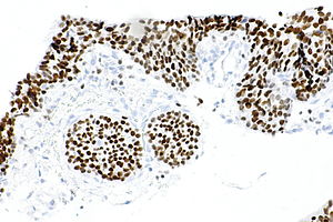
 GATA3 staining in benign urothelium. | |
| Similar stains | thrombomodulin, mammaglobin |
| Use | bladder versus prostate, bladder versus SCC, breast versus other, parathyroid versus thyroid |
| Subspeciality | Genitourinary pathology, Breast pathology |
| Normal staining pattern | nuclear |
| Positive | urothelial carcinoma, invasive ductal carcinoma of the breast, lobular breast carcinoma |
| Negative | prostatic carcinoma, squamous cell carcinoma of the lung |
GATA3 an immunostain that is increasingly used in genitourinary pathology.
Positive
- Urothelial carcinoma.[1]
- Breast carcinoma - more sensitive than GCDFP-15.[2]
- Chromophobe renal cell carcinoma ~50% of cases.[2]
- Trophoblastic tumours +ve[4] including choriocarcinoma.[5]
- Parathyroid - parathyroid hyperplasia, parathyroid adenoma, parathyroid carcinoma.[6]
- Salivary gland tumours [7]
- Pheochromocytoma (~70%) vs adrenocortical carcinoma (<10%[8]).
- Brenner tumours and Walthard cell rests.[9]
- Malignant mesotheliomas (58%[2]).
- Most T-lymphocytes.[10] Used as part of a wider panel of IHC to sub-type peripheral T-cell lymphoma, NOS.[11]
- Skin squamous cell carcinoma.[12]
- Clear cell tubulopapillary renal cell carcinoma.[13]
Images
Negative
See also
References
- ↑ 1.0 1.1 1.2 Chang A, Amin A, Gabrielson E, et al. (October 2012). "Utility of GATA3 immunohistochemistry in differentiating urothelial carcinoma from prostate adenocarcinoma and squamous cell carcinomas of the uterine cervix, anus, and lung". Am. J. Surg. Pathol. 36 (10): 1472–6. doi:10.1097/PAS.0b013e318260cde7. PMC 3444740. PMID 22982890. https://www.ncbi.nlm.nih.gov/pmc/articles/PMC3444740/.
- ↑ 2.0 2.1 2.2 Miettinen, M.; McCue, PA.; Sarlomo-Rikala, M.; Rys, J.; Czapiewski, P.; Wazny, K.; Langfort, R.; Waloszczyk, P. et al. (Jan 2014). "GATA3: a multispecific but potentially useful marker in surgical pathology: a systematic analysis of 2500 epithelial and nonepithelial tumors.". Am J Surg Pathol 38 (1): 13-22. doi:10.1097/PAS.0b013e3182a0218f. PMID 24145643.
- ↑ Ellis, CL.; Chang, AG.; Cimino-Mathews, A.; Argani, P.; Youssef, RF.; Kapur, P.; Montgomery, EA.; Epstein, JI. (Nov 2013). "GATA-3 immunohistochemistry in the differential diagnosis of adenocarcinoma of the urinary bladder.". Am J Surg Pathol 37 (11): 1756-60. doi:10.1097/PAS.0b013e31829cdba7. PMID 24061521.
- ↑ Mirkovic, J.; Elias, K.; Drapkin, R.; Barletta, JA.; Quade, B.; Hirsch, MS. (Nov 2015). "GATA3 expression in gestational trophoblastic tissues and tumours.". Histopathology 67 (5): 636-44. doi:10.1111/his.12681. PMID 25753145.
- ↑ Osman H, Cheng L, Ulbright TM, Idrees MT (February 2016). "The utility of CDX2, GATA3, and DOG1 in the diagnosis of testicular neoplasms: an immunohistochemical study of 109 cases". Hum Pathol 48: 18–24. doi:10.1016/j.humpath.2015.09.028. PMID 26772394.
- ↑ Ordóñez, NG.. "Value of GATA3 immunostaining in the diagnosis of parathyroid tumors.". Appl Immunohistochem Mol Morphol 22 (10): 756-61. doi:10.1097/PAI.0000000000000007. PMID 25046229.
- ↑ Schwartz, LE.; Begum, S.; Westra, WH.; Bishop, JA. (Dec 2013). "GATA3 immunohistochemical expression in salivary gland neoplasms.". Head Neck Pathol 7 (4): 311-5. doi:10.1007/s12105-013-0442-3. PMID 23604756.
- ↑ Perrino, CM.; Ho, A.; Dall, CP.; Zynger, DL. (Sep 2017). "Utility of GATA3 in the differential diagnosis of pheochromocytoma.". Histopathology 71 (3): 475-479. doi:10.1111/his.13229. PMID 28374498.
- ↑ Roma, AA.; Masand, RP. (Dec 2014). "Ovarian Brenner tumors and Walthard nests: a histologic and immunohistochemical study.". Hum Pathol 45 (12): 2417-22. doi:10.1016/j.humpath.2014.08.003. PMID 25281026.
- ↑ Ho IC, Tai TS, Pai SY (February 2009). "GATA3 and the T-cell lineage: essential functions before and after T-helper-2-cell differentiation". Nat. Rev. Immunol. 9 (2): 125–35. doi:10.1038/nri2476. PMC 2998182. PMID 19151747. https://www.ncbi.nlm.nih.gov/pmc/articles/PMC2998182/.
- ↑ Amador C, Greiner TC, Heavican TB, Smith LM, Galvis KT, Lone W, Bouska A, D'Amore F, Pedersen MB, Pileri S, Agostinelli C, Feldman AL, Rosenwald A, Ott G, Mottok A, Savage KJ, de Leval L, Gaulard P, Lim ST, Ong CK, Ondrejka SL, Song J, Campo E, Jaffe ES, Staudt LM, Rimsza LM, Vose J, Weisenburger DD, Chan WC, Iqbal J (December 2019). "Reproducing the molecular subclassification of peripheral T-cell lymphoma-NOS by immunohistochemistry". Blood 134 (24): 2159–2170. doi:10.1182/blood.2019000779. PMID 31562134.
- ↑ Mertens RB, de Peralta-Venturina MN, Balzer BL, Frishberg DP (December 2015). "GATA3 Expression in Normal Skin and in Benign and Malignant Epidermal and Cutaneous Adnexal Neoplasms". Am J Dermatopathol 37 (12): 885–91. doi:10.1097/DAD.0000000000000306. PMC 4894790. PMID 26595821. https://www.ncbi.nlm.nih.gov/pmc/articles/PMC4894790/.
- ↑ Mantilla JG, Antic T, Tretiakova M (August 2017). "GATA3 as a valuable marker to distinguish clear cell papillary renal cell carcinomas from morphologic mimics". Hum Pathol 66: 152–158. doi:10.1016/j.humpath.2017.06.016. PMID 28705707.
- ↑ Tian W, Dorn D, Wei S, Sanders RD, Matoso A, Shah RB, Gordetsky J (July 2017). "GATA3 expression in benign prostate glands with radiation atypia: a diagnostic pitfall". Histopathology 71 (1): 150–155. doi:10.1111/his.13214. PMID 28316088.
- ↑ "Aberrant GATA3 Staining in Prostatic Adenocarcinoma: A Potential Diagnostic Pitfall". Am J Surg Pathol 45 (3): 341–346. March 2021. doi:10.1097/PAS.0000000000001557. PMID 32769430.