Difference between revisions of "Ki-67"
Jump to navigation
Jump to search

(tweak) |
|||
| (4 intermediate revisions by the same user not shown) | |||
| Line 10: | Line 10: | ||
| Use = [[typical carcinoid]] versus [[atypical carcinoid]]), grading (gastrointestinal [[neuroendocrine tumours]]) | | Use = [[typical carcinoid]] versus [[atypical carcinoid]]), grading (gastrointestinal [[neuroendocrine tumours]]) | ||
| Subspecial = | | Subspecial = | ||
| Pattern = nuclear staining, membranous pattern | | Pattern = nuclear staining, occasionally membranous pattern ([[pneumocytoma]], [[hyalinizing trabecular tumour]]) | ||
| Positive = | | Positive = | ||
| Negative = | | Negative = | ||
| Other = | | Other = | ||
}} | }} | ||
'''Ki-67''' is a very commonly used [[immunostain]] that | '''Ki-67''' is a very commonly used [[immunostain]] that is reflective of proliferative activity. | ||
''MIB1'' redirects here; it is a commonly used antibody against Ki-67. | |||
==General== | ==General== | ||
| Line 22: | Line 24: | ||
===Membranous staining pattern=== | ===Membranous staining pattern=== | ||
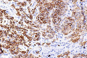
[[Image:Pneumocytoma - Ki67 -- high mag.jpg|right|thumb|Ki-67 showing a membranous pattern in [[pneumocytoma]].]] | |||
*[[Pneumocytoma]] has membranous pattern.<ref name=pmid23030396>{{Cite journal | last1 = Kim | first1 = BH. | last2 = Bae | first2 = YS. | last3 = Kim | first3 = SH. | last4 = Jeong | first4 = HJ. | last5 = Hong | first5 = SW. | last6 = Yoon | first6 = SO. | title = Usefulness of Ki-67 (MIB-1) immunostaining in the diagnosis of pulmonary sclerosing hemangiomas. | journal = APMIS | volume = 121 | issue = 2 | pages = 105-10 | month = Feb | year = 2013 | doi = 10.1111/j.1600-0463.2012.02945.x | PMID = 23030396 }}</ref> | *[[Pneumocytoma]] has membranous pattern.<ref name=pmid23030396>{{Cite journal | last1 = Kim | first1 = BH. | last2 = Bae | first2 = YS. | last3 = Kim | first3 = SH. | last4 = Jeong | first4 = HJ. | last5 = Hong | first5 = SW. | last6 = Yoon | first6 = SO. | title = Usefulness of Ki-67 (MIB-1) immunostaining in the diagnosis of pulmonary sclerosing hemangiomas. | journal = APMIS | volume = 121 | issue = 2 | pages = 105-10 | month = Feb | year = 2013 | doi = 10.1111/j.1600-0463.2012.02945.x | PMID = 23030396 }}</ref> | ||
*[[Hyalinizing trabecular tumour]].<ref name=pmid17525638>{{Cite journal | last1 = Leonardo | first1 = E. | last2 = Volante | first2 = M. | last3 = Barbareschi | first3 = M. | last4 = Cavazza | first4 = A. | last5 = Dei Tos | first5 = AP. | last6 = Bussolati | first6 = G. | last7 = Papotti | first7 = M. | title = Cell membrane reactivity of MIB-1 antibody to Ki67 in human tumors: fact or artifact? | journal = Appl Immunohistochem Mol Morphol | volume = 15 | issue = 2 | pages = 220-3 | month = Jun | year = 2007 | doi = 10.1097/01.pai.0000213122.66096.f0 | PMID = 17525638 }}</ref> | *[[Hyalinizing trabecular tumour]] - only with ''MIB1 antibody clone'' when processed at room temperature.<ref name=pmid17525638>{{Cite journal | last1 = Leonardo | first1 = E. | last2 = Volante | first2 = M. | last3 = Barbareschi | first3 = M. | last4 = Cavazza | first4 = A. | last5 = Dei Tos | first5 = AP. | last6 = Bussolati | first6 = G. | last7 = Papotti | first7 = M. | title = Cell membrane reactivity of MIB-1 antibody to Ki67 in human tumors: fact or artifact? | journal = Appl Immunohistochem Mol Morphol | volume = 15 | issue = 2 | pages = 220-3 | month = Jun | year = 2007 | doi = 10.1097/01.pai.0000213122.66096.f0 | PMID = 17525638 }}</ref> | ||
==See also== | ==See also== | ||
Latest revision as of 20:03, 7 July 2020
| Ki-67 | |
|---|---|
| Immunostain in short | |
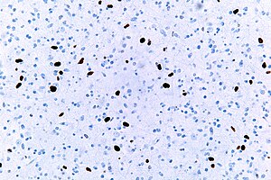
 Ki-67 staining in an anaplastic astrocytoma. | |
| Use | typical carcinoid versus atypical carcinoid), grading (gastrointestinal neuroendocrine tumours) |
| Normal staining pattern | nuclear staining, occasionally membranous pattern (pneumocytoma, hyalinizing trabecular tumour) |
Ki-67 is a very commonly used immunostain that is reflective of proliferative activity.
MIB1 redirects here; it is a commonly used antibody against Ki-67.
General
- Marker of proliferation.
- Usually nuclear staining.
Membranous staining pattern

Ki-67 showing a membranous pattern in pneumocytoma.
- Pneumocytoma has membranous pattern.[1]
- Hyalinizing trabecular tumour - only with MIB1 antibody clone when processed at room temperature.[2]
See also
References
- ↑ Kim, BH.; Bae, YS.; Kim, SH.; Jeong, HJ.; Hong, SW.; Yoon, SO. (Feb 2013). "Usefulness of Ki-67 (MIB-1) immunostaining in the diagnosis of pulmonary sclerosing hemangiomas.". APMIS 121 (2): 105-10. doi:10.1111/j.1600-0463.2012.02945.x. PMID 23030396.
- ↑ Leonardo, E.; Volante, M.; Barbareschi, M.; Cavazza, A.; Dei Tos, AP.; Bussolati, G.; Papotti, M. (Jun 2007). "Cell membrane reactivity of MIB-1 antibody to Ki67 in human tumors: fact or artifact?". Appl Immunohistochem Mol Morphol 15 (2): 220-3. doi:10.1097/01.pai.0000213122.66096.f0. PMID 17525638.