Difference between revisions of "Case 86"
Jump to navigation
Jump to search
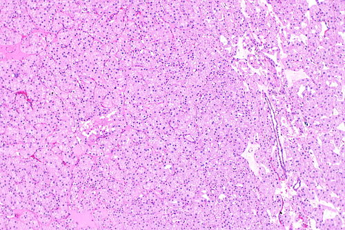
Low magnification. H&E stain.
(Created page with "===Provided clinical history=== 62 year old woman, mass lesion ===Site=== left kidney ===Primary image=== Image: Eosinophilic variant of chromophobe renal cell carcinoma -...") |
(more) |
||
| (One intermediate revision by the same user not shown) | |||
| Line 268: | Line 268: | ||
===Diagnosis=== | ===Diagnosis=== | ||
{{hidden|Diagnosis|<center>[[Chromophobe renal cell carcinoma]], | {{hidden|Diagnosis|<center>[[Chromophobe renal cell carcinoma]], eosinophilic variant</center> | ||
<br>Comment:<br> | |||
The main differential diagnosis is [[renal oncocytoma]]. [[Hale's colloidal iron]] (blue in ChRCC), and CK7 (strong membranous in ChRCC versus weak/patchy in oncocytoma) are quite useful. | |||
Morphologically, ChRCC typically has perinuclear halos (may be focal), frequent binucleation and wrinkled nuclear membranes (raisinoid nuclei).<ref name=pmid9844591>{{Cite journal | last1 = Tickoo | first1 = SK. | last2 = Amin | first2 = MB. | title = Discriminant nuclear features of renal oncocytoma and chromophobe renal cell carcinoma. Analysis of their potential utility in the differential diagnosis. | journal = Am J Clin Pathol | volume = 110 | issue = 6 | pages = 782-7 | month = Dec | year = 1998 | doi = | PMID = 9844591 }}</ref> | |||
===Reference=== | |||
{{Reflist|1}} | |||
}} | |||
<br> | <br> | ||
===Other cases=== | ===Other cases=== | ||
{{Cases navigation}} | {{Cases navigation}} | ||
Latest revision as of 04:20, 29 September 2015
Provided clinical history
62 year old woman, mass lesion
Site
left kidney
Primary image
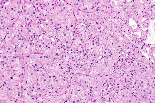
Intermediate magnification
|
|---|
 |
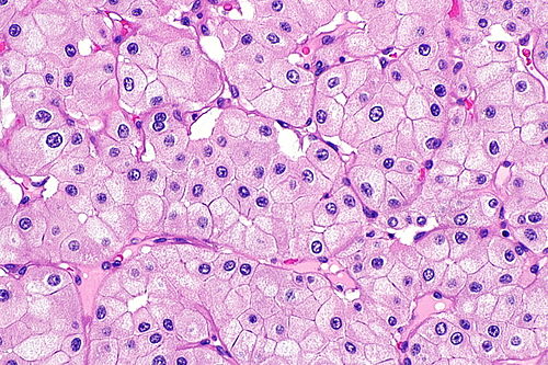
High magnification
|
|---|
 |
Differential diagnosis
Differential diagnosis
|
|---|
|
|
Additional tests
More history
More history
|
|---|
|
|
Ask a colleague
Ask a colleague
|
|---|
|
|
Stains
|
|
|
|
|
|
|---|
IHC
|
|
|
|
|
|
|---|
Molecular testing
Chromosomal translocations
|
|
|
|
|---|
Other molecular tests
|
|
|
|
|---|
Diagnosis
Diagnosis
|
|---|
|
Morphologically, ChRCC typically has perinuclear halos (may be focal), frequent binucleation and wrinkled nuclear membranes (raisinoid nuclei).[1] Reference |