Difference between revisions of "Invasive micropapillary carcinoma of the breast"
Jump to navigation
Jump to search
(+infobox) |
(gross) |
||
| (3 intermediate revisions by the same user not shown) | |||
| Line 1: | Line 1: | ||
{{ Infobox diagnosis | {{ Infobox diagnosis | ||
| Name = {{PAGENAME}} | | Name = {{PAGENAME}} | ||
| Image = | | Image = Breast MicropapillaryCarcinoma Invasive 2 PA.JPG | ||
| Width = | | Width = | ||
| Caption = Invasive micropapillary carcinoma. [[H&E stain]]. | | Caption = Invasive micropapillary carcinoma. [[H&E stain]]. | ||
| Line 14: | Line 14: | ||
| IF = | | IF = | ||
| Gross = | | Gross = | ||
| Grossing = | | Grossing = [[breast grossing]] | ||
| Staging = [[breast cancer staging]] | |||
| Site = [[breast]] - see ''[[invasive breast cancer]]'' | | Site = [[breast]] - see ''[[invasive breast cancer]]'' | ||
| Assdx = | | Assdx = | ||
| Line 45: | Line 46: | ||
*Abundant finely granular cytoplasm. | *Abundant finely granular cytoplasm. | ||
*Mucin cytoplasmic but not in the surrounding clear spaces. | *Mucin cytoplasmic but not in the surrounding clear spaces. | ||
*Nuclear atypia is moderate or severe. | *[[Nuclear atypia]] is moderate or severe. | ||
*Mixed(micropapillary + other) histological pattern common. | *Mixed(micropapillary + other) histological pattern common. | ||
*Can show [[ | *Can show [[psammoma bodies]] or other calcifications. | ||
DDX | DDX | ||
Latest revision as of 11:35, 8 September 2016
| Invasive micropapillary carcinoma of the breast | |
|---|---|
| Diagnosis in short | |
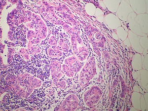
 Invasive micropapillary carcinoma. H&E stain. | |
|
| |
| LM | small micropapillary tufts of tumour cells or tubuloalveolar structures, central avascular stromal core, lear spaces/clefting around the small clusters of tumor cells, +/-lymphovascular invasion |
| LM DDx | IDC NST with micropapillary features, metastatic serous carcinoma |
| IHC | EMA +ve (periphery of nests) - described as inside-out pattern |
| Grossing notes | breast grossing |
| Staging | breast cancer staging |
| Site | breast - see invasive breast cancer |
|
| |
| Prevalence | rare |
| Prognosis | poor |
| Treatment | surgical excision |
Invasive micropapillary carcinoma of the breast, also micropapillary carcinoma, is a rare type of invasive breast cancer.
General
- Poor prognosis.
- Lymphovascular invasion common.[1]
Microscopic
Features:[2]
- Small micropapillary tufts of tumour cells or tubuloalveolar structures.
- Central avascular stromal core.
- Clear spaces/clefting around the small clusters of tumor cells - diffuse/through-out the tumour - key feature.
- Described as "small clusters of tumour lying within dilated vascular channel-like spaces".[3]
- Can appear sponge like or swiss cheese like.
- Abundant finely granular cytoplasm.
- Mucin cytoplasmic but not in the surrounding clear spaces.
- Nuclear atypia is moderate or severe.
- Mixed(micropapillary + other) histological pattern common.
- Can show psammoma bodies or other calcifications.
DDX
- Invasive mammary carcinoma of no special type with micropapillary features.
- Metastatic papillary serous carcinoma of the ovary (WT1, PAX8 positive).[4]
Note:
- Ductal carcinoma commonly has clefting... but it isn't diffuse.
- Even a minor component of this tumor type should be identified and reported due to the high rate of associated lymphatic invasion and nodal involvement.
- Skin involvement has been reported to be strongly correlated with a poor prognosis for this subtype.
- Micropapillary architectural is retained in node metastases, dermal lymphatic invasion, and recurrences.[5]
Images
www:
- Invasive micropapillary carcinoma (flickr.com/euthman).
- Invasive micropapillary carcinoma - poor quality image (breast-cancer.ca).[6]
IHC
- EMA +ve (periphery of nests); described as inside-out pattern.[3]
- E-cadherin +ve (centre of nests). (???)
- p63 +ve/-ve.
EMA limited to the cytoplasmic membrane oriented toward the stroma. E-cadherin absent on the cytoplasmic membrane oriented toward the stroma. Hypothesized to indicate an inversion of cell polarization and a disturbance in the cell adhesion molecules.[7]
See also
References
- ↑ Yu, JI.; Choi, DH.; Park, W.; Huh, SJ.; Cho, EY.; Lim, YH.; Ahn, JS.; Yang, JH. et al. (Jun 2010). "Differences in prognostic factors and patterns of failure between invasive micropapillary carcinoma and invasive ductal carcinoma of the breast: matched case-control study.". Breast 19 (3): 231-7. doi:10.1016/j.breast.2010.01.020. PMID 20304650.
- ↑ Pettinato, G.; Manivel, CJ.; Panico, L.; Sparano, L.; Petrella, G. (Jun 2004). "Invasive micropapillary carcinoma of the breast: clinicopathologic study of 62 cases of a poorly recognized variant with highly aggressive behavior.". Am J Clin Pathol 121 (6): 857-66. doi:10.1309/XTJ7-VHB4-9UD7-8X60. PMID 15198358.
- ↑ 3.0 3.1 Yamaguchi, R.; Tanaka, M.; Kondo, K.; Yokoyama, T.; Kaneko, Y.; Yamaguchi, M.; Ogata, Y.; Nakashima, O. et al. (Aug 2010). "Characteristic morphology of invasive micropapillary carcinoma of the breast: an immunohistochemical analysis.". Jpn J Clin Oncol 40 (8): 781-7. doi:10.1093/jjco/hyq056. PMID 20444748.
- ↑ Nonaka, D.; Chiriboga, L.; Soslow, RA. (Oct 2008). "Expression of pax8 as a useful marker in distinguishing ovarian carcinomas from mammary carcinomas.". Am J Surg Pathol 32 (10): 1566-71. doi:10.1097/PAS.0b013e31816d71ad. PMID 18724243.
- ↑ Pettinato, G.; Manivel, CJ.; Panico, L.; Sparano, L.; Petrella, G. (Jun 2004). "Invasive micropapillary carcinoma of the breast: clinicopathologic study of 62 cases of a poorly recognized variant with highly aggressive behavior.". Am J Clin Pathol 121 (6): 857-66. doi:10.1309/XTJ7-VHB4-9UD7-8X60. PMID 15198358.
- ↑ URL: http://www.breast-cancer.ca/type/micropapillary-breast-carcinoma.htm. Accessed on: 30 May 2012.
- ↑ Pettinato, G.; Manivel, CJ.; Panico, L.; Sparano, L.; Petrella, G. (Jun 2004). "Invasive micropapillary carcinoma of the breast: clinicopathologic study of 62 cases of a poorly recognized variant with highly aggressive behavior.". Am J Clin Pathol 121 (6): 857-66. doi:10.1309/XTJ7-VHB4-9UD7-8X60. PMID 15198358.

