Breast grossing
Jump to navigation
Jump to search

.jpg)
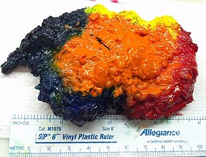
An inked breast lumpectomy specimen. (WC/Ed Uthman)
This article deals with breast grossing.
Introduction
- Lumpectomy = a common procedure for breast lesions that are small, typically have no skin.
- Mastectomy = removal of the breast, may include skeletal muscle (not common) or be skin sparing.[1]
Specimen opening
- Orientation:
- Lumpectomies are usually oriented with a short and long suture as per the surgeon; short is typically superior (aspect) and long is typically lateral (aspect).
- Mastectomies typically have tissue extending toward the axilla known as the "tail".
- The deep aspect in larger specimens can often be identified by the (flat) fascial plane.
- Inking - colours usually as per an institutional standard - see Protocol notes section.
- Slicing - medial to lateral.
Protocol
Identification:
- Specimen label: [description as per label].
- Specimen label and requisition: [match/do not match].
Specimen - type/size/characteristics:
- Specimen type: [total mastectomy/partial mastectomy].
- Specimen orientation: [short-superior, long-lateral, double deep].
- Surgical guidewire: [present/absent].
- Specimen size (superior-inferior, medial-lateral, anterior-posterior): [___ x ___ x ___] cm.
- Surface disruption/intactness: [intact/disrupted at (location) - defect measures ___ cm].
- Skin: [___ x ___ cm/absent].
- Axilla: [___ x ___ x ___ cm, [mass lesion ___x___x___ cm/mass lesion absent]/axillary tissue absent].
- Nipple: [___ length cm x ___ diameter cm, [unremarkable appearance/retracted]/nipple absent].
- Skeletal muscle: [present, [unremarkable appearance/fibrotic/suspicious for tumour/involved by tumour]/skeletal muscle absent].
- Inking code: [posterior-black, anterior-yellow, superior-blue, interior-red].‡
Slices:
- Slicing: [medial-to-lateral, parasagittal cuts].
- Number of slices: [number].
- Slices sent to x-ray: [yes/no].
- Calcifications: [present/not identified].
Tumour:
- Tumour location in slices: [___ to ___].
- Tumour size (superior-inferior, medial-lateral, anterior-posterior): [___ x ___ x ___] cm.
- Closest margin and distance: [___ margin, ___ cm].
- Distance to other margins: [anterior: [___ cm/not applicable], posterior: [___ cm/not applicable], superior [___ cm/not applicable], inferior [___ cm/not applicable], medial: [___ cm/not applicable], lateral: [___ cm/not applicable].
Other:
- Uninvolved parenchyma - appearance: [fibrous/fatty].
- Other findings: [none/description of other findings].
Sections:
- Margins - on edge if section can be taken with tumour and margin.
- Tumour - in total if small (<2 cm[2]).
Protocol notes
- ‡ There is no universally accepted inking protocol. Blue for superior and green for inferior is common, as the sky is blue and the grass is green.
- Hua[2] suggests: black = posterior, blue = superior, green = inferior, yellow = anterior, red = medial & lateral.
Staging
Main article: Breast cancer staging
The important cut-points (at the time of gross) for tumour staging are: 5, 10, 20, 50 mm.
Alternate approaches
See also
Related protocols
References
- ↑ Yu, P. (Aug 2016). "Breast reconstruction at the MD Anderson Cancer Center.". Gland Surg 5 (4): 416-21. doi:10.21037/gs.2016.05.03. PMID 27563563.
- ↑ 2.0 2.1 Huo, L. (Aug 2011). "A practical approach to grossing breast specimens.". Ann Diagn Pathol 15 (4): 291-301. doi:10.1016/j.anndiagpath.2011.03.005. PMID 21745648.