Difference between revisions of "Intraoperative consultation"
Jump to navigation
Jump to search
(→Surgeon-pathologist dialog: +rref) |
|||
| (14 intermediate revisions by the same user not shown) | |||
| Line 1: | Line 1: | ||
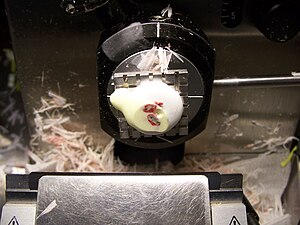
[[Image:Tissue for frozen section in cryostat.JPG|thumb|right|Tissue within a cryostat, as seen during an intraoperative consultation. (WC/000jaw)]] | |||
'''Intraoperative consultation''', also '''quick section''' and '''frozen section''', is when a surgeon requests an opinion during an operation so that they can appropriately manage a patient. It is abbreviated '''IC'''. Frozen section is often abbreviated '''FS'''. | '''Intraoperative consultation''', also '''quick section''' and '''frozen section''', is when a surgeon requests an opinion during an operation so that they can appropriately manage a patient. It is abbreviated '''IC'''. Frozen section is often abbreviated '''FS'''. | ||
| Line 62: | Line 63: | ||
| [[head and neck pathology]] | | [[head and neck pathology]] | ||
|- | |- | ||
| Whipple procedure | | [[Whipple procedure]] | ||
| margins | | margins | ||
| [[gastrointestinal pathology]] | | [[gastrointestinal pathology]] | ||
| Line 113: | Line 114: | ||
====Head and neck==== | ====Head and neck==== | ||
*Squamous cell carcinoma - margins. | *Squamous cell carcinoma - margins. | ||
*Thyroid nodule - diagnosis. | |||
*Parathyroid - confirm it is parathyroid. | |||
====Gastrointestinal tract==== | ====Gastrointestinal tract==== | ||
| Line 120: | Line 123: | ||
====Genitourinary tract==== | ====Genitourinary tract==== | ||
* | *Cystoprostatectomy/cystectomy - ureteral margins. | ||
====Pulmonary==== | ====Pulmonary==== | ||
| Line 130: | Line 133: | ||
*[[Brain tumour]] - diagnosis. | *[[Brain tumour]] - diagnosis. | ||
*Spinal tumour - diagnosis. | *Spinal tumour - diagnosis. | ||
====Prosthetic joint==== | ====Prosthetic joint==== | ||
| Line 214: | Line 214: | ||
*Myometrial involvement - after sectioning bivalving and sectioning (with a 5 mm interval). | *Myometrial involvement - after sectioning bivalving and sectioning (with a 5 mm interval). | ||
===Parathyroid gland=== | ===Parathyroid gland and thyroid gland=== | ||
{{Main|Parathyroid gland}} | {{Main|Parathyroid gland}} | ||
Possibilities: | Possibilities: | ||
*Parathyroid gland: | *Parathyroid gland: | ||
**Benign parathyroid gland. | **Benign parathyroid gland. | ||
**[[Parathyroid adenoma]]. | **[[Parathyroid adenoma]]. † | ||
**[[Parathyroid hyperplasia]]. | **[[Parathyroid hyperplasia]]. † | ||
**[[Parathyroid carcinoma]]. | **[[Parathyroid carcinoma]]. | ||
*Thyroid gland. | *Thyroid gland. | ||
**Thyroid usually follicular - though parathyroid occasionally is pseudofollicular. | |||
**Thyroid often has birefringent (calcium oxalate) crystals (60 of 80 cases) whereas parathyroid less often does (2 or 20 cases).<ref name=pmid24618617>{{cite journal |authors=Wong KS, Lewis JS, Gottipati S, Chernock RD |title=Utility of birefringent crystal identification by polarized light microscopy in distinguishing thyroid from parathyroid tissue on intraoperative frozen sections |journal=Am J Surg Pathol |volume=38 |issue=9 |pages=1212–9 |date=September 2014 |pmid=24618617 |doi=10.1097/PAS.0000000000000204 |url=}}</ref> | |||
*[[Lymph node]]. | *[[Lymph node]]. | ||
*Fibroadipose tissue. | *Fibroadipose tissue. | ||
| Line 237: | Line 239: | ||
====Sign out==== | ====Sign out==== | ||
<pre> | |||
Query Parathyroid Gland: | |||
- Hypercellular parathyroid tissue. | |||
</pre> | |||
<pre> | <pre> | ||
QUERY PARATHYROID GLAND: | QUERY PARATHYROID GLAND: | ||
- PARATHYROID GLAND. | - PARATHYROID GLAND. | ||
</pre> | </pre> | ||
===Whipple specimen=== | |||
====Sign out==== | |||
<pre> | |||
BILE DUCT MARGIN: | |||
- NEGATIVE FOR MALIGNANCY. | |||
</pre> | |||
===Skin specimens=== | |||
{{Main|Oriented skin ellipse grossing}} | |||
{{Main|Unoriented skin ellipse grossing}} | |||
{{Main|Grossing separately received oriented margins for a skin ellipse}} | |||
[[Cut-up]] at frozen section depends on how the specimen is received and its size. | |||
*Small skin specimens: the standard grossing procedure for fixed specimens. | |||
*Large skin specimens: inking is typically as per the routine process. It is useful to mark non-margin if the nearest margin is taken [[en face margin|en face]]. | |||
*Oriented margins should be grossed in a way that allows orientation by [[ink]]. Ideally, ink should allow one to identify the different specimens. See ''[[Grossing separately received oriented margins for a skin ellipse|grossing separately received oriented margins for a skin ellipse]]''. | |||
==See also== | ==See also== | ||
Latest revision as of 16:15, 15 December 2023
Intraoperative consultation, also quick section and frozen section, is when a surgeon requests an opinion during an operation so that they can appropriately manage a patient. It is abbreviated IC. Frozen section is often abbreviated FS.
Why intraoperative consultations are done
Reasons why IC are done:[1]
- Determine diagnosis & appropriate extent of operation ~ 50%.
- Margin status ~ 15%.
- Triage tissue ~ 10%.
- Inform family ~ 8%. †
- Sufficient tissue? ~ 8%.
- Treatment planning, e.g. insert a portacath ~ 3%.
Note:
- † Some don't consider this a good reason for an IC.[2]
Why intraoperative consultations may be refused
- Tissue is the issue - not enough of it.
- Infectious case and no back-up cryostat.
- Management - it won't make a difference.
- Diagnosis won't make a difference.
- Cannot make the diagnosis.
- Bone tumours.
Frozen section permanent section concordance
Main article: Quality
Categories by the Association of Directors of Anatomic and Surgical Pathology (ADASP):[3]
- Agreement
- Deferral - appropriate.
- Deferral – inappropriate
- Recommendation <=10% threshold.
- Disagreement – Minor.
- Disagreement – Major.
- Recommendation <=3% threshold.
Common specimens
Table of common FS specimens
| Specimen | Indication | Subspecialty |
|---|---|---|
| Pelvic mass | diagnosis | gynecologic pathology |
| Ovarian mass | diagnosis | gynecologic pathology |
| Uterine mass | diagnosis | gynecologic pathology |
| Sentinel lymph node | staging in vulvar melanoma | gynecologic pathology |
| Squamous cell carcinoma | margins | head and neck pathology |
| Whipple procedure | margins | gastrointestinal pathology |
| Liver resection (usu. metastatic disease) | margins | gastrointestinal pathology |
| Lower anterior resection | distal margin | gastrointestinal pathology |
| Cystectomy/cystoprostatectomy | ureteral margins | genitourinary pathology |
| Pneumonectomy (usu. cancer) | bronchus margin, lymph nodes for staging | pulmonary pathology |
| Brain tumour | diagnosis | neuropathology |
| Spinal tumour | diagnosis | neuropathology |
| Thyroid nodule | diagnosis | endocrine pathology |
| Prosthetic joint | query prosthetic joint infection | joint pathology |
| Parathyroid gland | identify parathyroid gland | endocrine pathology |
List of specimens
Gynecologic
- Pelvic mass - diagnosis (benign/borderline/malignant).
- Ovarian mass - diagnosis (benign/borderline/malignant).
- Uterine mass - diagnosis (benign/borderline/malignant).
- Sentinel lymph node - staging.
- Vulvar melanoma.
Head and neck
- Squamous cell carcinoma - margins.
- Thyroid nodule - diagnosis.
- Parathyroid - confirm it is parathyroid.
Gastrointestinal tract
- Whipple procedure - margins.
- Liver resection - margins.
- Lower anterior resection - distal margin.
Genitourinary tract
- Cystoprostatectomy/cystectomy - ureteral margins.
Pulmonary
- Pneumonectomy:
- Bronchus - margins.
- Lymph nodes - staging.
Neurologic
- Brain tumour - diagnosis.
- Spinal tumour - diagnosis.
Prosthetic joint
- Query prosthetic joint infection.
Surgeon-pathologist dialog
It should include:
- Identification:
- Pathology is calling - Dr. X is speaking.
- Patient identifiers - full name.
- Part(s) submitted.
- Diagnosis.
- Repeat of diagnosis from surgeon (known as read-back confirmation[4]).
- Additional requests?
Specific specimens - checklists
Ovarian mass
- Covers adnexal mass and pelvic mass.
Checklist
General:
- Specimen integrity: fragmented/intact/ruptured.
- Dimensions: ___ x ___ x ___ cm.
- Mass: ___ grams.
- Surface involvement: absent/present.
- Consistency: solid/cystic/solid and cystic.
Cystic:
- Type: unilocular, multilocular.
- Cyst content: mucinous/serous/hemorrhagic/purulent/chylous/other. ‡
- Papillary excrescences: absent/present.
- Necrosis: absent/present.
- Hair: absent/present.
Sections:
- Sample morphologically distinct areas - esp. solid areas, papillary excrescences.
Notes:
- ‡ Mucinous versus serous:
- Serous: low viscosity (flows with ease threw the grates typically seen at the bottom of sinks[5]).
- Mucinous: high viscosity (jello-like consistency).
DDx
| Feature | Dx/DDx | Notes |
|---|---|---|
| Unilocular cysts | follicular cyst, epithelial cyst, very rarely granulosa cell tumour (not typically diagnosed at FS) | no excrescences or solid areas dx = "simple cyst"; "shag carpet" appearance = serous borderline tumour |
| Sebaceous material or hair | teratoma | Rokitansky nodule present -> submit section; neuroepithelium = fleshy appearance on gross;[6]
immature teratoma not typically diagnosed at FS |
| Chocolate cyst | endometriosis | solid elements (fibrosis vs. clear cell & endometrioid carcinoma |
Endometrial carcinoma
General
- Diagnosis usually known before the surgery.
- The IC revolves around whether the lesion has a "high risk" of lymph node metastasis.
Factors that increase the risk of lymph node metastases:[7]
- Histology:
- Serous or clear cell.
- FIGO grade 3.
- Staging parameters - involvement of:
- Outer half of the myometrium.
- Uterine cervix.
- Adnexa.
Checklist
Gross assessment:
- Uterine cervix involvement (rare).
- Adnexal involvement (rare).
- Myometrial involvement - after sectioning bivalving and sectioning (with a 5 mm interval).
Parathyroid gland and thyroid gland
Main article: Parathyroid gland
Possibilities:
- Parathyroid gland:
- Benign parathyroid gland.
- Parathyroid adenoma. †
- Parathyroid hyperplasia. †
- Parathyroid carcinoma.
- Thyroid gland.
- Thyroid usually follicular - though parathyroid occasionally is pseudofollicular.
- Thyroid often has birefringent (calcium oxalate) crystals (60 of 80 cases) whereas parathyroid less often does (2 or 20 cases).[8]
- Lymph node.
- Fibroadipose tissue.
- Paraganglioma.
Notes:
- † It is not possible to determine which one without history or all parathyroid glands.
- Surgeons are pretty good at identifying parathyroid tissue ~ 94% accurate in one series.[9]
Checklist
- Weight.
- Dimensions.
- Appearance.
Sign out
Query Parathyroid Gland:
- Hypercellular parathyroid tissue.
QUERY PARATHYROID GLAND:
- PARATHYROID GLAND.
Whipple specimen
Sign out
BILE DUCT MARGIN:
- NEGATIVE FOR MALIGNANCY.
Skin specimens
Main article: Oriented skin ellipse grossing
Main article: Unoriented skin ellipse grossing
Cut-up at frozen section depends on how the specimen is received and its size.
- Small skin specimens: the standard grossing procedure for fixed specimens.
- Large skin specimens: inking is typically as per the routine process. It is useful to mark non-margin if the nearest margin is taken en face.
- Oriented margins should be grossed in a way that allows orientation by ink. Ideally, ink should allow one to identify the different specimens. See grossing separately received oriented margins for a skin ellipse.
See also
References
- ↑ Zarbo, RJ.; Schmidt, WA.; Bachner, P.; Howanitz, PJ.; Meier, FA.; Schifman, RB.; Boone, DJ.; Herron, RM. (Jan 1996). "Indications and immediate patient outcomes of pathology intraoperative consultations. College of American Pathologists/Centers for Disease Control and Prevention Outcomes Working Group Study.". Arch Pathol Lab Med 120 (1): 19-25. PMID 8554440.
- ↑ Taxy, J.; Husain, A; Montag, A. (2009). Biopsy Interpretation: The Frozen Section (1st ed.). Lippincott Williams & Wilkins. pp. 5. ISBN 978-0781767798.
- ↑ URL: http://www.adasp.org/papers/position/QualityAssurance.htm. Accessed on: 2 March 2012.
- ↑ Nakhleh, RE. (Nov 2011). "Quality in surgical pathology communication and reporting.". Arch Pathol Lab Med 135 (11): 1394-7. doi:10.5858/arpa.2011-0192-RA. PMID 22032564.
- ↑ Taxy, J.; Husain, A; Montag, A. (2009). Biopsy Interpretation: The Frozen Section (1st ed.). Lippincott Williams & Wilkins. pp. 33. ISBN 978-0781767798.
- ↑ Taxy, J.; Husain, A; Montag, A. (2009). Biopsy Interpretation: The Frozen Section (1st ed.). Lippincott Williams & Wilkins. pp. 34. ISBN 978-0781767798.
- ↑ Taxy, J.; Husain, A; Montag, A. (2009). Biopsy Interpretation: The Frozen Section (1st ed.). Lippincott Williams & Wilkins. pp. 43. ISBN 978-0781767798.
- ↑ Wong KS, Lewis JS, Gottipati S, Chernock RD (September 2014). "Utility of birefringent crystal identification by polarized light microscopy in distinguishing thyroid from parathyroid tissue on intraoperative frozen sections". Am J Surg Pathol 38 (9): 1212–9. doi:10.1097/PAS.0000000000000204. PMID 24618617.
- ↑ Dewan, AK.; Kapadia, SB.; Hollenbeak, CS.; Stack, BC. (Dec 2005). "Is routine frozen section necessary for parathyroid surgery?". Otolaryngol Head Neck Surg 133 (6): 857-62. doi:10.1016/j.otohns.2005.05.001. PMID 16360503.
