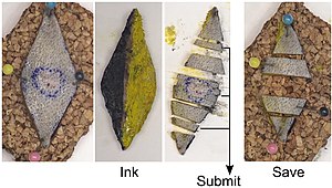
Unoriented skin ellipse grossing
Jump to navigation
Jump to search
The article deals with small unoriented skin ellipse grossing.
Punch biopsies and oriented skin ellipses are dealt with separately.
Introduction
These specimens are very common.
Protocol
- Name and patient identifiers on the requisition match the specimen container.
- Specimen labelled as: "[ ]".
- Specimen received in: [formalin/fresh].
- Type: unoriented portion of skin measuring [ ] x [ ] cm (in the plane of surface), by [ ] cm (depth).
- Inking: resection margin blue. †
- Lesion: [ brown ] colour, [ diffuse / patchy] with a [ regular / irregular ] border.
- Lesion dimensions: [ ] x [ ] cm (in the plane of surface), by [ ] cm (depth).
- Margins: [ ] peripheral cm, [ ] deep cm.
Serially sectioned with cuts perpendicular to the long axis:
- Block A1 - tips.
- Block A2 - remainder of specimen.
Protocol notes
- † One should avoid black ink if there is any suspicion of melanoma or if the lesion is pigmented. This can be remember by black is bad and green is good!
- In general, green and blue are the preferred marking ink colours as they are easier to see at the time of embedding.[1]
Alternate approaches
See also
Related protocols
References
- ↑ Lester, Susan Carole (2010). Manual of Surgical Pathology (3rd ed.). Saunders. pp. 312. ISBN 978-0-323-06516-0.
