Difference between revisions of "Inflammatory pseudopolyp"
Jump to navigation
Jump to search
| (2 intermediate revisions by the same user not shown) | |||
| Line 58: | Line 58: | ||
**[[Solitary rectal ulcer]]. | **[[Solitary rectal ulcer]]. | ||
Images: | ===Images=== | ||
<gallery> | |||
Image: Inflammatory polyp -- very low mag.jpg | IP - very low mag. | |||
Image: Inflammatory polyp -- low mag.jpg | IP - low mag. | |||
Image: Inflammatory polyp -- intermed mag.jpg | IP - intermed. mag. | |||
Image: Inflammatory polyp -- high mag.jpg | IP - high mag. | |||
</gallery> | |||
www: | |||
*[http://www.humpath.com/spip.php?article8234&id_document=18554 Pseudopolyp (humpath.com)]. | *[http://www.humpath.com/spip.php?article8234&id_document=18554 Pseudopolyp (humpath.com)]. | ||
*[http://missinglink.ucsf.edu/lm/IDS_106_LowerGI/Lower_GI_histo_small/24-UC-pseudoplp.jpg Pseudopolyp (ucsf.edu)]. | *[http://missinglink.ucsf.edu/lm/IDS_106_LowerGI/Lower_GI_histo_small/24-UC-pseudoplp.jpg Pseudopolyp (ucsf.edu)]. | ||
| Line 72: | Line 79: | ||
- INFLAMED POLYPOID FRAGMENT OF COLORECTAL-TYPE MUCOSA. | - INFLAMED POLYPOID FRAGMENT OF COLORECTAL-TYPE MUCOSA. | ||
-- NEGATIVE FOR DYSPLASIA. | -- NEGATIVE FOR DYSPLASIA. | ||
</pre> | |||
===Diverticular disease-associated=== | |||
<pre> | |||
Polyp, Sigmoid Colon, Polypectomy: | |||
- Colonic mucosa with ulceration, acute inflammation and granulation tissue. | |||
- NEGATIVE for dysplasia. | |||
Comment: | |||
This may represent a polyp seen in the context of diverticular disease. Other | |||
considerations include ischemia, idiopathic inflammation and infections. | |||
Clinical correlation is suggested. | |||
</pre> | |||
====Block letters==== | |||
<pre> | |||
POLYP (AT EDGE OF DIVERTICULUM), SIGMOID COLON, POLYPECTOMY: | |||
- GRANULATION TISSUE AND SCANT BENIGN EPITHELIUM. | |||
- NO EVIDENCE OF DYSPLASIA. | |||
</pre> | </pre> | ||
===Micro=== | ===Micro=== | ||
The sections show a fragment of colorectal mucosa with focal ulceration, acute inflammation and a well-vascularized stroma with plump stromal cells. Occasional stromal cells have nuclear hyperchromasia. | The sections show a fragment of colorectal mucosa with focal ulceration, acute inflammation and a well-vascularized stroma with plump stromal cells. Occasional stromal cells have nuclear hyperchromasia. | ||
====Alternate==== | |||
The sections show a fragment of tissue with scant benign epithelium, acute and chronic | |||
inflammation (neutrophils and plasma cells predominantly), abundant blood vessels with | |||
reactive endothelial cells and plump stromal cells. Occasional stromal cells have nuclear hyperchromasia but do not show significant atypia. | |||
==See also== | ==See also== | ||
Latest revision as of 16:14, 20 January 2016
| Inflammatory pseudopolyp | |
|---|---|
| Diagnosis in short | |
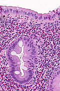
 Inflammatory polyp. H&E stain. | |
|
| |
| LM | polypoid shape, inflammatory cells - esp. neutrophils |
| LM DDx | juvenile polyp, mucosal prolapse, adenomatous polyps |
| Site | colon, rectum, others |
|
| |
| Clinical history | +/-inflammatory bowel disease |
| Prevalence | not common |
| Prognosis | benign |
| Clin. DDx | other types of polyps |
| Inflammatory pseudopolyp | |
|---|---|
| External resources | |
| EHVSC | 9992 |
Inflammatory pseudopolyp is a benign polypoid lesion usually seen in the context of inflammatory bowel disease.
It is also referred to as inflammatory polyp.
General
- Not a true polyp.
- The label inflammatory pseudopolyp = inflammatory bowel disease (IBD).
- If there is no history of IBD... reconsider the diagnosis.
Microscopic
Features:
- Polypoid shape.
- Inflammation - esp. neutrophils - key feature.
Negatives:
- No nuclear atypia.
- May have focal nuclear hyperchromasia and nuclear enlargement.
- No dilated glands.
DDx:
Images
www:
Sign out
SIGMOID COLON POLYP, PERI-DIVERTICULAR, BIOPSY:
- INFLAMMATORY PSEUDOPOLYP.
POLYP, DESCENDING COLON, BIOPSY:
- INFLAMED POLYPOID FRAGMENT OF COLORECTAL-TYPE MUCOSA.
-- NEGATIVE FOR DYSPLASIA.
Diverticular disease-associated
Polyp, Sigmoid Colon, Polypectomy:
- Colonic mucosa with ulceration, acute inflammation and granulation tissue.
- NEGATIVE for dysplasia.

Comment:
This may represent a polyp seen in the context of diverticular disease. Other
considerations include ischemia, idiopathic inflammation and infections.
Clinical correlation is suggested.
Block letters
 POLYP (AT EDGE OF DIVERTICULUM), SIGMOID COLON, POLYPECTOMY:
- GRANULATION TISSUE AND SCANT BENIGN EPITHELIUM.
- NO EVIDENCE OF DYSPLASIA.
Micro
The sections show a fragment of colorectal mucosa with focal ulceration, acute inflammation and a well-vascularized stroma with plump stromal cells. Occasional stromal cells have nuclear hyperchromasia.
Alternate
The sections show a fragment of tissue with scant benign epithelium, acute and chronic inflammation (neutrophils and plasma cells predominantly), abundant blood vessels with reactive endothelial cells and plump stromal cells. Occasional stromal cells have nuclear hyperchromasia but do not show significant atypia.
See also
References
- ↑ Aust, DE.; Rüschoff, J. (Jul 2011). "[Polyps of the colorectum: non-neoplastic and non-hamartomatous].". Pathologe 32 (4): 297-302. doi:10.1007/s00292-011-1435-1. PMID 21607734.


