Kaposi sarcoma
(Redirected from Kaposi's sarcoma)
Jump to navigation
Jump to search
| Kaposi sarcoma | |
|---|---|
| Diagnosis in short | |
 Kaposi sarcoma. H&E stain. | |
|
| |
| LM | vascular lesion (abundant RBCs), +/-"promontory sign", +/-spindle cells with minimal nuclear atypia, RBC extravasation, +/-intracytoplasmic hyaline globules, +/-hemosiderin deposits, +/-plasma cells |
| Subtypes | classic, endemic, immunosuppression-associated or transplant-associated, AIDS-associated |
| LM DDx | angiosarcoma, Masson's hemangioma (intravascular papillary endothelial hyperplasia), benign lymphangioendothelioma |
| IHC | HHV-8 +ve, CD31 +ve, CD34 +ve |
| Gross | red or brown patches, plaques or nodules |
| Site | skin, others |
|
| |
| Associated Dx | +/-HIV infection or immunoincompentence |
| Prevalence | uncommon |
| Prognosis | dependent on subtype |
Kaposi sarcoma, abbreviated KS, is an uncommon vascular tumour that is often associated with HIV/AIDS.
General
- Caused by Human herpesvirus-8 (HHV-8).
- In the North American context, it is often associated with immunodeficiency, e.g. HIV/AIDS.
Interesting note:
- It has been said that KS is not really a sarcoma.[1]
Stages
It is seen in different stages:[2][3]
- Patch stage.
- Plaque stage.
- Nodular stage.
- Exophytic stage.
- Infiltrative stage.
- Lymphadenopathic stage.
Note:
- The first three are the classic ones.
Type or form
Classically divided into four types:[4][5][6]
- Classic = old men Mediterranean or Ashkenazi Jew.
- Endemic = African infants and young males.
- Immunosuppression-associated or transplant-associated - iatrogenic.
- AIDS-associated.
Gross
- Skin: red or brown patches, plaques or nodules.
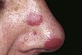
KS. (WC)

Microscopic
Features:[7]
- Vascular lesion (abundant RBCs) with:
- +/-"Promontory sign" - small vessel protruding into an abnormal vascular space.[8]
- Not pathognomonic for KS.[9]
- +/-Spindle cells with minimal nuclear atypia.
- RBC extravasation - very useful - important feature.[10]
- +/-"Promontory sign" - small vessel protruding into an abnormal vascular space.[8]
- +/-Intracytoplasmic hyaline globules - uncommon - one usu. needs to search for 'em.[11]
- Pale pink globs (that are paler than RBCs) - important feature.
- +/-Hemosiderin deposits.
- +/-Plasma cells.[12]
Notes:
- Hyaline globules have a DDx (hepatocellular carcinoma, lung adenocarcinoma, chondrosarcomas + others).[11]
DDx:
- Angiosarcoma - have many mitoses, nuclear atypia, RBC extravasation not common.
- Masson's hemangioma - AKA intravascular papillary endothelial hyperplasia.
- Benign lymphangioendothelioma.[13]
- Histologically very similar.[14]
- Cavernous hemangioma.[15]
Images
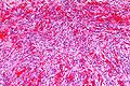
Kaposi sacoma - high mag. (WC)
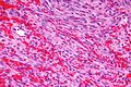
Kaposi sarcoma - intermed. mag. (WC)

www:
Stains
- PAS +ve -- hyaline globules.
- Hale's colloidal iron stain +ve.[16]
IHC
Features:[15]
See also
References
- ↑ Pérez, A.; Sánchez, JL.; Almodóvar, PI. (Oct 2003). "Kaposi's sarcoma is not a neoplasm let alone a sarcoma.". Int J Dermatol 42 (10): 844-5. PMID 14521707.
- ↑ URL: http://www.histopathology-india.net/KS.htm. Accessed on: 31 January 2010.
- ↑ URL: http://emedicine.medscape.com/article/1083998-clinical#a0217. Accessed on: 17 November 2011.
- ↑ Szajerka, T.; Jablecki, J.. "Kaposi's sarcoma revisited.". AIDS Rev 9 (4): 230-6. PMID 18219366.
- ↑ Morand, JJ.; Lightburn, E.; Simon, F.; Patte, JH. (Apr 2007). "[Update on Kaposi's sarcoma].". Med Trop (Mars) 67 (2): 123-30. PMID 17691428.
- ↑ Antman, K.; Chang, Y. (Apr 2000). "Kaposi's sarcoma.". N Engl J Med 342 (14): 1027-38. doi:10.1056/NEJM200004063421407. PMID 10749966.
- ↑ Klatt, Edward C. (2006). Robbins and Cotran Atlas of Pathology (1st ed.). Saunders. pp. 23. ISBN 978-1416002741.
- ↑ Lazova R, McNiff JM, Glusac EJ, Godic A (April 2009). "Promontory sign--present in patch and plaque stage of angiosarcoma!". Am J Dermatopathol 31 (2): 132–6. doi:10.1097/DAD.0b013e3181951045. PMID 19318797.
- ↑ Fernandez-Flores A, Rodriguez R (June 2010). "Promontory Sign in a Reactive Benign Vascular Proliferation". Am J Dermatopathol. doi:10.1097/DAD.0b013e3181cf0ae5. PMID 20577080.
- ↑ Kato, H.; Hamada, T.; Tsuji, T.; Baba, T.; Seki, J.; Kobayashi, Y. (Jul 1990). "Kaposi's sarcoma: a light and electron microscopic study.". J Dermatol 17 (7): 414-22. PMID 2229644.
- ↑ 11.0 11.1 del Rosario AD, Bui HX, Singh J, Ginsburg R, Ross JS (December 1994). "Intracytoplasmic eosinophilic hyaline globules in cartilaginous neoplasms: a surgical, pathological, ultrastructural, and electron probe x-ray microanalytic study". Hum. Pathol. 25 (12): 1283–9. PMID 7528163.
- ↑ Douglas, JL.; Gustin, JK.; Dezube, B.; Pantanowitz, JL.; Moses, AV. (Sep 2007). "Kaposi's sarcoma: a model of both malignancy and chronic inflammation.". Panminerva Med 49 (3): 119-38. PMID 17912148.
- ↑ Guillou, L.; Fletcher, CD. (Aug 2000). "Benign lymphangioendothelioma (acquired progressive lymphangioma): a lesion not to be confused with well-differentiated angiosarcoma and patch stage Kaposi's sarcoma: clinicopathologic analysis of a series.". Am J Surg Pathol 24 (8): 1047-57. PMID 10935645.
- ↑ URL: http://path.upmc.edu/cases/case134/dx.html. Accessed on: 5 January 2012.
- ↑ 15.0 15.1 Onak Kandemir N, Barut F, Doğan Gün B, Solak Tekin N, Hallaç Keser S, Oğuz Özdamar S (2013). "Cavernous hemangioma-like kaposi sarcoma: histomorphologic features and differential diagnosis". Case Rep Med 2013: 959812. doi:10.1155/2013/959812. PMC 3800618. PMID 24187557. https://www.ncbi.nlm.nih.gov/pmc/articles/PMC3800618/.
- ↑ 16.0 16.1 Wada DA, Perkins SL, Tripp S, Coffin CM, Florell SR (February 2007). "Human herpesvirus 8 and iron staining are useful in differentiating Kaposi sarcoma from interstitial granuloma annulare". Am. J. Clin. Pathol. 127 (2): 263–70. doi:10.1309/GMH9CENH4909AWVB. PMID 17210517.
- ↑ Miettinen M1, Wang ZF, Paetau A, Tan SH, Dobi A, Srivastava S, Sesterhenn I. (march 2011). "ERG transcription factor as an immunohistochemical marker for vascular endothelial tumors and prostatic carcinoma.". Am J Surg Pathol. 2011 Mar;35(3):432-41. 35 (3): 432-41. PMID 21317715. http://www.ncbi.nlm.nih.gov/pubmed/21317715.