Intracystic papillary carcinoma of the breast
Intracystic papillary carcinoma of the breast, also known as encapsulated papillary carcinoma of the breast (abbreviated EPC), is an uncommon type of breast cancer with a very good prognosis.
| Intracystic papillary carcinoma of the breast | |
|---|---|
| Diagnosis in short | |
|
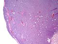
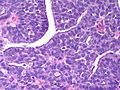
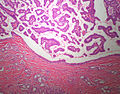
Template:Px Intracystic Papillary Breast Carcinoma. H&E stain. | |
|
| |
| LM | Papillary lesion within a cyst |
| LM DDx | Intraductal papilloma, papillary DCIS, Invasive papillary breast carcinoma |
| Site | breast |
|
| |
| Signs | +/-bloody discharge from nipple |
| Prevalence | Rare |
| Prognosis | very good |
| Clin. DDx | other breast tumours |
| Treatment | surgical |
It should not be confused with the invasive papillary carcinoma of the breast, a more aggressive tumour of the breast.
General
Microscopic
Features:
- Lesion confined to a cyst.
- May have a thick fibrous capsule
- The involved space is not lined by myoepithelial cells.
- The cyst contains an abnormal epithelial proliferation with cribriform, solid or papillary architecture.
- Loss of myoepithelial cells within the epithelial proliferation is a key feature.
- Scattered large cells with pale eosinophilic cytoplasm may be observed[3].
- These cells are so-called globoid cells or clear cells and are immunoreactive for GCDFP-15.
- They should not be mistaken for myoepithelial cells.
- Neoplastic epithelial cells:
- Nuclear atypia - including: nucleoli, nuclear pleomorphism.
Notes:
- Many potential pitfalls with papillary breast lesions on needle core biopsy.
- Complete excision is recommended.[4]
- Adequately and carefully sample the specimen to exclude an invasive component.
- Report only the size of the invasive component (if present) to prevent over-estimation of tumor stage.
- Intraductal papilloma.
- Absent or scant stroma favors papillary carcinoma over papilloma.
- Is there a single cell or dual cell population in the lesion?
- ER staining will be heterologous in a benign lesion.
- Myoepithelial markers (calponin/p63/SMA +ve)s hould be positive in a benign lesion.
- Papillary ductal carcinoma in situ.
- Papillary DCIS shows myoepithelial cells (calponin/p63/SMA +ve) at the periphery of the involved spaces
- But papillary DCIS should be negative for myoepithelial cells within the focus of DCIS
- Papillary intracystic carcinoma does not show myoepithelial cells at the periphery of the involved spaces
- Invasive papillary carcinoma of the breast.
- Similar architecture but no cystic space, frankly invasive.
- Very rare.
- Invasive carcinoma arising in association with papillary intracystic carcinoma
- Epithelial entrapment in the encysting fibrous tissue should not be interpreted as invasion.
- Carcinoma must be seen in the breast tissue outside the encysting fibrous tissue.
- Infiltrating carcinoma is usually of the 'no special type' variety.
- Adenoid cystic carcinoma of the breast
- The solid variant looks basaloid - solid adenoid cystic carcinoma or a 'basal-like' carcinoma should be considered in these cases.
Images
- Breast PapillaryCarcinomaEncysted 3 PA.JPG
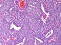
Breast - Intracystic Papillary Carcinoma - Medium power (SKB)
- Breast PapillaryCarcinomaEncysted 2 PA.JPG
Breast - Intracystic Papillary Carcinoma - Medium power (SKB)
- Breast PapillaryCarcinomaEncysted PA.JPG
Breast - Intracystic Papillary Carcinoma - High power (SKB)
Breast - Papillary Intracystic Carcinoma, Solid Variant - low power (SKB)
Breast - Papillary Intracystic Carcinoma, Solid Variant - medium power (SKB)
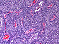
Breast - Papillary Intracystic Carcinoma, Solid Variant - medium power (SKB)
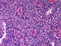
Breast - Papillary Intracystic Carcinoma, Solid Variant - high power (SKB)
Breast - Papillary Intracystic Carcinoma, Solid Variant - high power (SKB)
Breast - Intracystic papillary adenocarcinoma (top) with associated invasive ductal carcinoma (bottom) (SKB)


IHC
- Calponin/p63/SMA/CK5-6.
- Loss of myoepithelial cells within the tumour.
- Loss of myoepithelial cells at the cyst wall.
- ER - Homogeneous staining of the epithelial proliferation.
References
- ↑ Rakha, EA.; Gandhi, N.; Climent, F.; van Deurzen, CH.; Haider, SA.; Dunk, L.; Lee, AH.; Macmillan, D. et al. (Aug 2011). "Encapsulated papillary carcinoma of the breast: an invasive tumor with excellent prognosis.". Am J Surg Pathol 35 (8): 1093-103. doi:10.1097/PAS.0b013e31821b3f65. PMID 21753694.
- ↑ Rodríguez, MC.; Secades, AL.; Angulo, JM. (Nov 2010). "Best cases from the AFIP: intracystic papillary carcinoma of the breast.". Radiographics 30 (7): 2021-7. doi:10.1148/rg.307105003. PMID 21057133.
- ↑ Collins, LC.; Schnitt, SJ. (Jan 2008). "Papillary lesions of the breast: selected diagnostic and management issues.". Histopathology 52 (1): 20-9. doi:10.1111/j.1365-2559.2007.02898.x. PMID 18171414.
- ↑ Rizzo, M.; Linebarger, J.; Lowe, MC.; Pan, L.; Gabram, SG.; Vasquez, L.; Cohen, MA.; Mosunjac, M. (Mar 2012). "Management of papillary breast lesions diagnosed on core-needle biopsy: clinical pathologic and radiologic analysis of 276 cases with surgical follow-up.". J Am Coll Surg 214 (3): 280-7. doi:10.1016/j.jamcollsurg.2011.12.005. PMID 22244207.
- ↑ Collins, LC.; Schnitt, SJ. (Jan 2008). "Papillary lesions of the breast: selected diagnostic and management issues.". Histopathology 52 (1): 20-9. doi:10.1111/j.1365-2559.2007.02898.x. PMID 18171414.
- ↑ Pathmanathan, N.; Albertini, AF.; Provan, PJ.; Milliken, JS.; Salisbury, EL.; Bilous, AM.; Byth, K.; Balleine, RL. (Jul 2010). "Diagnostic evaluation of papillary lesions of the breast on core biopsy.". Mod Pathol 23 (7): 1021-8. doi:10.1038/modpathol.2010.81. PMID 20473278.