Osteosarcoma
(Redirected from Chondroblastic osteosarcoma)
Jump to navigation
Jump to search
| Osteosarcoma | |
|---|---|
| Diagnosis in short | |
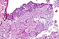
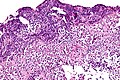
 Osteosarcoma. H&E stain. | |
|
| |
| LM | cells with malignant features (e.g. nuclear membrane irregularities, marked nuclear size differences, mitoses) surrounded by delicate strands of osteoid |
| Subtypes | conventional osteosarcoma (osteoblastic osteosarcoma, fibroblastic osteosarcoma, chondroblastic osteosarcoma), small cell osteosarcoma, telangiectatic osteosarcoma, parosteal osteosarcoma, periosteal osteosarcoma, low-grade central osteosarcoma, high-grade surface osteosarcoma, secondary osteosarcoma, gnathic osteosarcoma |
| LM DDx | chondrosarcoma, phosphaturic mesenchymal tumour, mixed connective tissue type, undifferentiated pleomorphic sarcoma (for fibroblastic osteosarcoma), aneurysmal bone cyst (for telangiectatic osteosarcoma), fibrous dysplasia, small round cell tumours (for small cell osteosarcoma) |
| Site | bone |
|
| |
| Syndromes | Li-Fraumeni syndrome |
|
| |
| Prevalence | uncommon |
| Clin. DDx | aneurysmal bone cyst,fibrous dysplasia, Ewing sarcoma |
Osteosarcoma, also known as osteogenic sarcoma, is a malignant bone tumour. It is grouped with the chondro-osseous tumours.
General
- Most common malignant bone tumour in children.
- May be seen in the context of Li-Fraumeni syndrome.
Trivia:
- Terry Fox was afflicited by this tumour.
Definition
- Tumour that makes osteoid.
- Osteoid = (extracellular) organic component of bone, normally produced by osteoblasts (cells which make bone matrix).
Gross
Classic locations:[1]
- Distal femur ~ 45%.
- Proximal tibia ~ 20%.
- Proximal humerous ~ 15%.
Microscopic
Features:
- Cells with malignant features (e.g. nuclear membrane irregularities, marked nuclear size differences, mitoses) surrounded by delicate strands of osteoid.
- Osteoid on H&E: pink, homogenous, "glassy".
- Tumours typically very cellular - when compared to normal bone.
- +/-Large (multinucleated) osteoclast-like giant cells.[2]
DDx:
Images
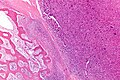
Osteosarcoma - low mag. (WC)
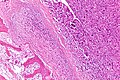
Osteosarcoma - intermed. mag. (WC)
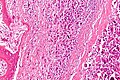
Osteosarcoma - high mag. (WC)
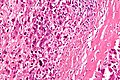
Osteosarcoma - very high mag. (WC)
Small cell osteosarcoma - intermed. mag. (WC)
Small cell osteosarcoma - high mag. (WC)


www:
Subtypes
- Conventional osteosarcoma (high grade).
- Osteoblastic osteosarcoma.
- Fibroblastic osteosarcoma.
- Chondroblastic osteosarcoma.
- Small cell osteosarcoma.
- Telangiectatic osteosarcoma.
- Parosteal osteosarcoma.
- Periosteal osteosarcoma.
- Low-grade central osteosarcoma.
- High-grade surface osteosarcoma.
- Secondary osteosarcoma.
- Gnathic osteosarcoma - jaw bones - usually chondroblastic.
How to remember:
- Convention FOC = fibroblastic, osteogenic, chondroblastic.
- Low-grade central.
- High-grade surface.
- Parosteal.
- Periosteal.
- Small cell.
- Secondary.
- Telangiectatic.
Chondroblastic osteosarcoma
- Chondroid matrix present - may be prominent; osteoid may be a minor component.
- May be confused with chondrosarcoma.
Fibroblastic osteosarcoma
- Undifferentiated pleomorphic sarcoma-like/MFH-like.
Images:
Low-grade central osteosarcoma
- Well-formed bone.
- Usu. minimal nuclear atypia.
DDx:
Telangiectatic osteosarcoma
Important radiologic DDx:
Parosteal osteosarcoma
DDx:
Periosteal osteosarcoma
- Intermediate grade.[10]
Small cell osteosarcoma
- May mimic (other) small round cell tumours.
Secondary osteosarcoma
Arises in the context of something else - causes:
- Paget disease of the bone (~80% of secondary osteosarcomas)
- Radiation (~15% of secondary osteosarcomas)).[11]
- Prognosis often poor.[10]
Images:
See also
References
- ↑ Greenwald, J.; Heng, M. (2007). Toronto Notes for Medical Students 2007 (2007 ed.). The Toronto Notes Inc. for Medical Students Inc.. pp. OR43. ISBN 978-0968592878.
- ↑ Papalas JA, Balmer NN, Wallace C, Sangueeza OP (June 2009). "Ossifying dermatofibroma with osteoclast-like giant cells: report of a case and literature review". Am J Dermatopathol 31 (4): 379-83. doi:10.1097/DAD.0b013e3181966747. PMID 19461244.
- ↑ Papandreou, C.; Skopelitou, A.; Kappes, G.; Daouaher, H. (2010). "Primary osteosarcoma of the urinary bladder treated with external radiotherapy in a patient with a history of transitional cell carcinoma: a case report.". J Med Case Rep 4: 70. doi:10.1186/1752-1947-4-70. PMC 2843711. PMID 20181254. https://www.ncbi.nlm.nih.gov/pmc/articles/PMC2843711/.
- ↑ Humphrey, Peter A; Dehner, Louis P; Pfeifer, John D (2008). The Washington Manual of Surgical Pathology (1st ed.). Lippincott Williams & Wilkins. pp. 638. ISBN 978-0781765275.
- ↑ URL: http://bestpractice.bmj.com/best-practice/monograph/780/basics/classification.html. Accessed on: 7 April 2011.
- ↑ Inwards, CY (2001). "Low-grade central osteosarcoma versus fibrous dysplasia". Pathology Case Reviews 6 (1): 22-27. http://journals.lww.com/pathologycasereviews/Fulltext/2001/01000/Low_Grade_Central_Osteosarcoma_Versus_Fibrous.5.aspx.
- ↑ Patibandla, MR.; Uppin, SG.; Thotakura, AK.; Panigrahi, MK.; Challa, S.. "Primary telangiectatic osteosarcoma of occipital bone: a case report and review of literature.". Neurol India 59 (1): 117-9. doi:10.4103/0028-3886.76891. PMID 21339678.
- ↑ Weiss, A.; Khoury, JD.; Hoffer, FA.; Wu, J.; Billups, CA.; Heck, RK.; Quintana, J.; Poe, D. et al. (Apr 2007). "Telangiectatic osteosarcoma: the St. Jude Children's Research Hospital's experience.". Cancer 109 (8): 1627-37. doi:10.1002/cncr.22574. PMID 17351949.
- ↑ The International Agency for Research on Cancer (Editors: Fletcher, C.D.M.; Unni, K. Krishnan; Mertens, F.) (2006). Pathology and Genetics of Tumours of Soft Tissue and Bone (IARC WHO Classification of Tumours) (3rd ed.). World Health Organization. pp. 279. ISBN 978-9283224136.
- ↑ 10.0 10.1 10.2 Carrle, D.; Bielack, SS. (Dec 2006). "Current strategies of chemotherapy in osteosarcoma.". Int Orthop 30 (6): 445-51. doi:10.1007/s00264-006-0192-x. PMC 3172747. PMID 16896870. https://www.ncbi.nlm.nih.gov/pmc/articles/PMC3172747/.
- ↑ URL: http://www.rsna.org/REG/publications/rg/afip/privateM/1997/0017/0005/1205/6.htm. Accessed on: 8 April 2011.