Vascular disease
(Redirected from Atherosclerosis)
Jump to navigation
Jump to search
The article covers vascular disease, i.e. diseases of blood vessels. These keep vascular surgeons and cardiac surgeon busy.
Vasculitides are covered in a separate article called vasculitides.
Normal blood vessels
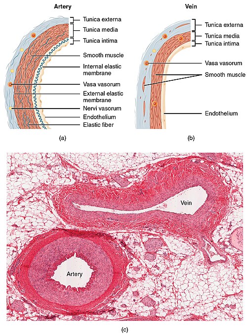
Comparing arteries and veins:[1]
| Feature | Artery | Vein |
|---|---|---|
| Internal elastic lamina (IEL) | prominent/thick, usu. complete | thin & incomplete |
| External elastic lamina (EEL) | present, thick | absent |
| Shape | circular / lumen wide open | collapsed |
| Wall thickness | thick | thin |
Great vessels
When things go wrong here, you see a cardiac surgeon.
Atherosclerosis
General
- A leading cause of death, esp. in the Western world.
- May have multi-system manifestations.
Location and associated pathology:
- Coronary artery atherosclerosis (AKA coronary artery disease) -> myocardial infarction +/-coronary thrombosis.
- Atherosclerotic peripheral vascular disease -> leg amputations.
- Carotid artery atherosclerosis -> thrombotic stroke.
- Superior mesenteric artery atherosclerosis -> ischemic enteritis or ischemic colitis or ischemic enterocolitis.
- Penile artery atherosclerosis -> impotence.
Clinical risk factors:
- Age.
- Blood pressure (high) - modifiable (antihypertensives).
- Cholesterol - modifiable (statins, diet).
- Diabetes mellitus - modifiable (hypoglycemic medications, diet, lifestyle).
- Smoking - modifiable (cessation).
- Family history.
Microscopic
Features:
- Intimal hyperplasia.
- Lipid deposition.
- Foamy macrophages within intima & media.
- Cholesterol clefts
- Luminal narrowing.
Notes:
- Considered "complex" if any of the following are present:[2]
- Calcifications.
- Thrombosis.
- Haemorrhage.
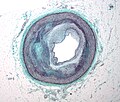
Image
Stains
- Elastic trichrome stain or Movat stain - highlights duplication of internal elastic lamina, allows on to identify with ease intimal thickening.
Aortic dissection
- Abbreviated AoD.
Main article: Aortic dissection
Cystic medial degeneration
Main article: Cystic medial degeneration
Medial calcific sclerosis
AKA Moenckeberg medial calcific sclerosis, calcific medial sclerosis of Monckeberg, and Monckeberg's arteriosclerosis.
Main article: Medial calcific sclerosis
Hyperplastic arteriolosclerosis
General
- Associated with:[4]
- Malignant hypertension.
- Scleroderma.
- May be a consequence of thrombotic microangiopathy.[citation needed]
Note:
- Hyperplasia = proliferation of cells.
Microscopic
Features:[5]
- Onion-skin appearance of intima & media due to:
- Intimal hyperplasia.
- Smooth muscle hyperplasia.
Image: Hyperplastic arteriolosclerosis (utah.edu).
Fibromuscular dysplasia
- Abbreviated FMD.
General
Etiology:
- Unknown, possibly genetic.
Gender:
- Women > men.
- May be seen in virtually any artery.
- Reported as a cause of sudden death with involvement of the artery supplying the AV node.[6]
Gross/radiologic
- Segmental - thinning and thickening.[7]
Classical locations:[7]
- Renal artery - leading to hypertension.
- Carotid artery.
Microscopic
Features:[7]
- Smooth muscle hyperplasia - key feature.
- Elastic fibre fragmentation.
- Luminal narrowing.
Images:
Stains
- Elastic trichrome or Movat stain - to demonstrate elastic fibre fragmentation.
Thromboangiitis obliterans
Main article: Thromboangiitis obliterans
Thrombosis
- See also: Cerebral venous thrombosis.
General
Definition:
- Blood clot formation within a vessel.
Complications:
- Embolism - see: Pulmonary thromboembolism.
Risk factors:
- The classic pimping question is what "Virchow's triad?"
- Stasis, hypercoagulability, endothelial injury.
- A long list is found in: risk factors for VTE.
Gross
Microscopic
Features:
- Lines of Zahn.
- Fibrin - pink acellular stuff on a H&E stain.
Image
Cholesterol embolism
- Abbreviated CE.
Main article: Cholesterol embolism
Coarctation of the aorta
- AKA aortic coarctation.
General
- Uncommon.
Classification:
- Preductal.
- Postductal.
Associations:
Clinical
Presentation:[10]
- Heart failure.
- Hypertension - esp. upper extremity vs. lower extremity.
Gross
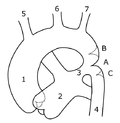
- Narrowing (stenosis) of the aorta proximal or distal to the ductus arteriosis.
Image
Intracranial berry aneurysm
Main article: Berry aneurysm
See also
References
- ↑ URL: http://www.lab.anhb.uwa.edu.au/mb140/corepages/vascular/vascular.htm. Accessed on: 13 January 2011.
- ↑ Klatt, Edward C. (2006). Robbins and Cotran Atlas of Pathology (1st ed.). Saunders. pp. 4. ISBN 978-1416002741.
- ↑ URL: http://emedicine.medscape.com/article/756835-overview. Accessed on: 12 August 2010.
- ↑ URL: http://library.med.utah.edu/WebPath/IMMHTML/IMM028.html. Accessed on: 11 May 2011.
- ↑ Klatt, Edward C. (2006). Robbins and Cotran Atlas of Pathology (1st ed.). Saunders. pp. 7. ISBN 978-1416002741.
- ↑ 6.0 6.1 Lee, S.; Chae, J.; Cho, Y. (Dec 2006). "Causes of sudden death related to sexual activity: results of a medicolegal postmortem study from 2001 to 2005.". J Korean Med Sci 21 (6): 995-9. PMID 17179675.
- ↑ 7.0 7.1 7.2 Hata, D. (Sep 2001). "Fibromuscular dysplasia.". Intern Med 40 (9): 978-9. PMID 11579971.
- ↑ Braverman, AC.; Güven, H.; Beardslee, MA.; Makan, M.; Kates, AM.; Moon, MR. (Sep 2005). "The bicuspid aortic valve.". Curr Probl Cardiol 30 (9): 470-522. doi:10.1016/j.cpcardiol.2005.06.002. PMID 16129122.
- ↑ Hjerrild, BE.; Mortensen, KH.; Sørensen, KE.; Pedersen, EM.; Andersen, NH.; Lundorf, E.; Hansen, KW.; Hørlyck, A. et al. (2010). "Thoracic aortopathy in Turner syndrome and the influence of bicuspid aortic valves and blood pressure: a CMR study.". J Cardiovasc Magn Reson 12: 12. doi:10.1186/1532-429X-12-12. PMID 20222980.
- ↑ Peres, A.; Martins, JD.; Paramés, F.; Gil, R.; Matias, C.; Franco, J.; Freitas, I.; Trigo, C. et al. (Jan 2010). "Isolated aortic coarctation: experience in 100 consecutive patients.". Rev Port Cardiol 29 (1): 23-35. PMID 20391897.


