Difference between revisions of "Viruses"
(redirect) |
|||
| (68 intermediate revisions by 2 users not shown) | |||
| Line 1: | Line 1: | ||
# | This article deals with '''viruses'''. The more general topic of infective things is dealt with in [[microorganisms]]. | ||
Many viruses afflict humans. Only a few of them can be diagnosed histologically. | |||
[[cancer viruses|Several viruses cause cancer]] and seen directly or indirectly by pathologists frequently. | |||
==Viral inclusions - types== | |||
Cowdry types:<ref>URL: [http://www.pathconsultddx.com/pathCon/largeImage?pii=S1559-8675%2806%2970864-6&figureId=fig3&ecomponentId=mmc3 http://www.pathconsultddx.com/pathCon/largeImage?pii=S1559-8675%2806%2970864-6&figureId=fig3&ecomponentId=mmc3]. Accessed: 12 January 2010.</ref> | |||
*Cowdry type A inclusion:<ref>URL: [http://www.whonamedit.com/synd.cfm/3495.html http://www.whonamedit.com/synd.cfm/3495.html]. Accessed on: 22 January 2010.</ref> | |||
**Round eosinophilic material surrounded by a clear halo. | |||
*Cowdry type B inclusion:<ref>[http://www.whonamedit.com/synd.cfm/3496.html http://www.whonamedit.com/synd.cfm/3496.html]. Accessed on: 22 January 2010.</ref> | |||
**Neuropathology thingy. (???) | |||
Images: | |||
*[http://www.daff.gov.au/animal-plant-health/pests-diseases-weeds/aquatic_animal_diseases_significant_to_australia_identification_field_guide/diseases_of_crustaceans/viral_diseases_of_crustaceans/infectious_hypodermal_and_haematopoietic_necrosis/histological_page_for_infectious_hypodermal_and_haematopoietic_necrosis Cowdry A inclusion (daff.gov.au)]. | |||
*[http://focosi.altervista.org/pathohomoprocess_regressive.html Cowdry type A & type B inclusions (altervista.org)]. | |||
=Viruses= | |||
==Herpes simplex virus== | |||
:''In the context of gynecologic cytopathology see: [[Gynecologic_cytopathology#Herpes_simplex_virus]]''. | |||
*Abbreviated ''HSV''. | |||
===General=== | |||
Several subtypes: | |||
*Canker sores - usually HSV-1. | |||
*Genital herpes - usually HSV-2. | |||
===Histology/cytology=== | |||
Features:<ref>SM. 11 January 2010.</ref> | |||
*Clear "ground glass" nuclei. | |||
**Rim of peripheral chromatin. | |||
*Nuclear inclusions. | |||
*Multinucleation with nuclear molding, i.e. multiple nuclei that touch over a large surface area. | |||
Mnemonic - 3 Ms: Margination, Multinucleation, Molding. | |||
====Images==== | |||
www: | |||
*[http://www.virology.org/sbpgphoto2.html Herpes simplex virus - multinucleation (virology.org)]. | |||
<gallery> | |||
Image:Herpes_simplex_virus_pap_test.jpg | HSV on a Pap test - showing multinucleation. (WC) | |||
Image:Herpes_esophagitis_-_very_high_mag.jpg | HSV esophagitis - very high mag. (WC) | |||
Image:Herpes_esophagitis_-_intermed_mag.jpg | HSV esophagitis - intermed. mag. (WC) | |||
</gallery> | |||
===IHC=== | |||
*HSV-1 +ve (cytoplasmic and strong nuclear). | |||
*HSV-2 +ve. | |||
Images: | |||
*[http://path.upmc.edu/cases/case120/images/d-1.jpg HSV-1 staining (upmc.edu)].<ref>URL: [http://path.upmc.edu/cases/case120/dx.html http://path.upmc.edu/cases/case120/dx.html]. Accessed on: 28 February 2013.</ref> | |||
*[http://www.antibodies-online.com/media/57/images/anti-Herpes+Simplex+Virus+1+HSV1+antibody_original_rp018.jpg HSV-1 staining (antibodies-online.com)].<ref>URL: [http://www.antibodies-online.com/antibody/100405/anti-Herpes+Simplex+Virus+1+HSV1/ http://www.antibodies-online.com/antibody/100405/anti-Herpes+Simplex+Virus+1+HSV1/]. Accessed on: 28 February 2013.</ref> | |||
==Cytomegalovirus== | |||
*Abbreviated ''CMV''. | |||
:''For pneumonia caused by CMV - see [[Cytomegalovirus pneumonia]]''. | |||
:''For colitis caused by CMV - see [[Cytomegalovirus colitis]]''. | |||
===General=== | |||
*The name comes from the microscopic appearance. | |||
*One of the [[TORCH infections]]. | |||
**May cause [[fetal hydrops]] and intracerebral hemorrhage.<ref name=pmid18417974>{{Cite journal | last1 = Tongsong | first1 = T. | last2 = Sukpan | first2 = K. | last3 = Wanapirak | first3 = C. | last4 = Phadungkiatwattna | first4 = P. | title = Fetal cytomegalovirus infection associated with cerebral hemorrhage, hydrops fetalis, and echogenic bowel: case report. | journal = Fetal Diagn Ther | volume = 23 | issue = 3 | pages = 169-72 | month = | year = 2008 | doi = 10.1159/000116737 | PMID = 18417974 }}</ref> | |||
===Microscopic=== | |||
Features: | |||
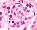
*Very large nucleus (as the name implies) with clearing. | |||
**Classically described as owl's eye-like. | |||
*Granular cytoplasmic inclusions (red on H&E sections). | |||
Notes: | |||
*Classically in endothelial cells. | |||
**In the context of [[esophagus|esophageal ulcers]], it is therefore useful to biopsy the base of the ulcer - if this is suspected. | |||
====Images==== | |||
<gallery> | |||
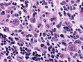
Image:CMV_placentitis2_mini.jpg | CMV [[acute villitis|placentitis]]. (WC) | |||
File:Cmv neuronal inclusions.jpg | CMV [[encephalitis]]. (WC) | |||
</gallery> | |||
www: | |||
*[http://www.asm.org/division/c/photo/cmv1.jpg CMV - owl's eye-like (asm.org)]. | |||
*[http://path.upmc.edu/cases/case149.html CMV - case 1 - several images (upmc.edu)]. | |||
*[http://path.upmc.edu/cases/case481.html CMV - case 2 - several images (upmc.edu)]. | |||
===IHC=== | |||
*IHC for CMV is available - highlights granular cytoplasmic inclusions; increases [[sensitivity]]. | |||
==Human papillomavirus== | |||
*Abbreviated ''HPV''. | |||
{{Main|Human papillomavirus}} | |||
==Adenovirus== | |||
===General=== | |||
*Common in kids - usually a mild respiratory infection with fever and pharyngitis. | |||
**Can cause post-infectious [[bronchiolitis obliterans]].<ref name=pmid20717912>{{Cite journal | last1 = Aguerre | first1 = V. | last2 = Castaños | first2 = C. | last3 = Pena | first3 = HG. | last4 = Grenoville | first4 = M. | last5 = Murtagh | first5 = P. | title = Postinfectious bronchiolitis obliterans in children: clinical and pulmonary function findings. | journal = Pediatr Pulmonol | volume = 45 | issue = 12 | pages = 1180-5 | month = Dec | year = 2010 | doi = 10.1002/ppul.21304 | PMID = 20717912 }}</ref> | |||
**May be seen in the context of [[adenovirus appendicitis|(adenovirus) appendicitis]]. | |||
===Microscopic=== | |||
Features: | |||
*"Smudge" cells<ref>URL: [http://www.pathguy.com/lectures/infect.htm http://www.pathguy.com/lectures/infect.htm]. Accessed on: 8 July 2010.</ref> - black/blue blob ~ 10-15 micrometers. (???) | |||
Notes: | |||
*May be morphologically similar to ''[[CMV]]'', ''[[HSV]]'', ''[[VZV]]'' inclusions. | |||
Images: | |||
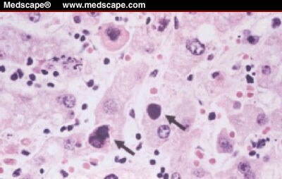
*[http://img.medscape.com/fullsize/migrated/438/534/cc438534.haur.fig1.jpg Adenovirus (medscape.com)].<ref>URL:[http://www.medscape.com/viewarticle/438534_2 http://www.medscape.com/viewarticle/438534_2]. Accessed on: 8 July 2010.</ref> | |||
*[http://wiki.medpedia.com/Image:Ab14.jpg?filetimestamp=20091014175858 Smudge cell (medpedia.com)]. | |||
*[http://www.flickr.com/photos/ckrishnan/3746778145/in/photostream/ Necrosis in germinal centre - low mag. (flickr.com)]. | |||
*[http://www.flickr.com/photos/ckrishnan/3746778007/in/photostream/ Viral inclusions - high mag. (flickr.com)]. | |||
*[http://www.flickr.com/photos/ckrishnan/3747567554/in/photostream/ IHC for adenovirus (flickr.com)] | |||
*[http://path.upmc.edu/cases/case620.html Adenovirus encephalitis - several images (upmc.edu)]. | |||
==Parvovirus== | |||
*[[AKA]] ''Parvovirus B19''. | |||
===General=== | |||
*Most significant in pregnant women. | |||
**Parvovirus attacks the nucleated RBCs of the fetus - causes an ''aplastic [[anemia]]''. | |||
*May cause ''[[collapsing glomerulopathy]]''.<ref name=pmid12704581>{{Cite journal | last1 = Schwimmer | first1 = JA. | last2 = Markowitz | first2 = GS. | last3 = Valeri | first3 = A. | last4 = Appel | first4 = GB. | title = Collapsing glomerulopathy. | journal = Semin Nephrol | volume = 23 | issue = 2 | pages = 209-18 | month = Mar | year = 2003 | doi = 10.1053/snep.2003.50019 | PMID = 12704581 }}</ref> | |||
Trivia: | |||
*First described in 1975.<ref name=pmid46024>{{Cite journal | last1 = Cossart | first1 = YE. | last2 = Field | first2 = AM. | last3 = Cant | first3 = B. | last4 = Widdows | first4 = D. | title = Parvovirus-like particles in human sera. | journal = Lancet | volume = 1 | issue = 7898 | pages = 72-3 | month = Jan | year = 1975 | doi = | PMID = 46024 }}</ref> | |||
**The "B19" part comes from the label for the specimen.<ref name="pmid17304869">{{cite journal |author=Servey JT, Reamy BV, Hodge J |title=Clinical presentations of parvovirus B19 infection |journal=Am Fam Physician |volume=75 |issue=3 |pages=373–6 |year=2007 |month=February |pmid=17304869 |doi= |url=http://www.aafp.org/afp/991001ap/1455.html}}</ref> | |||
===Microscopic=== | |||
Features: | |||
*Glassy (red) nuclear inclusions.<ref>URL: [http://www.pathguy.com/lectures/infect.htm http://www.pathguy.com/lectures/infect.htm]. Accessed on: 8 July 2010.</ref> | |||
*Nuclear enlargement. | |||
====Images==== | |||
<gallery> | |||
Image:Parvovirus_infection_-_cropped_2_-_very_high_mag.jpg | Parvovirus - version 1 - very high mag. (WC) | |||
Image:Parvovirus_infection_-_cropped_1_-_very_high_mag.jpg | Parvovirus - version 2 - very high mag. (WC) | |||
</gallery> | |||
www: | |||
*[http://info.fujita-hu.ac.jp/~tsutsumi/photo/photo210-1.htm Parvovirus (fujita-hu.ac.jp)].<ref>URL:[http://info.fujita-hu.ac.jp/~tsutsumi/case/case210.htm http://info.fujita-hu.ac.jp/~tsutsumi/case/case210.htm]. Accessed on: 8 February 2011.</ref> | |||
*[http://www.scielo.br/img/revistas/rimtsp/v49n2/07f1a.jpg Parvovirus - placenta - (scielo.br)].<ref>URL: [http://www.scielo.br/scielo.php?pid=S0036-46652007000200007&script=sci_arttext http://www.scielo.br/scielo.php?pid=S0036-46652007000200007&script=sci_arttext]. Accessed on: 18 August 2011.</ref> | |||
*[http://www.fujita-hu.ac.jp/~tsutsumi/case/case219.htm Parvovirus - several images (fujita-hu.ac.jp)]. | |||
==Epstein-Barr virus== | |||
{{Main|Epstein-Barr virus}} | |||
==Polyomavirus== | |||
{{Main|Polyomavirus}} | |||
==Human herpesvirus-8== | |||
{{Main|Human herpesvirus-8}} | |||
==West Nile virus== | |||
*Abbreviated ''WNV''. | |||
===General=== | |||
*Uncommon pathologen. | |||
Clinical: | |||
*Fever. | |||
*Muscle weakness. | |||
===Microscopic=== | |||
Features:<ref>{{Cite journal | last1 = Sampson | first1 = BA. | last2 = Ambrosi | first2 = C. | last3 = Charlot | first3 = A. | last4 = Reiber | first4 = K. | last5 = Veress | first5 = JF. | last6 = Armbrustmacher | first6 = V. | title = The pathology of human West Nile Virus infection. | journal = Hum Pathol | volume = 31 | issue = 5 | pages = 527-31 | month = May | year = 2000 | doi = | PMID = 10836291 }}</ref> | |||
*Perivascular clusters in grey and white matter: | |||
**Mononuclear infiltrates (lymphocytes, plasma cells). | |||
**Microglial nodules (macrophage clusters). | |||
==Measles virus== | |||
===General=== | |||
*Causes ''Measles''. | |||
**Should '''not''' be confused with ''Rubella'' ([[AKA]] ''German measles''). | |||
*Uncommon due to widespread MMR vaccine. | |||
**However increasing in the last years most likely due to insufficient vaccination. | |||
*May develop weeks to years after infection. | |||
*Illness may be complicated by ''subacute sclerosing panencephalitis'' (SSPE) - a chronic neurodegenerative condition.<ref>URL: [http://path.upmc.edu/cases/case595/dx.html http://path.upmc.edu/cases/case595/dx.html]. Accessed on: 26 January 2012.</ref> | |||
===Microscopic=== | |||
Features: | |||
*+/-Intranuclear Cowdry type A inclusions. | |||
**Glassy (pink) nucleus. | |||
*Lymphocytes and macrophages (microglial cells). | |||
**May be mild in in measles inclusion body encephalitis. | |||
*Multinucleated cells. | |||
*Microglial nodules. | |||
*Demyelination. | |||
*Gliosis. | |||
Notes: | |||
*Measles inclusions are intranuclear. RSV inclusions are intracytoplasmic.{{fact}} | |||
====Images==== | |||
<gallery> | |||
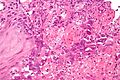
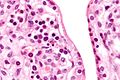
Image:Morbillo.jpg | Measles pneumonia. (WC/CDC) | |||
</gallery> | |||
*[http://path.upmc.edu/cases/case595.html SSPE - several images (upmc.edu)]. | |||
==Rabies virus== | |||
===General=== | |||
*Causes rabies. | |||
Virus affects:<ref>{{Ref APBR|424 Q36}}</ref> | |||
*Cerebral cortex. | |||
*Hippocamus pyramidal cells. | |||
*Purkinje cells. | |||
===Microscopic=== | |||
Features: | |||
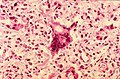
*[[Negri bodies]]: | |||
**Dense-appearing eosinophilic cytoplasmic bodies with a pale halo. | |||
====Images==== | |||
<gallery> | |||
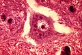
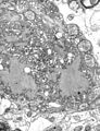
Image:Rabies_encephalitis_Negri_bodies_PHIL_3377_lores.jpg | Negri bodies. (WC/CDC) | |||
Image:Rabies_Virus_EM_PHIL_1876.JPG | Negri bodies - EM. (WC) | |||
File:Rabies negri bodies brain.jpg | Negri bodies in cerebellar Purkinje cells. (WC/CDC). | |||
</gallery> | |||
www: | |||
*[http://www.nature.com/modpathol/journal/v18/n1/fig_tab/3800274f1.html#figure-title Rabies encephalitis (nature.com)].<ref name=pmid15389258>{{Cite journal | last1 = Nuovo | first1 = GJ. | last2 = Defaria | first2 = DL. | last3 = Chanona-Vilchi | first3 = JG. | last4 = Zhang | first4 = Y. | title = Molecular detection of rabies encephalitis and correlation with cytokine expression. | journal = Mod Pathol | volume = 18 | issue = 1 | pages = 62-7 | month = Jan | year = 2005 | doi = 10.1038/modpathol.3800274 | PMID = 15389258 | url = http://www.nature.com/modpathol/journal/v18/n1/full/3800274a.html}}</ref> | |||
=See also= | |||
*[[Microorganisms]]. | |||
*[[Basics]]. | |||
*[[HIV]]. | |||
=References= | |||
{{Reflist|2}} | |||
[[Category:Microorganisms]] | |||
Latest revision as of 15:43, 9 December 2021
This article deals with viruses. The more general topic of infective things is dealt with in microorganisms. Many viruses afflict humans. Only a few of them can be diagnosed histologically.
Several viruses cause cancer and seen directly or indirectly by pathologists frequently.
Viral inclusions - types
Cowdry types:[1]
- Cowdry type A inclusion:[2]
- Round eosinophilic material surrounded by a clear halo.
- Cowdry type B inclusion:[3]
- Neuropathology thingy. (???)
Images:
Viruses
Herpes simplex virus
- In the context of gynecologic cytopathology see: Gynecologic_cytopathology#Herpes_simplex_virus.
- Abbreviated HSV.
General
Several subtypes:
- Canker sores - usually HSV-1.
- Genital herpes - usually HSV-2.
Histology/cytology
Features:[4]
- Clear "ground glass" nuclei.
- Rim of peripheral chromatin.
- Nuclear inclusions.
- Multinucleation with nuclear molding, i.e. multiple nuclei that touch over a large surface area.
Mnemonic - 3 Ms: Margination, Multinucleation, Molding.
Images
www:
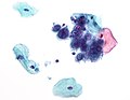
HSV on a Pap test - showing multinucleation. (WC)
HSV esophagitis - very high mag. (WC)
HSV esophagitis - intermed. mag. (WC)


IHC
- HSV-1 +ve (cytoplasmic and strong nuclear).
- HSV-2 +ve.
Images:
Cytomegalovirus
- Abbreviated CMV.
- For pneumonia caused by CMV - see Cytomegalovirus pneumonia.
- For colitis caused by CMV - see Cytomegalovirus colitis.
General
- The name comes from the microscopic appearance.
- One of the TORCH infections.
- May cause fetal hydrops and intracerebral hemorrhage.[7]
Microscopic
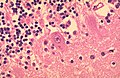
Features:
- Very large nucleus (as the name implies) with clearing.
- Classically described as owl's eye-like.
- Granular cytoplasmic inclusions (red on H&E sections).
Notes:
- Classically in endothelial cells.
- In the context of esophageal ulcers, it is therefore useful to biopsy the base of the ulcer - if this is suspected.
Images
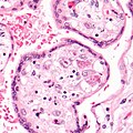
CMV placentitis. (WC)
CMV encephalitis. (WC)


www:
- CMV - owl's eye-like (asm.org).
- CMV - case 1 - several images (upmc.edu).
- CMV - case 2 - several images (upmc.edu).
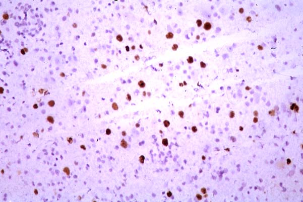
IHC
- IHC for CMV is available - highlights granular cytoplasmic inclusions; increases sensitivity.
Human papillomavirus
- Abbreviated HPV.
Adenovirus
General
- Common in kids - usually a mild respiratory infection with fever and pharyngitis.
- Can cause post-infectious bronchiolitis obliterans.[8]
- May be seen in the context of (adenovirus) appendicitis.
Microscopic
Features:
- "Smudge" cells[9] - black/blue blob ~ 10-15 micrometers. (???)
Notes:
Images:
- Adenovirus (medscape.com).[10]
- Smudge cell (medpedia.com).
- Necrosis in germinal centre - low mag. (flickr.com).
- Viral inclusions - high mag. (flickr.com).
- IHC for adenovirus (flickr.com)
- Adenovirus encephalitis - several images (upmc.edu).
Parvovirus
- AKA Parvovirus B19.
General
- Most significant in pregnant women.
- Parvovirus attacks the nucleated RBCs of the fetus - causes an aplastic anemia.
- May cause collapsing glomerulopathy.[11]
Trivia:
Microscopic
Features:
- Glassy (red) nuclear inclusions.[14]
- Nuclear enlargement.
Images
Parvovirus - version 1 - very high mag. (WC)
Parvovirus - version 2 - very high mag. (WC)


www:
- Parvovirus (fujita-hu.ac.jp).[15]
- Parvovirus - placenta - (scielo.br).[16]
- Parvovirus - several images (fujita-hu.ac.jp).
Epstein-Barr virus
Polyomavirus
Human herpesvirus-8
West Nile virus
- Abbreviated WNV.
General
- Uncommon pathologen.
Clinical:
- Fever.
- Muscle weakness.
Microscopic
Features:[17]
- Perivascular clusters in grey and white matter:
- Mononuclear infiltrates (lymphocytes, plasma cells).
- Microglial nodules (macrophage clusters).
Measles virus
General
- Causes Measles.
- Should not be confused with Rubella (AKA German measles).
- Uncommon due to widespread MMR vaccine.
- However increasing in the last years most likely due to insufficient vaccination.
- May develop weeks to years after infection.
- Illness may be complicated by subacute sclerosing panencephalitis (SSPE) - a chronic neurodegenerative condition.[18]
Microscopic
Features:
- +/-Intranuclear Cowdry type A inclusions.
- Glassy (pink) nucleus.
- Lymphocytes and macrophages (microglial cells).
- May be mild in in measles inclusion body encephalitis.
- Multinucleated cells.
- Microglial nodules.
- Demyelination.
- Gliosis.
Notes:
- Measles inclusions are intranuclear. RSV inclusions are intracytoplasmic.[citation needed]
Images
Measles pneumonia. (WC/CDC)

Rabies virus
General
- Causes rabies.
Virus affects:[19]
- Cerebral cortex.
- Hippocamus pyramidal cells.
- Purkinje cells.
Microscopic
Features:
- Negri bodies:
- Dense-appearing eosinophilic cytoplasmic bodies with a pale halo.
Images
Negri bodies. (WC/CDC)
Negri bodies - EM. (WC)
Negri bodies in cerebellar Purkinje cells. (WC/CDC).


www:
See also
References
- ↑ URL: http://www.pathconsultddx.com/pathCon/largeImage?pii=S1559-8675%2806%2970864-6&figureId=fig3&ecomponentId=mmc3. Accessed: 12 January 2010.
- ↑ URL: http://www.whonamedit.com/synd.cfm/3495.html. Accessed on: 22 January 2010.
- ↑ http://www.whonamedit.com/synd.cfm/3496.html. Accessed on: 22 January 2010.
- ↑ SM. 11 January 2010.
- ↑ URL: http://path.upmc.edu/cases/case120/dx.html. Accessed on: 28 February 2013.
- ↑ URL: http://www.antibodies-online.com/antibody/100405/anti-Herpes+Simplex+Virus+1+HSV1/. Accessed on: 28 February 2013.
- ↑ Tongsong, T.; Sukpan, K.; Wanapirak, C.; Phadungkiatwattna, P. (2008). "Fetal cytomegalovirus infection associated with cerebral hemorrhage, hydrops fetalis, and echogenic bowel: case report.". Fetal Diagn Ther 23 (3): 169-72. doi:10.1159/000116737. PMID 18417974.
- ↑ Aguerre, V.; Castaños, C.; Pena, HG.; Grenoville, M.; Murtagh, P. (Dec 2010). "Postinfectious bronchiolitis obliterans in children: clinical and pulmonary function findings.". Pediatr Pulmonol 45 (12): 1180-5. doi:10.1002/ppul.21304. PMID 20717912.
- ↑ URL: http://www.pathguy.com/lectures/infect.htm. Accessed on: 8 July 2010.
- ↑ URL:http://www.medscape.com/viewarticle/438534_2. Accessed on: 8 July 2010.
- ↑ Schwimmer, JA.; Markowitz, GS.; Valeri, A.; Appel, GB. (Mar 2003). "Collapsing glomerulopathy.". Semin Nephrol 23 (2): 209-18. doi:10.1053/snep.2003.50019. PMID 12704581.
- ↑ Cossart, YE.; Field, AM.; Cant, B.; Widdows, D. (Jan 1975). "Parvovirus-like particles in human sera.". Lancet 1 (7898): 72-3. PMID 46024.
- ↑ Servey JT, Reamy BV, Hodge J (February 2007). "Clinical presentations of parvovirus B19 infection". Am Fam Physician 75 (3): 373–6. PMID 17304869. http://www.aafp.org/afp/991001ap/1455.html.
- ↑ URL: http://www.pathguy.com/lectures/infect.htm. Accessed on: 8 July 2010.
- ↑ URL:http://info.fujita-hu.ac.jp/~tsutsumi/case/case210.htm. Accessed on: 8 February 2011.
- ↑ URL: http://www.scielo.br/scielo.php?pid=S0036-46652007000200007&script=sci_arttext. Accessed on: 18 August 2011.
- ↑ Sampson, BA.; Ambrosi, C.; Charlot, A.; Reiber, K.; Veress, JF.; Armbrustmacher, V. (May 2000). "The pathology of human West Nile Virus infection.". Hum Pathol 31 (5): 527-31. PMID 10836291.
- ↑ URL: http://path.upmc.edu/cases/case595/dx.html. Accessed on: 26 January 2012.
- ↑ Lefkowitch, Jay H. (2006). Anatomic Pathology Board Review (1st ed.). Saunders. pp. 424 Q36. ISBN 978-1416025887.
- ↑ Nuovo, GJ.; Defaria, DL.; Chanona-Vilchi, JG.; Zhang, Y. (Jan 2005). "Molecular detection of rabies encephalitis and correlation with cytokine expression.". Mod Pathol 18 (1): 62-7. doi:10.1038/modpathol.3800274. PMID 15389258. http://www.nature.com/modpathol/journal/v18/n1/full/3800274a.html.