Difference between revisions of "Urinary bladder"
Jump to navigation
Jump to search
(→IHC) |
(→Images) |
||
| Line 57: | Line 57: | ||
====Images==== | ====Images==== | ||
<gallery> | <gallery> | ||
Image:Urachal_carcinoma_-_very_low_mag.jpg | UC - very low mag. (WC) | |||
Image:Urachal_carcinoma_-_low_mag.jpg | UC - low mag. (WC) | Image:Urachal_carcinoma_-_low_mag.jpg | UC - low mag. (WC) | ||
Image:Urachal_carcinoma_-_high_mag.jpg | UC - high mag. (WC) | Image:Urachal_carcinoma_-_high_mag.jpg | UC - high mag. (WC) | ||
Revision as of 04:13, 26 November 2013
The urinary bladder stores urine until one has to go wee-wee.
It is commonly afflicted by cancer.
A well-know mimicker of cancer is malakoplakia.[1]
Normal
Microscopic
- Muscularis mucosae - thin, discontinuous.
- Fat - many be in lamina propria.
Note:
- On TURBT - don't ever call T3.
Urinary bladder cancer
The most common type of cancer to affect the bladder is urothelial carcinoma. This is covered in the urothelium article.
Risk factors for bladder cancer SEX LIC:
- Schistomsoma haematobium - esp. squamous cell carcinoma.[2]
- EXtrophy of the bladder.
- Lithiasis.
- Indwelling catheter or chronic Inflammation.[2]
- Cyclophosphamide.
DDx:
- Urothelial carcinoma - most common in the Western world.
- Squamous cell carcinoma of the urinary bladder - most common in areas with Schistosoma.
- Adenocarcinoma - see urachal carcinoma.
Squamous cell carcinoma of the urinary bladder
Main article: Squamous cell carcinoma
General
- Strong association with Schistosoma haematobium.[2]
- Common in areas with S. haematobium.
- Uncommon in areas without S. haematobium.
Microscopic
Features:
- See squamous cell carcinoma article.
Urachal carcinoma
General
- Rare.[3]
- Classically - dome of bladder lesion.
- Younger <55 years-old.
Microscopic
Patterns
- Enteric - looks like colonic adenocarcinoma.
- Mucinous.
- Signet ring.
DDx:
- Metastatic adenocarcinoma/adenocarcinoma extending from another structure, e.g. colorectal adenocarcinoma.
- Adenocarcinoma arising from the urinary bladder.
Images
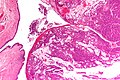
UC - very low mag. (WC)
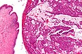
UC - low mag. (WC)
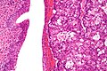
UC - high mag. (WC)


IHC
Features:[4]
- CK20 +ve.
- CK7 +ve/-ve.
- CK34betaE12 +ve/-ve.
- Beta-catenin -- usu cytoplasmic/membranous +ve.
Others:[5]
- p63 -ve (+ve in only 3%).
UC versus CRC -- not absolute but useful:
- CK34betaE12 +ve in UC (-ve in CRC).
- Beta-catenin -ve nuclei in UC (+ve nuclei in CRC).
Urinary bladder infarct
Main article: Infarction
General
- Case report rare - as the organ has many colaterals.[6]
- May be seen in association with pseudocarcinomatous urothelial hyperplasia.[7]
Microscopic
Features:[6]
- Necrosis without liquefaction.
- Outlines of cells visible.
- No nuclei present.
Note:
- Liquefaction implies an infectious etiology.[6]
Rhabdomyosarcoma
Main article: Rhabdomyosarcoma
See also
References
- ↑ Wong-You-Cheong JJ, Woodward PJ, Manning MA, Davis CJ (2006). "From the archives of the AFIP: Inflammatory and nonneoplastic bladder masses: radiologic-pathologic correlation". Radiographics 26 (6): 1847–68. doi:10.1148/rg.266065126. PMID 17102055.
- ↑ 2.0 2.1 2.2 Michaud, DS.. "Chronic inflammation and bladder cancer.". Urol Oncol 25 (3): 260-8. doi:10.1016/j.urolonc.2006.10.002. PMID 17483025.
- ↑ Ashley, RA.; Inman, BA.; Sebo, TJ.; Leibovich, BC.; Blute, ML.; Kwon, ED.; Zincke, H. (Aug 2006). "Urachal carcinoma: clinicopathologic features and long-term outcomes of an aggressive malignancy.". Cancer 107 (4): 712-20. doi:10.1002/cncr.22060. PMID 16826585.
- ↑ Gopalan, A.; Sharp, DS.; Fine, SW.; Tickoo, SK.; Herr, HW.; Reuter, VE.; Olgac, S. (May 2009). "Urachal carcinoma: a clinicopathologic analysis of 24 cases with outcome correlation.". Am J Surg Pathol 33 (5): 659-68. doi:10.1097/PAS.0b013e31819aa4ae. PMID 19252435.
- ↑ Paner, GP.; McKenney, JK.; Barkan, GA.; Yao, JL.; Frankel, WL.; Sebo, TJ.; Shen, SS.; Jimenez, RE. (Jun 2011). "Immunohistochemical analysis in a morphologic spectrum of urachal epithelial neoplasms: diagnostic implications and pitfalls.". Am J Surg Pathol 35 (6): 787-98. doi:10.1097/PAS.0b013e3182189c11. PMID 21572312.
- ↑ 6.0 6.1 6.2 Nino-Murcia, M.; Friedland, GW. (1988). "Bladder infarct.". Urol Radiol 9 (4): 234-6. PMID 3394185.
- ↑ Kryvenko, ON.; Epstein, JI. (Jun 2013). "Pseudocarcinomatous urothelial hyperplasia of the bladder: clinical findings and followup of 70 patients.". J Urol 189 (6): 2083-6. doi:10.1016/j.juro.2012.12.005. PMID 23228381.