Difference between revisions of "Staghorn vessels"
Jump to navigation
Jump to search
(create) |
|||
| (5 intermediate revisions by the same user not shown) | |||
| Line 1: | Line 1: | ||
'''Staghorn vessels''' are a non-specific finding seen in a number of tumours.<ref name=pmid8545589>{{Cite journal | last1 = Nappi | first1 = O. | last2 = Ritter | first2 = JH. | last3 = Pettinato | first3 = G. | last4 = Wick | first4 = MR. | title = Hemangiopericytoma: histopathological pattern or clinicopathologic entity? | journal = Semin Diagn Pathol | volume = 12 | issue = 3 | pages = 221-32 | month = Aug | year = 1995 | doi = | PMID = 8545589 }}</ref> | '''Staghorn vessels''', also known as '''[[hemangiopericytoma]]-like vessels''' (abbreviated '''HPC-like vessels'''), are a non-specific finding seen in a number of tumours.<ref name=pmid8545589>{{Cite journal | last1 = Nappi | first1 = O. | last2 = Ritter | first2 = JH. | last3 = Pettinato | first3 = G. | last4 = Wick | first4 = MR. | title = Hemangiopericytoma: histopathological pattern or clinicopathologic entity? | journal = Semin Diagn Pathol | volume = 12 | issue = 3 | pages = 221-32 | month = Aug | year = 1995 | doi = | PMID = 8545589 }}</ref> | ||
==Microscopic== | |||
Features: | |||
*Small branching vessels: | |||
**"Antler-like" or "staghorn-like" appearance. | |||
===Images=== | |||
<gallery> | |||
Image:Solitary_fibrous_tumour_high_mag.jpg | SFT - high mag. (WC/Nephron) | |||
Image:Monophasic_synovial_sarcoma_-_intermed_mag.jpg | Synovial sarcoma - intermed. mag. (WC/Nephron) | |||
</gallery> | |||
Gross: | |||
*[http://commons.wikimedia.org/wiki/File:Staghorn_coral.jpg Staghorn coral (WC)]. | |||
*[http://commons.wikimedia.org/wiki/File:Staghorn_Fungus_in_Millington_Wood_-_geograph.org.uk_-_638148.jpg Staghorn fungus (WC)]. | |||
*[http://commons.wikimedia.org/wiki/File:PSM_V09_D778_Deer_antler_dimensions_1.jpg Antlers (WC)]. | |||
==Differential diagnosis== | |||
Entities in which staghorn vessels are seen: | Entities in which staghorn vessels are seen: | ||
*[[Solitary fibrous tumour]]/[[hemangiopericytoma]].<ref name=pmid8545589/> | *[[Solitary fibrous tumour]]/[[hemangiopericytoma]].<ref name=pmid8545589/> | ||
| Line 17: | Line 33: | ||
*Sarcomatoid carcinoma. | *Sarcomatoid carcinoma. | ||
*[[Malignant melanoma]]. | *[[Malignant melanoma]]. | ||
*[[Glomus tumour]].<ref name=pmid19952942>{{Cite journal | last1 = Slone | first1 = SP. | last2 = Moore | first2 = GD. | last3 = Parker | first3 = LP. | last4 = Rickard | first4 = KA. | last5 = Nixdorf-Miller | first5 = AS. | title = Glomus tumor of the ovary masquerading as granulosa cell tumor: case report. | journal = Int J Gynecol Pathol | volume = 29 | issue = 1 | pages = 24-6 | month = Jan | year = 2010 | doi = 10.1097/PGP.0b013e3181b0b771 | PMID = 19952942 }}</ref> | |||
==References== | ==References== | ||
{{Reflist| | {{Reflist|2}} | ||
[[Category:Stuff]] | [[Category:Stuff]] | ||
Latest revision as of 19:53, 12 June 2014
Staghorn vessels, also known as hemangiopericytoma-like vessels (abbreviated HPC-like vessels), are a non-specific finding seen in a number of tumours.[1]
Microscopic
Features:
- Small branching vessels:
- "Antler-like" or "staghorn-like" appearance.
Images
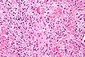
SFT - high mag. (WC/Nephron)
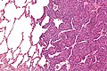
Synovial sarcoma - intermed. mag. (WC/Nephron)


Gross:
Differential diagnosis
Entities in which staghorn vessels are seen:
- Solitary fibrous tumour/hemangiopericytoma.[1]
- Malignant peripheral nerve sheath tumour (MPNST).[1]
- Synovial sarcoma.[1]
- Myofibroma.[1]
Others:[1]
- Mesenchymal chondrosarcoma.
- Infantile fibrosarcoma.
- Pleomorphic undifferentiated sarcoma.
- Leiomyosarcoma.
- Endometrial stromal sarcoma.
- Malignant mesothelioma.
- Thymoma
- Sarcomatoid carcinoma.
- Malignant melanoma.
- Glomus tumour.[2]
References
- ↑ 1.0 1.1 1.2 1.3 1.4 1.5 Nappi, O.; Ritter, JH.; Pettinato, G.; Wick, MR. (Aug 1995). "Hemangiopericytoma: histopathological pattern or clinicopathologic entity?". Semin Diagn Pathol 12 (3): 221-32. PMID 8545589.
- ↑ Slone, SP.; Moore, GD.; Parker, LP.; Rickard, KA.; Nixdorf-Miller, AS. (Jan 2010). "Glomus tumor of the ovary masquerading as granulosa cell tumor: case report.". Int J Gynecol Pathol 29 (1): 24-6. doi:10.1097/PGP.0b013e3181b0b771. PMID 19952942.