Pleomorphic undifferentiated sarcoma
| Pleomorphic undifferentiated sarcoma | |
|---|---|
| Diagnosis in short | |
 Pleomorphic undifferentiated sarcoma. H&E stain. | |
|
| |
| Synonyms | undifferentiated pleomorphic sarcoma (UPS), malignant fibrous histiocytoma (MFH) |
|
| |
| LM | storiform pattern (AKA patternless pattern), marked nuclear pleomorphism, mitoses, necrosis (common), mix of spindle cells and epithelioid cells, deep to skin |
| LM DDx | atypical fibroxanthoma (superficial), dedifferentiated liposarcoma, leiomyosarcoma,metaplastic carcinoma, malignant melanoma, rhabdomyosarcoma, synovial sarcoma,undifferentiated endometrial sarcoma, others |
| Site | deep soft tissue - trunk and extremities |
|
| |
| Prevalence | uncommon overall |
| Prognosis | poor |
| Treatment | surgery if feasible |
Pleomorphic undifferentiated sarcoma, abbreviated PUS, is an undifferentiated malignant soft tissue lesion.
It is also known as undifferentiated pleomorphic sarcoma (abbreviated UPS). Previously, it was known as malignant fibrous histiocytoma, commonly abbreviated MFH.[1]
General
- Common sarcoma.
- Usually deep tissue of the trunk and extremities.
- A diagnosis of exclusion[2] / wastebasket for unclassifiable high grade sarcomas.
Microscopic
Features:[3]
- Storiform pattern (AKA patternless pattern) - key feature.
- Marked nuclear pleomorphism key feature.
- Variation is nuclear size, nuclear shape and nuclear staining (esp. hyperchromasia).
- Mitoses - abundant; atypical mitoses common.
- Necrosis (common).
- Mix of spindle cells and epithelioid cells.
- Deep to skin - important.
Other findings:
- +/-Giant cells (see subclassification).
- +/-Inflammation (see subclassification).
- Neutrophils.
- Eosinophils.
Notes:
- Superficial lesions with the morphology of PUS are called by some atypical fibroxanthomas (AFXs).
DDx:
- Atypical fibroxanthoma (AFX) - superficial skin.
- Dedifferentiated liposarcoma.
- Leiomyosarcoma.
- Metaplastic carcinoma.
- Malignant melanoma.
- Rhabdomyosarcoma.
- Synovial sarcoma.
- Undifferentiated endometrial sarcoma - uterus.
- Others.
Images
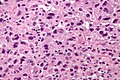
PUS - high mag. (WC/Nephron)
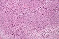
PUS - intermed. mag. (WC/Nephron)


Pleomorphic sarcoma in thigh of late middle aged man. A. Tumor haphazardly spreads and invades and surrounds degenerated skeletal muscle fibers. B. Bizarre, sometimes multinucleated cells are seen. C. In other areas, spindle cells predominate. D. Invasion of adipose tissue by tumor should not be interpreted as proof of dedifferentiated liposarcoma; lipoblasts should be seen. Vital is remembering this is a diagnosis of exclusion, with negative keratin, CD34, CD30, S100, and CD31 stains being suggested.
Subclassification
Pleomorphic sarcoma (PS) is subclassified the following way:[4]
- PS with giant cells.
- PS with inflammation.
- PUS (not otherwise specified) - wastebasket diagnosis; if neither of the above two apply.
IHC
Exclusionary stains - should be negative:
- AE1/AE3.
- p63.
- Myogenin.
- S-100.
- HMB-45.
Usually negative, may be positive:[5]
- Desmin.
- SMA.
Commonly positive:
- CD68.[5]
- Vimentin.
See also
References
- ↑ URL: http://sarcomahelp.org/learning_center/mfh.html. Accessed on: 8 April 2011.
- ↑ Matushansky I, Charytonowicz E, Mills J, Siddiqi S, Hricik T, Cordon-Cardo C (August 2009). "MFH classification: differentiating undifferentiated pleomorphic sarcoma in the 21st Century". Expert Rev Anticancer Ther 9 (8): 1135–44. doi:10.1586/era.09.76. PMC 3000413. PMID 19671033. https://www.ncbi.nlm.nih.gov/pmc/articles/PMC3000413/.
- ↑ Humphrey, Peter A; Dehner, Louis P; Pfeifer, John D (2008). The Washington Manual of Surgical Pathology (1st ed.). Lippincott Williams & Wilkins. pp. 613. ISBN 978-0781765275.
- ↑ Humphrey, Peter A; Dehner, Louis P; Pfeifer, John D (2008). The Washington Manual of Surgical Pathology (1st ed.). Lippincott Williams & Wilkins. pp. 613-4. ISBN 978-0781765275.
- ↑ 5.0 5.1 Humphrey, Peter A; Dehner, Louis P; Pfeifer, John D (2008). The Washington Manual of Surgical Pathology (1st ed.). Lippincott Williams & Wilkins. pp. 613. ISBN 978-0781765275.