Difference between revisions of "Squamous cell carcinoma of the lung"
Jump to navigation
Jump to search
(+images) |
|||
| Line 1: | Line 1: | ||
{{ Infobox diagnosis | {{ Infobox diagnosis | ||
| Name = {{PAGENAME}} | | Name = {{PAGENAME}} | ||
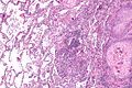
| Image = | | Image = Lung squamous carcinoma -- intermed mag.jpg | ||
| Width = | | Width = | ||
| Caption = Squamous cell carcinoma of the lung. [[ | | Caption = Squamous cell carcinoma of the lung. [[H&E stain]]. | ||
| Synonyms = squamous carcinoma of the lung | | Synonyms = squamous carcinoma of the lung | ||
| Micro = | | Micro = | ||
| Line 59: | Line 59: | ||
*+/-Small nucleolus. | *+/-Small nucleolus. | ||
*Intracellular bridges - classic. | *Intracellular bridges - classic. | ||
Note: | |||
*[[Lymphovascular invasion]] (LVI) is relatively common in small tumours. In one series of NSLC tumours less than 2 cm the prevalence of LVI was 16%.<ref>{{cite journal |author=Tao H, Hayashi T, Sano F, ''et al.'' |title=Prognostic impact of lymphovascular invasion compared with that of visceral pleural invasion in patients with pN0 non-small-cell lung cancer and a tumor diameter of 2 cm or smaller |journal=J. Surg. Res. |volume=185 |issue=1 |pages=250–4 |year=2013 |month=November |pmid=23830361 |doi=10.1016/j.jss.2013.05.104 |url=}}</ref> | |||
**Unlike in [[lung adenocarcinoma]], LVI in lung SCC does ''not'' seem to increase the risk of distant metastases and death.<ref name=pmid22617241>{{cite journal |author=Higgins KA, Chino JP, Ready N, ''et al.'' |title=Lymphovascular invasion in non-small-cell lung cancer: implications for staging and adjuvant therapy |journal=J Thorac Oncol |volume=7 |issue=7 |pages=1141–7 |year=2012 |month=July |pmid=22617241 |doi=10.1097/JTO.0b013e3182519a42 |url=}}</ref> | |||
DDx: | DDx: | ||
| Line 66: | Line 70: | ||
*[[Small cell carcinoma of the lung]] - for ''basaloid squamous cell carcinoma''. | *[[Small cell carcinoma of the lung]] - for ''basaloid squamous cell carcinoma''. | ||
===Images=== | |||
<gallery> | |||
Image: Lung squamous carcinoma -- very low mag.jpg | Lung SCC - very low mag. (WC) | |||
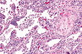
Image: Lung squamous carcinoma -- low mag.jpg | Lung SCC - low mag. (WC) | |||
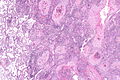
Image: Lung squamous carcinoma -- intermed mag.jpg | Lung SCC - intermed. mag. (WC) | |||
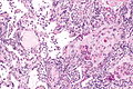
Image: Lung squamous carcinoma -- high mag.jpg | Lung SCC - high mag. (WC) | |||
Image: Lung squamous carcinoma - alt -- low mag.jpg | Lung SCC - low mag. (WC) | |||
Image: Lung squamous carcinoma - alt -- intermed mag.jpg | Lung SCC - intermed. mag. (WC) | |||
</gallery> | |||
====Cytology==== | |||
<gallery> | |||
Image: Squamous carcinoma lung cytology.gif | Lung SCC - cytology. (WC) | |||
</gallery> | |||
==IHC== | ==IHC== | ||
Revision as of 05:59, 9 January 2016
| Squamous cell carcinoma of the lung | |
|---|---|
| Diagnosis in short | |
 Squamous cell carcinoma of the lung. H&E stain. | |
|
| |
| Synonyms | squamous carcinoma of the lung |
| LM DDx | lung adenocarcinoma, non-small cell lung carcinoma, metastatic squamous cell carcinoma, others |
| IHC | p40 +ve, p63 +ve, TTF-1 -ve, CK7 -ve |
| Staging | lung cancer staging |
| Site | lung - see lung tumours |
|
| |
| Clinical history | smoking |
| Symptoms | +/-hemoptysis |
| Prevalence | common |
| Blood work | serum calcium elevated |
| Radiology | typically a mass assoc. with a large airway, +/-spiculated, +/-cavitation |
| Prognosis | usually poor |
| Clin. DDx | other lung tumours - esp. small cell carcinoma of the lung |
| Treatment | surgical resection if possible |
Squamous cell carcinoma of the lung, also lung squamous cell carcinoma, is a common malignant lung tumour that is associated with smoking.
It is also known as squamous carcinoma of the lung and lung squamous carcinoma.
Squamous cell carcinoma can be abbreviated SCC; however, this can be confusing as small cell carcinoma is sometimes abbreviated as such.
General
- Strong association with smoking.
- May be treated with surgery.
Clinical:
- May be associated with elevated serum calcium.[1]
- +/-Hemoptysis.
Gross
- Lung mass - usually centrally located, i.e. associated with a large airway.
Image
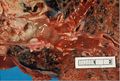
Squamous carcinoma of the lung. (WC)

Microscopic
Features:
- Central nucleus.
- Dense appearing cytoplasm, usu. eosinophilic.
- +/-Small nucleolus.
- Intracellular bridges - classic.
Note:
- Lymphovascular invasion (LVI) is relatively common in small tumours. In one series of NSLC tumours less than 2 cm the prevalence of LVI was 16%.[2]
- Unlike in lung adenocarcinoma, LVI in lung SCC does not seem to increase the risk of distant metastases and death.[3]
DDx:
- Metastatic squamous cell carcinoma.
- Adenocarcinoma of the lung.
- Non-small cell lung carcinoma - diagnosis should be avoided if possible.
- Small cell carcinoma of the lung - for basaloid squamous cell carcinoma.
Images
Lung SCC - very low mag. (WC)
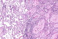
Lung SCC - low mag. (WC)
Lung SCC - intermed. mag. (WC)

Lung SCC - high mag. (WC)
Lung SCC - low mag. (WC)
Lung SCC - intermed. mag. (WC)


Cytology
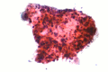
Lung SCC - cytology. (WC)

IHC
- p40 +ve.[4]
- p63 +ve -- less specific.
- Calponin -ve.
- CK5/6 +ve.
Others:[5]
- CK7 -ve.
- CK20 -ve.
- TTF-1 -ve.
- Positive in adenocarcinoma of the lung.
SCC versus adenocarcinoma:
- p40 +ve.
- CK5/6 +ve.
- TTF-1 -ve.
- Napsin -ve.
Sign out
LUNG, RIGHT UPPER LOBE, BIOPSY:
- INVASIVE SQUAMOUS CELL CARCINOMA.

COMMENT:
The tumour stains as follows:
POSITIVE: p40, CK5/6.
NEGATIVE: TTF-1, napsin.
Resection
LUNG, RIGHT UPPER LOBE, LOBECTOMY:
- SQUAMOUS CELL CARCINOMA, MODERATELY DIFFERENTIATED, pT2b, pN0.
-- MARGINS NEGATIVE.
-- PLEASE SEE TUMOUR SUMMARY.
See also
References
- ↑ Campbell, JH.; Ralston, S.; Boyle, IT.; Banham, SW. (May 1991). "Symptomatic hypercalcaemia in lung cancer.". Respir Med 85 (3): 223-7. PMID 1831917.
- ↑ Tao H, Hayashi T, Sano F, et al. (November 2013). "Prognostic impact of lymphovascular invasion compared with that of visceral pleural invasion in patients with pN0 non-small-cell lung cancer and a tumor diameter of 2 cm or smaller". J. Surg. Res. 185 (1): 250–4. doi:10.1016/j.jss.2013.05.104. PMID 23830361.
- ↑ Higgins KA, Chino JP, Ready N, et al. (July 2012). "Lymphovascular invasion in non-small-cell lung cancer: implications for staging and adjuvant therapy". J Thorac Oncol 7 (7): 1141–7. doi:10.1097/JTO.0b013e3182519a42. PMID 22617241.
- ↑ Bishop, JA.; Teruya-Feldstein, J.; Westra, WH.; Pelosi, G.; Travis, WD.; Rekhtman, N. (Mar 2012). "p40 (ΔNp63) is superior to p63 for the diagnosis of pulmonary squamous cell carcinoma.". Mod Pathol 25 (3): 405-15. doi:10.1038/modpathol.2011.173. PMID 22056955.
- ↑ Montezuma, D.; Azevedo, R.; Lopes, P.; Vieira, R.; Cunha, AL.; Henrique, R. (Dec 2013). "A panel of four immunohistochemical markers (CK7, CK20, TTF-1, and p63) allows accurate diagnosis of primary and metastatic lung carcinoma on biopsy specimens.". Virchows Arch 463 (6): 749-54. doi:10.1007/s00428-013-1488-z. PMID 24126803.