Difference between revisions of "Sarcoidosis"
| (10 intermediate revisions by 2 users not shown) | |||
| Line 1: | Line 1: | ||
{{ Infobox diagnosis | {{ Infobox diagnosis | ||
| Name = {{PAGENAME}} | | Name = {{PAGENAME}} | ||
| Image = Sarcoidosis (1) lymph node biopsy.jpg | | Image = Sarcoidosis (1) lymph node biopsy.jpg | ||
| Width = | | Width = | ||
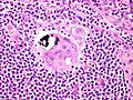
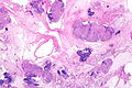
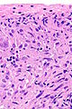
| Caption = Sarcoidosis-like granulomas in a lymph node. [[H&E stain]]. | | Caption = Sarcoidosis-like granulomas in a lymph node. [[H&E stain]]. | ||
| Line 10: | Line 10: | ||
| IHC = | | IHC = | ||
| EM = | | EM = | ||
| Molecular = | | Molecular = PCR for [[tuberculosis]] -ve | ||
| IF = | | IF = | ||
| Gross = | | Gross = | ||
| Line 49: | Line 49: | ||
*Carcinomatosis - interstitial pattern.<ref>URL: [http://www.radiologyassistant.nl/en/46b480a6e4bdc http://www.radiologyassistant.nl/en/46b480a6e4bdc]. Accessed on: 23 May 2010.</ref> | *Carcinomatosis - interstitial pattern.<ref>URL: [http://www.radiologyassistant.nl/en/46b480a6e4bdc http://www.radiologyassistant.nl/en/46b480a6e4bdc]. Accessed on: 23 May 2010.</ref> | ||
*Lymphoma - bilateral lymphadenopathy +/- mediastinal lymphadenopathy.<ref name=pmid23207258>{{Cite journal | last1 = Boujaoude | first1 = Z. | last2 = Dahdel | first2 = M. | last3 = Pratter | first3 = M. | last4 = Kass | first4 = J. | title = Endobronchial ultrasound with transbronchial needle aspiration in the diagnosis of bilateral hilar and mediastinal lymphadenopathy. | journal = J Bronchology Interv Pulmonol | volume = 19 | issue = 1 | pages = 19-23 | month = Jan | year = 2012 | doi = 10.1097/LBR.0b013e3182442b89 | PMID = 23207258 }}</ref> | *Lymphoma - bilateral lymphadenopathy +/- mediastinal lymphadenopathy.<ref name=pmid23207258>{{Cite journal | last1 = Boujaoude | first1 = Z. | last2 = Dahdel | first2 = M. | last3 = Pratter | first3 = M. | last4 = Kass | first4 = J. | title = Endobronchial ultrasound with transbronchial needle aspiration in the diagnosis of bilateral hilar and mediastinal lymphadenopathy. | journal = J Bronchology Interv Pulmonol | volume = 19 | issue = 1 | pages = 19-23 | month = Jan | year = 2012 | doi = 10.1097/LBR.0b013e3182442b89 | PMID = 23207258 }}</ref> | ||
<gallery> | |||
File:Sarcoidosis - Bilateral hilar lymphadenoathy (6076995153).jpg | Markedly enlarged bilaterally hilar lymph nodes in stage 1 sarcoidosis (WC/Yale Rosen) | |||
File:Sarcoidosis - Fibrosis (6076822244).jpg | Fibrosis (WC/Yale Rosen) | |||
</gallery> | |||
==Microscopic== | ==Microscopic== | ||
| Line 55: | Line 60: | ||
**Negative for microorganisms with special stains. | **Negative for microorganisms with special stains. | ||
*Usu. minimal (lymphoid) inflammation; sarcoid granulomas are known as "naked granulomas".<ref name=pmid18948765>{{Cite journal | last1 = Brinster | first1 = NK. | title = Dermatopathology for the surgical pathologist: a pattern-based approach to the diagnosis of inflammatory skin disorders (part II). | journal = Adv Anat Pathol | volume = 15 | issue = 6 | pages = 350-69 | month = Nov | year = 2008 | doi = 10.1097/PAP.0b013e31818b1ac6 | PMID = 18948765 }}</ref><ref name=pmid24138972>{{Cite journal | last1 = Noiles | first1 = K. | last2 = Beleznay | first2 = K. | last3 = Crawford | first3 = RI. | last4 = Au | first4 = S. | title = Sarcoidosis can present with necrotizing granulomas histologically: two cases of ulcerated sarcoidosis and review of the literature. | journal = J Cutan Med Surg | volume = 17 | issue = 6 | pages = 377-83 | month = | year = | doi = | PMID = 24138972 }}</ref> | *Usu. minimal (lymphoid) inflammation; sarcoid granulomas are known as "naked granulomas".<ref name=pmid18948765>{{Cite journal | last1 = Brinster | first1 = NK. | title = Dermatopathology for the surgical pathologist: a pattern-based approach to the diagnosis of inflammatory skin disorders (part II). | journal = Adv Anat Pathol | volume = 15 | issue = 6 | pages = 350-69 | month = Nov | year = 2008 | doi = 10.1097/PAP.0b013e31818b1ac6 | PMID = 18948765 }}</ref><ref name=pmid24138972>{{Cite journal | last1 = Noiles | first1 = K. | last2 = Beleznay | first2 = K. | last3 = Crawford | first3 = RI. | last4 = Au | first4 = S. | title = Sarcoidosis can present with necrotizing granulomas histologically: two cases of ulcerated sarcoidosis and review of the literature. | journal = J Cutan Med Surg | volume = 17 | issue = 6 | pages = 377-83 | month = | year = | doi = | PMID = 24138972 }}</ref> | ||
*In lung: | *In lung: | ||
**Interstitial location +/- centrilobular. | |||
Notes: | Notes: | ||
| Line 62: | Line 68: | ||
DDx: | DDx: | ||
*Infections. | *Infections. | ||
**Fungal. | |||
**Mycobacterial, e.g. [[tuberculosis]]. | |||
**Tertiary [[syphilis]].<ref name=pmid19249139>{{Cite journal | last1 = Hervier | first1 = B. | last2 = Wastiaux | first2 = H. | last3 = Freour | first3 = T. | last4 = Masseau | first4 = A. | last5 = Corvec | first5 = S. | last6 = Armingeat | first6 = T. | last7 = Hamidou | first7 = M. | title = [Sarcoidosis-like granulomatosis revealing a tertiary syphilis]. | journal = Rev Med Interne | volume = 30 | issue = 9 | pages = 806-8 | month = Sep | year = 2009 | doi = 10.1016/j.revmed.2009.01.003 | PMID = 19249139 }}</ref> | **Tertiary [[syphilis]].<ref name=pmid19249139>{{Cite journal | last1 = Hervier | first1 = B. | last2 = Wastiaux | first2 = H. | last3 = Freour | first3 = T. | last4 = Masseau | first4 = A. | last5 = Corvec | first5 = S. | last6 = Armingeat | first6 = T. | last7 = Hamidou | first7 = M. | title = [Sarcoidosis-like granulomatosis revealing a tertiary syphilis]. | journal = Rev Med Interne | volume = 30 | issue = 9 | pages = 806-8 | month = Sep | year = 2009 | doi = 10.1016/j.revmed.2009.01.003 | PMID = 19249139 }}</ref> | ||
*[[Hypersensitivity pneumonitis]] - usually poorly-formed granulomas, centrilobular, not paraseptal.{{fact}} | *[[Hypersensitivity pneumonitis]] - usually poorly-formed granulomas, centrilobular, not paraseptal.{{fact}} | ||
| Line 71: | Line 79: | ||
**All testicular tumours<ref name=pmid18496978>{{Cite journal | last1 = Paparel | first1 = P. | last2 = Devonec | first2 = M. | last3 = Perrin | first3 = P. | last4 = Ruffion | first4 = A. | last5 = Decaussin-Petrucci | first5 = M. | last6 = Akin | first6 = O. | last7 = Sheinfeld | first7 = J. | last8 = Guillonneau | first8 = B. | title = Association between sarcoidosis and testicular carcinoma: a diagnostic pitfall. | journal = Sarcoidosis Vasc Diffuse Lung Dis | volume = 24 | issue = 2 | pages = 95-101 | month = Sep | year = 2007 | doi = | PMID = 18496978 }}</ref> - esp. [[seminoma]].<ref name=pmid17240627>{{Cite journal | last1 = Jankilevich | first1 = G. | last2 = Mendizabal | first2 = J. | last3 = Massa | first3 = MA. | last4 = Pedernera | first4 = A. | last5 = Galmes | first5 = M. | last6 = Spizzamiglio | first6 = N. | title = [Mediastinal sarcoidal reaction in follow up for seminoma]. | journal = Medicina (B Aires) | volume = 66 | issue = 6 | pages = 552-4 | month = | year = 2006 | doi = | PMID = 17240627 }}</ref> | **All testicular tumours<ref name=pmid18496978>{{Cite journal | last1 = Paparel | first1 = P. | last2 = Devonec | first2 = M. | last3 = Perrin | first3 = P. | last4 = Ruffion | first4 = A. | last5 = Decaussin-Petrucci | first5 = M. | last6 = Akin | first6 = O. | last7 = Sheinfeld | first7 = J. | last8 = Guillonneau | first8 = B. | title = Association between sarcoidosis and testicular carcinoma: a diagnostic pitfall. | journal = Sarcoidosis Vasc Diffuse Lung Dis | volume = 24 | issue = 2 | pages = 95-101 | month = Sep | year = 2007 | doi = | PMID = 18496978 }}</ref> - esp. [[seminoma]].<ref name=pmid17240627>{{Cite journal | last1 = Jankilevich | first1 = G. | last2 = Mendizabal | first2 = J. | last3 = Massa | first3 = MA. | last4 = Pedernera | first4 = A. | last5 = Galmes | first5 = M. | last6 = Spizzamiglio | first6 = N. | title = [Mediastinal sarcoidal reaction in follow up for seminoma]. | journal = Medicina (B Aires) | volume = 66 | issue = 6 | pages = 552-4 | month = | year = 2006 | doi = | PMID = 17240627 }}</ref> | ||
**[[Lung adenocarcinoma]].<ref name=pmid22499972>{{Cite journal | last1 = Tao | first1 = H. | last2 = Yamamoto | first2 = H. | last3 = Matsuda | first3 = E. | last4 = Sano | first4 = F. | last5 = Okabe | first5 = K. | last6 = Sugi | first6 = K. | title = Severe bronchoconstriction due to sarcoid-like reaction to lung cancer. | journal = Asian Cardiovasc Thorac Ann | volume = 20 | issue = 2 | pages = 199-201 | month = Apr | year = 2012 | doi = 10.1177/0218492311431493 | PMID = 22499972 }}</ref> | **[[Lung adenocarcinoma]].<ref name=pmid22499972>{{Cite journal | last1 = Tao | first1 = H. | last2 = Yamamoto | first2 = H. | last3 = Matsuda | first3 = E. | last4 = Sano | first4 = F. | last5 = Okabe | first5 = K. | last6 = Sugi | first6 = K. | title = Severe bronchoconstriction due to sarcoid-like reaction to lung cancer. | journal = Asian Cardiovasc Thorac Ann | volume = 20 | issue = 2 | pages = 199-201 | month = Apr | year = 2012 | doi = 10.1177/0218492311431493 | PMID = 22499972 }}</ref> | ||
===Images=== | ===Images=== | ||
<gallery> | <gallery> | ||
Image:Asteroid_body_intermed_mag.jpg | Sarcoidosis - lung. (WC) | Image:Asteroid_body_intermed_mag.jpg | Sarcoidosis - lung. (WC) | ||
Image:Asteroid_body_very_high_mag.jpg | Granulomata in sarcoidosis with asteroid bodies. (WC) | Image:Asteroid_body_very_high_mag.jpg | Granulomata in sarcoidosis with [[asteroid bodies]]. (WC) | ||
File:Sarcoidosis - Schaumann body (6151514639).jpg | [[Schaumann body]] in sarcoidosis. (WC/Yale Rosen) | |||
File:Sarcoidosis - Fibrosis of granulomas (6148634442).jpg | Fibrosis in sarcoid granulomas. (WC/Yale Rosen) | |||
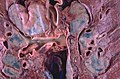
File:Sarkoidosis muscle.jpg | Muscle involvement in sarcoidosis. (WC/jensflorian) | |||
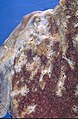
File:Sarcoidosis_histology_skin_involvement.jpg | Skin involvement in sarcoidosis. (WC/jensflorian) | |||
</gallery> | |||
====Case==== | |||
<gallery> | |||
Image: Sarcoidosis - lung FNA -- very low mag.jpg | Sarcoidosis lung - very low mag. (WC) | |||
Image: Sarcoidosis - lung FNA -- low mag.jpg | Sarcoidosis lung - low mag. (WC) | |||
Image: Sarcoidosis - lung FNA -- intermed mag.jpg | Sarcoidosis lung - intermed. mag. (WC) | |||
Image: Sarcoidosis - lung FNA -- high mag.jpg | Sarcoidosis lung - high mag. (WC) | |||
Image: Sarcoidosis - lung FNA -- very high mag.jpg | Sarcoidosis lung - very high mag. (WC) | |||
</gallery> | </gallery> | ||
www | ====www==== | ||
*[http://path.upmc.edu/cases/case412.html Sarcoidosis - several images (upmc.edu)]. | *[http://path.upmc.edu/cases/case412.html Sarcoidosis - several images (upmc.edu)]. | ||
*[http://path.upmc.edu/cases/case517.html Neurosarcoidosis - several images (upmc.edu)]. | *[http://path.upmc.edu/cases/case517.html Neurosarcoidosis - several images (upmc.edu)]. | ||
| Line 89: | Line 108: | ||
Note: | Note: | ||
*Must be done to exclude infection. | *Must be done to exclude infection. | ||
==Molecular== | |||
*PCR for Tuberculosis -ve. | |||
==Sign out== | ==Sign out== | ||
| Line 112: | Line 134: | ||
Sarcoidosis is favoured based on the morphology of the granulomas and the | Sarcoidosis is favoured based on the morphology of the granulomas and the | ||
lack of microorganisms with special stains (ZN, PASF, GMS). | lack of microorganisms with special stains (ZN, PASF, GMS). | ||
A serum ACE level should be considered, if not already done. | |||
Clinical and radiologic correlation is required. | Clinical and radiologic correlation is required. | ||
| Line 119: | Line 143: | ||
*[[Medical lung diseases]]. | *[[Medical lung diseases]]. | ||
*[[Cardiac sarcoidosis]]. | *[[Cardiac sarcoidosis]]. | ||
*[[Melkersson-Rosenthal syndrome]]. | |||
==References== | ==References== | ||
Latest revision as of 13:25, 18 October 2021
| Sarcoidosis | |
|---|---|
| Diagnosis in short | |
_lymph_node_biopsy.jpg) Sarcoidosis-like granulomas in a lymph node. H&E stain. | |
|
| |
| LM | well-formed granulomas often with few surrounding lymphocytes ("naked"), usually non-necrotizing |
| LM DDx | fungal infections, MAC, tuberculosis, other infections, drug reactions, reactive process to malignancy |
| Stains | AFB -ve, GMS -ve, PASD -ve |
| Molecular | PCR for tuberculosis -ve |
| Site | lung, hilar lymph nodes of the lung, skin, heart, other sites |
|
| |
| Prevalence | uncommon |
| Blood work | +/-ACE elevated |
| Radiology | +/-bilateral hilar lymphadenopathy (very common), +/-interstitial pattern, +/- pulmonary infiltrates, +/-cystic/bullous changes |
| Clin. DDx | lymphoma, metastatic carcinoma, Wegener's granulomatosis, others |
Sarcoidosis is non-necrotizing granulomatous disease of unknown etiology. It classically associated with (pulmonary) hilar lymphadenopathy. It may be found in almost any organ.
This article covers the topic in general and focuses on the lung aspects. Cardiac sarcoidosis is dealt with separately.
General
- Diagnosis of exclusion - infection, neoplasm, and drugs must be excluded.
- Uncommon.
- Afflicits skin ~25% of the time.[1]
Serology:
- Angiotensin-converting enzyme (ACE) - used for diagnosis and to monitor activity.[2][3]
- Elevated in approximately 65% of patients in one series.[4]
Gross
- Lungs - classic location.[5]
- Bilateral hilar lymphadenopathy.
DDx lungs - radiologic:
- Carcinomatosis - interstitial pattern.[6]
- Lymphoma - bilateral lymphadenopathy +/- mediastinal lymphadenopathy.[7]
Markedly enlarged bilaterally hilar lymph nodes in stage 1 sarcoidosis (WC/Yale Rosen)
Fibrosis (WC/Yale Rosen)
.jpg)
.jpg)
Microscopic
Features:
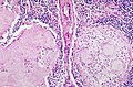
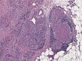
- Granulomata, well-formed, non-necrotizing. ‡
- Negative for microorganisms with special stains.
- Usu. minimal (lymphoid) inflammation; sarcoid granulomas are known as "naked granulomas".[8][1]
- In lung:
- Interstitial location +/- centrilobular.
Notes:
- ‡ Reported with necrosis - uncommon.[1]
DDx:
- Infections.
- Fungal.
- Mycobacterial, e.g. tuberculosis.
- Tertiary syphilis.[9]
- Hypersensitivity pneumonitis - usually poorly-formed granulomas, centrilobular, not paraseptal.[citation needed]
- Reaction to treatment/drug.
- Common variable immunodeficiency.[12]
- Reactive changes associated with tumours:
- All testicular tumours[13] - esp. seminoma.[14]
- Lung adenocarcinoma.[15]
Images
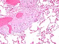
Sarcoidosis - lung. (WC)
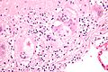
Granulomata in sarcoidosis with asteroid bodies. (WC)
Schaumann body in sarcoidosis. (WC/Yale Rosen)
Fibrosis in sarcoid granulomas. (WC/Yale Rosen)
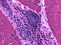
Muscle involvement in sarcoidosis. (WC/jensflorian)
Skin involvement in sarcoidosis. (WC/jensflorian)


.jpg)
.jpg)


Case
Sarcoidosis lung - very low mag. (WC)
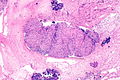
Sarcoidosis lung - low mag. (WC)
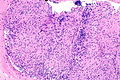
Sarcoidosis lung - intermed. mag. (WC)
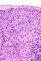
Sarcoidosis lung - high mag. (WC)
Sarcoidosis lung - very high mag. (WC)


www
Stains
Note:
- Must be done to exclude infection.
Molecular
- PCR for Tuberculosis -ve.
Sign out
- Should be something like sarcoid-like granulomas and clinical correlation required.
A. RIGHT LOWER LOBE LUNG, WEDGE RESECTION:
- GRANULOMATOUS INFLAMMATION, NON-NECROTIZING (SARCOID-LIKE), SEE COMMENT.

B. RIGHT MIDDLE LOBE LUNG, WEDGE RESECTION:
- GRANULOMATOUS INFLAMMATION, NON-NECROTIZING (SARCOID-LIKE), SEE COMMENT.

C. RIGHT LOWER LOBE LUNG, WEDGE RESECTION:
- GRANULOMATOUS INFLAMMATION, NON-NECROTIZING (SARCOID-LIKE), SEE COMMENT.

COMMENT:
The sections show multiple sarcoid-like granulomas with both centrilobular and
septal involvement. There is a slight upper lobe predominance of the disease.

The main histomorphologic differential diagnoses are: sarcoidosis,
infectious granulomatous pneumonia.

Sarcoidosis is favoured based on the morphology of the granulomas and the
lack of microorganisms with special stains (ZN, PASF, GMS).

A serum ACE level should be considered, if not already done.

Clinical and radiologic correlation is required.
See also
References
- ↑ 1.0 1.1 1.2 Noiles, K.; Beleznay, K.; Crawford, RI.; Au, S.. "Sarcoidosis can present with necrotizing granulomas histologically: two cases of ulcerated sarcoidosis and review of the literature.". J Cutan Med Surg 17 (6): 377-83. PMID 24138972.
- ↑ Kaura, V.; Kaura, NV.; Kaura, BN.; Kaura, CS.. "Angiotensin-converting enzyme inhibitors in the treatment of sarcoidosis and association with ACE gene polymorphism: case series.". Indian J Chest Dis Allied Sci 55 (2): 105-7. PMID 24047001.
- ↑ Stouten, K.; Werken, MV.; Tchetverikov, I.; Saboerali, M.; Vermeer, HJ.; Castel, R.; Verheijen, FM. (Jul 2013). "Extreme elevation of serum angiotensin-converting enzyme (ACE) activity: always consider familial ACE hyperactivity.". Ann Clin Biochem. doi:10.1177/0004563213489812. PMID 23897103.
- ↑ Shorr, AF.; Torrington, KG.; Parker, JM. (Aug 1997). "Serum angiotensin converting enzyme does not correlate with radiographic stage at initial diagnosis of sarcoidosis.". Respir Med 91 (7): 399-401. PMID 9327039.
- ↑ Rao, DA.; Dellaripa, PF. (May 2013). "Extrapulmonary manifestations of sarcoidosis.". Rheum Dis Clin North Am 39 (2): 277-97. doi:10.1016/j.rdc.2013.02.007. PMID 23597964.
- ↑ URL: http://www.radiologyassistant.nl/en/46b480a6e4bdc. Accessed on: 23 May 2010.
- ↑ Boujaoude, Z.; Dahdel, M.; Pratter, M.; Kass, J. (Jan 2012). "Endobronchial ultrasound with transbronchial needle aspiration in the diagnosis of bilateral hilar and mediastinal lymphadenopathy.". J Bronchology Interv Pulmonol 19 (1): 19-23. doi:10.1097/LBR.0b013e3182442b89. PMID 23207258.
- ↑ Brinster, NK. (Nov 2008). "Dermatopathology for the surgical pathologist: a pattern-based approach to the diagnosis of inflammatory skin disorders (part II).". Adv Anat Pathol 15 (6): 350-69. doi:10.1097/PAP.0b013e31818b1ac6. PMID 18948765.
- ↑ Hervier, B.; Wastiaux, H.; Freour, T.; Masseau, A.; Corvec, S.; Armingeat, T.; Hamidou, M. (Sep 2009). "[Sarcoidosis-like granulomatosis revealing a tertiary syphilis].". Rev Med Interne 30 (9): 806-8. doi:10.1016/j.revmed.2009.01.003. PMID 19249139.
- ↑ Tong, D.; Manolios, N.; Howe, G.; Spencer, D. (Jan 2012). "New onset sarcoid-like granulomatosis developing during anti-TNF therapy: an under-recognised complication.". Intern Med J 42 (1): 89-94. PMID 22389903.
- ↑ Reule, RB.; North, JP. (Nov 2013). "Cutaneous and pulmonary sarcoidosis-like reaction associated with ipilimumab.". J Am Acad Dermatol 69 (5): e272-3. doi:10.1016/j.jaad.2013.07.028. PMID 24124863.
- ↑ Vultaggio, A.; Matucci, A.; Parronchi, P.; Rossi, O.; Filì, L.; Giudizi, MG.; Palandri, F.; Agostini, C. et al. (Sep 2007). "Association between sarcoidosis-like disease and common variable immunodeficiency (CVI): a new CVI variant showing an activation of the immune system.". Sarcoidosis Vasc Diffuse Lung Dis 24 (2): 127-33. PMID 18496983.
- ↑ Paparel, P.; Devonec, M.; Perrin, P.; Ruffion, A.; Decaussin-Petrucci, M.; Akin, O.; Sheinfeld, J.; Guillonneau, B. (Sep 2007). "Association between sarcoidosis and testicular carcinoma: a diagnostic pitfall.". Sarcoidosis Vasc Diffuse Lung Dis 24 (2): 95-101. PMID 18496978.
- ↑ Jankilevich, G.; Mendizabal, J.; Massa, MA.; Pedernera, A.; Galmes, M.; Spizzamiglio, N. (2006). "[Mediastinal sarcoidal reaction in follow up for seminoma].". Medicina (B Aires) 66 (6): 552-4. PMID 17240627.
- ↑ Tao, H.; Yamamoto, H.; Matsuda, E.; Sano, F.; Okabe, K.; Sugi, K. (Apr 2012). "Severe bronchoconstriction due to sarcoid-like reaction to lung cancer.". Asian Cardiovasc Thorac Ann 20 (2): 199-201. doi:10.1177/0218492311431493. PMID 22499972.