Difference between revisions of "Radiation colitis"
Jump to navigation
Jump to search
m |
|||
| (4 intermediate revisions by the same user not shown) | |||
| Line 1: | Line 1: | ||
'''Radiation [[colitis]]''' is inflammation of the [[colon]] due to radiation. It is usually iatrogenic. This article also covers '''radiation proctitis'''. | {{ Infobox diagnosis | ||
| Name = {{PAGENAME}} | |||
| Image = Radiation_proctitis_-_2_alt_--_high_mag.jpg | |||
| Width = | |||
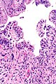
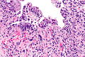
| Caption = Radiation proctitis. [[H&E stain]]. | |||
| Synonyms = | |||
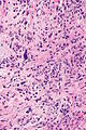
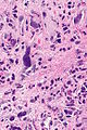
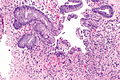
| Micro = ''acute'': mucosal changes ([[necrosis]] of epithelium, "ghost cells" = cells without nuclei, hemorrhage), submucosa edema with neutrophilic infiltrate, +/-fibrin thrombi; ''chronic'': nuclear atypia - esp. of the stromal cells, fibrosis - esp. of the submucosa | |||
| Subtypes = | |||
| LMDDx = [[inflammatory bowel disease]], [[Infectious colitis]], [[ischemic colitis]], pseudosarcomatous stromal changes, [[sarcoma]] | |||
| Stains = | |||
| IHC = | |||
| EM = | |||
| Molecular = | |||
| IF = | |||
| Gross = | |||
| Grossing = | |||
| Site = [[colon]] and [[rectum]] (''radiation proctitis'') | |||
| Assdx = | |||
| Syndromes = | |||
| Clinicalhx = history of radiation, history of [[cancer]] | |||
| Signs = | |||
| Symptoms = | |||
| Prevalence = uncommon | |||
| Bloodwork = | |||
| Rads = | |||
| Endoscopy = | |||
| Prognosis = | |||
| Other = | |||
| ClinDDx = cancer recurrence, colitis/proctitis (idiopathic, infection, ischemic, radiation) | |||
| Tx = | |||
}} | |||
'''Radiation [[colitis]]''' is inflammation of the [[colon]] due to radiation. It is usually iatrogenic. This article also covers '''radiation proctitis''' (abbreviated '''RP'''). | |||
==General== | ==General== | ||
| Line 14: | Line 45: | ||
*Telangiectatic lesions. | *Telangiectatic lesions. | ||
Image | ===Image=== | ||
*[http://www.nature.com/nrgastro/journal/v5/n1/fig_tab/ncpgasthep1005_F4.html#figure-title Telangiectatic lesions (nature.com)].<ref name=pmid18174905>{{Cite journal | last1 = Nielsen | first1 = OH. | last2 = Vainer | first2 = B. | last3 = Rask-Madsen | first3 = J. | title = Non-IBD and noninfectious colitis. | journal = Nat Clin Pract Gastroenterol Hepatol | volume = 5 | issue = 1 | pages = 28-39 | month = Jan | year = 2008 | doi = 10.1038/ncpgasthep1005 | PMID = 18174905 }}</ref> | *[http://www.nature.com/nrgastro/journal/v5/n1/fig_tab/ncpgasthep1005_F4.html#figure-title Telangiectatic lesions (nature.com)].<ref name=pmid18174905>{{Cite journal | last1 = Nielsen | first1 = OH. | last2 = Vainer | first2 = B. | last3 = Rask-Madsen | first3 = J. | title = Non-IBD and noninfectious colitis. | journal = Nat Clin Pract Gastroenterol Hepatol | volume = 5 | issue = 1 | pages = 28-39 | month = Jan | year = 2008 | doi = 10.1038/ncpgasthep1005 | PMID = 18174905 }}</ref> | ||
==Microscopic== | ==Microscopic== | ||
| Line 35: | Line 66: | ||
*Infectious colitis. | *Infectious colitis. | ||
Images: | ===Images=== | ||
<gallery> | |||
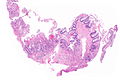
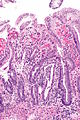
Image: Radiation proctitis -- very low mag.jpg | RP - very low mag. | |||
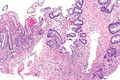
Image: Radiation proctitis -- low mag.jpg | RP - low mag. | |||
Image: Radiation proctitis - alt -- low mag.jpg | RP - low mag. | |||
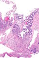
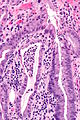
Image: Radiation proctitis -- intermed mag.jpg | RP - intermed. mag. | |||
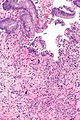
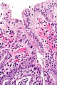
Image: Radiation proctitis -- high mag.jpg | RP - high mag. | |||
Image: Radiation proctitis -- very high mag.jpg | RP - very high mag. | |||
Image: Radiation proctitis - 2 -- intermed mag.jpg | RP - intermed. mag. | |||
Image: Radiation proctitis - 2 -- high mag.jpg | RP - high mag. | |||
Image: Radiation proctitis - 2 alt -- high mag.jpg | RP - high mag. | |||
Image: Proctitis with reactive changes -- intermed mag.jpg | PRC - intermed. mag. | |||
Image: Proctitis with reactive changes -- high mag.jpg | PRC - high mag. | |||
Image: Proctitis with reactive changes - alt -- high mag.jpg | PRC - high mag. | |||
</gallery> | |||
www: | |||
*[http://gut.bmj.com/content/41/3/354/F2.large.jpg Radiation colitis - rat model (bmj.com)].<ref name=pmid9378391/> | *[http://gut.bmj.com/content/41/3/354/F2.large.jpg Radiation colitis - rat model (bmj.com)].<ref name=pmid9378391/> | ||
*[http://www.nature.com/nrgastro/journal/v5/n1/fig_tab/ncpgasthep1005_F5.html#figure-title Radiation colitis (nature.com)].<ref name=pmid18174905>{{Cite journal | last1 = Nielsen | first1 = OH. | last2 = Vainer | first2 = B. | last3 = Rask-Madsen | first3 = J. | title = Non-IBD and noninfectious colitis. | journal = Nat Clin Pract Gastroenterol Hepatol | volume = 5 | issue = 1 | pages = 28-39 | month = Jan | year = 2008 | doi = 10.1038/ncpgasthep1005 | PMID = 18174905 }}</ref> | *[http://www.nature.com/nrgastro/journal/v5/n1/fig_tab/ncpgasthep1005_F5.html#figure-title Radiation colitis (nature.com)].<ref name=pmid18174905>{{Cite journal | last1 = Nielsen | first1 = OH. | last2 = Vainer | first2 = B. | last3 = Rask-Madsen | first3 = J. | title = Non-IBD and noninfectious colitis. | journal = Nat Clin Pract Gastroenterol Hepatol | volume = 5 | issue = 1 | pages = 28-39 | month = Jan | year = 2008 | doi = 10.1038/ncpgasthep1005 | PMID = 18174905 }}</ref> | ||
==Sign out== | |||
<pre> | |||
RECTUM, BIOPSY: | |||
- RECTAL MUCOSA WITH ACTIVE INFLAMMATION, ULCERATION AND | |||
REGENERATIVE CHANGES. | |||
- LARGE ATYPICAL STROMAL CELLS COMPATIBLE WITH RADIATION CHANGES. | |||
- NO EVIDENCE OF DYSPLASIA AND NO EVIDENCE OF MALIGNANCY. | |||
COMMENT: | |||
The immunostains done (pankeratin, p53, Ki-67) are in keeping | |||
with regeneration and radiation changes. | |||
</pre> | |||
==See also== | ==See also== | ||
Latest revision as of 04:56, 28 December 2014
| Radiation colitis | |
|---|---|
| Diagnosis in short | |
 Radiation proctitis. H&E stain. | |
|
| |
| LM | acute: mucosal changes (necrosis of epithelium, "ghost cells" = cells without nuclei, hemorrhage), submucosa edema with neutrophilic infiltrate, +/-fibrin thrombi; chronic: nuclear atypia - esp. of the stromal cells, fibrosis - esp. of the submucosa |
| LM DDx | inflammatory bowel disease, Infectious colitis, ischemic colitis, pseudosarcomatous stromal changes, sarcoma |
| Site | colon and rectum (radiation proctitis) |
|
| |
| Clinical history | history of radiation, history of cancer |
| Prevalence | uncommon |
| Clin. DDx | cancer recurrence, colitis/proctitis (idiopathic, infection, ischemic, radiation) |
Radiation colitis is inflammation of the colon due to radiation. It is usually iatrogenic. This article also covers radiation proctitis (abbreviated RP).
General
- Diagnosis should be supported by the clinical history.
General DDx for a colitis:
- Idiopathic, e.g. inflammatory bowel disease.
- Infection.
- Ischemia.
- Radiation.
Gross
- Superficial bowel wall injury - shallow ulceration.[1]
- Telangiectatic lesions.
Image
Microscopic
Features - acute:[1]
- Mucosal changes:
- Necrosis of epithelium.
- "Ghost cells" = cells without nuclei.
- Hemorrhage.
- Necrosis of epithelium.
- Submucosa edema with neutrophilic infiltrate.
- +/-Fibrin thrombi.
Features - chronic:
- Nuclear atypia - esp. of the stromal cells.
- The epithelium is shed and regenerates... therefore usually does not have the changes.
- Fibrosis - esp. of the submucosa.
DDx:
- Ischemic colitis.
- Inflammatory bowel disease.
- Infectious colitis.
Images
RP - very low mag.
RP - low mag.
RP - low mag.
RP - intermed. mag.
RP - high mag.
RP - very high mag.
RP - intermed. mag.
RP - high mag.
RP - high mag.
PRC - intermed. mag.
PRC - high mag.
PRC - high mag.


www:
Sign out
RECTUM, BIOPSY:
- RECTAL MUCOSA WITH ACTIVE INFLAMMATION, ULCERATION AND
  REGENERATIVE CHANGES.
- LARGE ATYPICAL STROMAL CELLS COMPATIBLE WITH RADIATION CHANGES.
- NO EVIDENCE OF DYSPLASIA AND NO EVIDENCE OF MALIGNANCY.

COMMENT:
The immunostains done (pankeratin, p53, Ki-67) are in keeping
with regeneration and radiation changes.
See also
References
- ↑ 1.0 1.1 1.2 Yano, Y.; Yao, H.; Aoyagi, K.; Kawakubo, K.; Nakamura, S.; Doi, K.; Ibayashi, S.; Fujishima, M. (Sep 1997). "Photochemically induced colonic ischaemic lesions: a new model of ischaemic colitis in rats.". Gut 41 (3): 354-7. PMID 9378391.
- ↑ 2.0 2.1 Nielsen, OH.; Vainer, B.; Rask-Madsen, J. (Jan 2008). "Non-IBD and noninfectious colitis.". Nat Clin Pract Gastroenterol Hepatol 5 (1): 28-39. doi:10.1038/ncpgasthep1005. PMID 18174905.