Difference between revisions of "Pneumonia"
Jump to navigation
Jump to search
(+CMV pneumonia) |
|||
| Line 47: | Line 47: | ||
===Gross pathology=== | ===Gross pathology=== | ||
*Consolidation (the lung parenchyma is firm) - best appreciated by running a finger over the cut surface of the lung with a small-to-moderate amount of pressure. | *Consolidation (the lung parenchyma is firm) - best appreciated by running a finger over the cut surface of the lung with a small-to-moderate amount of pressure. | ||
Lobar pneumnia is classically described in four stages:<ref>{{Ref AoGP|92}}</ref><ref>URL: [http://www.histopathology-india.net/Lobar_Pneumonia.htm http://www.histopathology-india.net/Lobar_Pneumonia.htm]. Accessed on: 27 February 2012.</ref> | |||
#Congestion - day 1-2. | |||
#Red hepatization - day 2-4. | |||
#Gray hepatization - day 4-6. | |||
#Resolution - day 6+. | |||
Note: | |||
*The above is considered historical. In the age of antibiotics, lobar pneumonia is uncommon. | |||
===Microscopic=== | ===Microscopic=== | ||
Revision as of 02:17, 28 February 2012
Pneumonia is inflammation of the lung and grouped with the medical lung diseases.
There are various types of pneumonia.
Infectious pnemonia
Anatomical classification of pneumonia
- Generally, not used by clinicians.
- Use of the terms without qualification is discouraged... as they do not make explicit the etiology.
Bronchopneumonia
- Multiple foci of (acute) inflammation involving the bronchi.
- This is the most common form of (infectious) pneumonia.
Lobar pneumonia
- Pneumonia that involves a whole lobe.
- Rarely seen in areas where antibiotic treatments are widely available.
Acute infectious pneumonia
General
- This is seen by pathologists, in autopsy, from time-to-time.
Most common cause:
- Streptococcus pneumoniae.[1]
The top three community acquired (acute) pneumonia:[2]
- Streptococcuc pneumonia.
- Haemophilus influenzae.
- Moraxella catarrhalis.
Other community acquired pneumonia:[1]
- S. aureus.
- Legionaella pneumophila.
- Klebsiella pneumoniae.
- Pseudomonas.
Hospital-acquired pneumonia:[1]
- Gram-negative rods.
- Staphylococcus aureus.
Radiologic correlate
- Air space disease.
Gross pathology
- Consolidation (the lung parenchyma is firm) - best appreciated by running a finger over the cut surface of the lung with a small-to-moderate amount of pressure.
Lobar pneumnia is classically described in four stages:[3][4]
- Congestion - day 1-2.
- Red hepatization - day 2-4.
- Gray hepatization - day 4-6.
- Resolution - day 6+.
Note:
- The above is considered historical. In the age of antibiotics, lobar pneumonia is uncommon.
Microscopic
Features:
- Alveoli packed with PMNs.
- +/-Clusters of bacteria - small dots or rods.
Image: Normal alveoli & pneumonia (WC).
Stains
- Gram stain -- to type the bacteria.
Chronic infectious pneumonia
General
Common microorganisms:[1]
- Nocardia.
- Actinomyces.
- Mycobacterium tuberculosis.
- Atypical mycobacterium, e.g. Mycobacterium avium-intracellulare.
- Histoplasma capsulatum.
- Coccidioides immitis.
- Blastomyces dermatitidis.
Note:
- All of the later ones are granulomatous.
Microscopic
Features:
- Inflammation.
- +/-Granulomas.
Aspiration pneumonia
General
- Usually seen in the context of a toxin and/or pathology that affects the swallowing and cough reflexes.[5]
- Stroke.
- Multiple sclerosis.
- Alcohol.
- The microorganisms involved are usually different than in other causes of acute pneumonia.
Gross
- More common in the right lung.
- Right main stem bronchus is more vertical.
Microscopic
Features:
- +/-Foreign body giant cells.
- Microorganisms.
Images:
Cytomegalovirus pneumonia
Main article: Cytomegalovirus
General
- Immunodeficiency.
- Critical illness.[6]
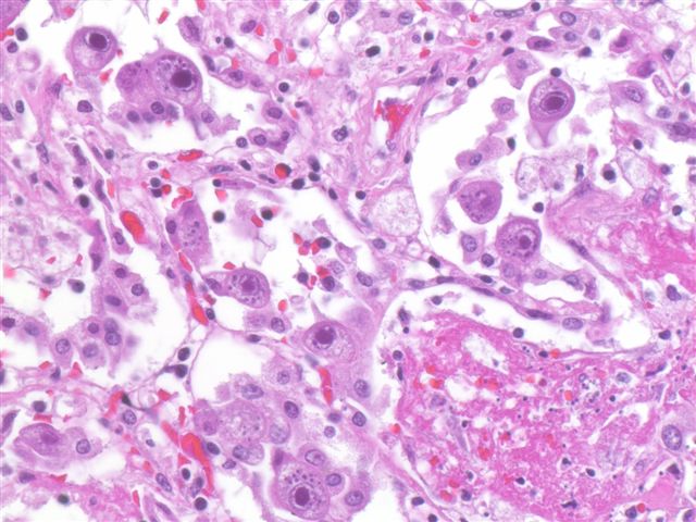
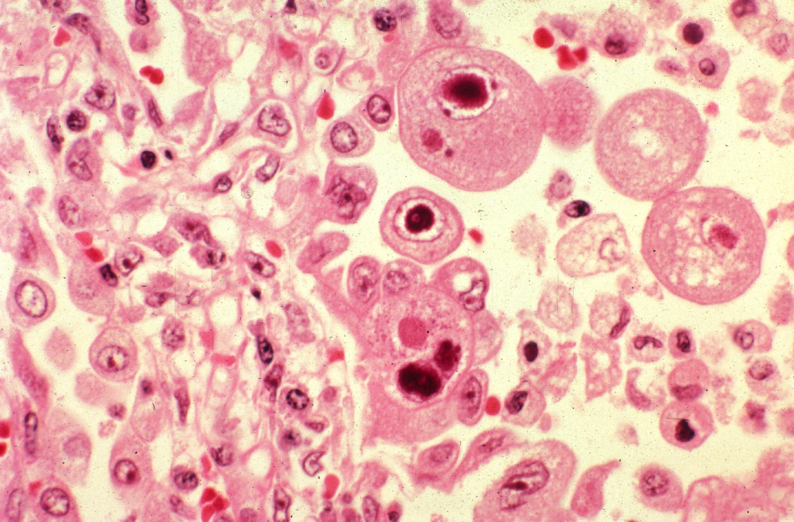
Microscopic
Features:
- CMV nuclear changes:
- Large red nucleus with a pale halo.
- Eosinophilic granular cytoplasmic inclusions.
Images:
IHC
- CMV +ve -- cytoplasmic inclusions, large nucleus.
Diffuse lung diseases
Main article: Diffuse lung disease
- AKA idiopathic interstitial pneumonia.
Histologic pattern:
- Organizing pneumonia.
- Usual interstitial pneumonia.
- Nonspecific interstitial pneumonia.
- Lymphocytic interstitial pneumonia.
- Desquamative interstitial pneumonia.
- Diffuse alveolar damage.
See also
References
- ↑ 1.0 1.1 1.2 1.3 Kumar, Vinay; Abbas, Abul K.; Fausto, Nelson; Aster, Jon (2009). Robbins and Cotran pathologic basis of disease (8th ed.). Elsevier Saunders. pp. 711. ISBN 978-1416031215.
- ↑ Nicolau, D. (Sep 2002). "Clinical and economic implications of antimicrobial resistance for the management of community-acquired respiratory tract infections.". J Antimicrob Chemother 50 Suppl S1: 61-70. PMID 12239229.
- ↑ Rose, Alan G. (2008). Atlas of Gross Pathology with Histologic Correlation (1st ed.). Cambridge University Press. pp. 92. ISBN 978-0521868792.
- ↑ URL: http://www.histopathology-india.net/Lobar_Pneumonia.htm. Accessed on: 27 February 2012.
- ↑ Ohrui, T. (Sep 2005). "Preventive strategies for aspiration pneumonia in elderly disabled persons.". Tohoku J Exp Med 207 (1): 3-12. PMID 16082150.
- ↑ Limaye, AP.; Boeckh, M. (Nov 2010). "CMV in critically ill patients: pathogen or bystander?". Rev Med Virol 20 (6): 372-9. doi:10.1002/rmv.664. PMID 20931610.
- ↑ URL: http://www.pathologyoutlines.com/topic/lungnontumorCMV.html. Accessed on: 23 January 2012.