Difference between revisions of "Placenta"
(→Microscopy: wikify) |
(re-order/group) |
||
| Line 1: | Line 1: | ||
The '''placenta''' feeds the developing baby, breathes for it and disposes of its waste. | The '''placenta''' feeds the developing baby, breathes for it and disposes of its waste. | ||
==Normal== | =Clinical= | ||
==Examination of the placenta== | |||
*Most placentas are ''not'' examined by a pathologist. | |||
Indications for exam by a pathologist: | |||
*Abnormalities in the: | |||
*#Fetus: | |||
*#*Bad fetal outcome. | |||
*#*Suspected or known congenital abnormalities ''or'' chromosomal abnormalities. | |||
*#Mother: | |||
*#*Infection/suspected infection. | |||
*#*Pre-term labour. | |||
*#*Maternal disease (e.g. SLE, coagulopathy). | |||
*#*Complicated pregnancy (preclampsia, pregnancy induced hypertension, gestational diabetes). | |||
*#Placenta: | |||
*#*Unusual gross characteristics. | |||
==Bleeding in late pregnancy== | |||
DDx of bleeding in late pregnancy: | |||
*Placental abruption (most common). | |||
*Placenta previa. | |||
*Vasa previa (fetus losing blood). | |||
==Clinical screening tests== | |||
*PAPP-A - low values seen in aneuploidy.<ref>URL: [http://www.ncbi.nlm.nih.gov/sites/entrez?Db=gene&Cmd=ShowDetailView&TermToSearch=5069 http://www.ncbi.nlm.nih.gov/sites/entrez?Db=gene&Cmd=ShowDetailView&TermToSearch=5069]. Accessed on: 7 July 2010.</ref> | |||
{{main|Pregnancy}} | |||
=Normal histology= | |||
==Amnion== | |||
General: | |||
*Next to fetus, surrounds amniotic fluid, avascular. | |||
Characteristics: | |||
*Characterized by a single layer of cells.<ref name=Ref_H4P2_974>{{Ref H4P2|974}}</ref> | *Characterized by a single layer of cells.<ref name=Ref_H4P2_974>{{Ref H4P2|974}}</ref> | ||
**Cuboidal/squamoid shape. | **Cuboidal/squamoid shape. | ||
| Line 12: | Line 44: | ||
*'Fibroblastic layer'.<ref name=Ref_H4P2_974>{{Ref H4P2|974}}</ref> | *'Fibroblastic layer'.<ref name=Ref_H4P2_974>{{Ref H4P2|974}}</ref> | ||
Chorion | ==Chorion== | ||
General: | |||
*Surrounds amnion. | |||
Characteristics: | |||
*Layers:<ref name=Ref_H4P2_977>{{Ref H4P2|977}}</ref> | *Layers:<ref name=Ref_H4P2_977>{{Ref H4P2|977}}</ref> | ||
**'Reticular layer' - cellular (inner aspect). | **'Reticular layer' - cellular (inner aspect). | ||
| Line 21: | Line 57: | ||
**Beneath of the "trophoblastic X cells" is ''decidua'' (mnemonic ''NEW'' = nucleus central, eosinophilic, well-defined cell border), which is maternal tissue. | **Beneath of the "trophoblastic X cells" is ''decidua'' (mnemonic ''NEW'' = nucleus central, eosinophilic, well-defined cell border), which is maternal tissue. | ||
== | ==Common terms== | ||
*Chorionic plate - fetal aspect of placenta. | *Chorionic plate - fetal aspect of placenta. | ||
*Basal plate - maternal aspect of placenta. | *Basal plate - maternal aspect of placenta. | ||
| Line 27: | Line 63: | ||
**Place to look for maternal vessels. | **Place to look for maternal vessels. | ||
== | =Grossing= | ||
This is often very quick. The gross is quite important, as some things cannot be diagnosed microscopically. | |||
==General== | |||
*Dimensions: | *Dimensions: | ||
**Disc. | **Disc. | ||
| Line 50: | Line 88: | ||
**Maternal surface - are the cotyledons intact? | **Maternal surface - are the cotyledons intact? | ||
==Sections== | |||
*Cord two sections. | *Cord two sections. | ||
*Cord at insertion. | *Cord at insertion. | ||
| Line 56: | Line 94: | ||
*Placenta - full thickness (maternal and fetal surface). | *Placenta - full thickness (maternal and fetal surface). | ||
==Placental membranes== | |||
Appearance:<ref name=Ref_Lester461>{{Ref Lester|461}}</ref> | Appearance:<ref name=Ref_Lester461>{{Ref Lester|461}}</ref> | ||
*Normal - shiny. | *Normal - shiny. | ||
| Line 66: | Line 104: | ||
**Gross: - (single) yellow patch or yellow nodules . | **Gross: - (single) yellow patch or yellow nodules . | ||
==Sign-out | ==Placental mass== | ||
Placental mass by gestational age:<ref>AFIP Placental pathol. ISBN: 1-881041-89-1. P.312</ref> | |||
{| class="wikitable" | |||
|Gest. Age/Percentile ||'''25%''' ||'''50%''' ||'''75%''' | |||
|- | |||
|'''32 weeks''' ||275 g ||318 g ||377 g | |||
|- | |||
|'''36 weeks''' ||369 g ||440 g ||508 g | |||
|- | |||
|'''40 weeks''' ||440 g ||501 g ||572 g | |||
|- | |||
|} | |||
===Linear regression - placental mass-gestational age=== | |||
Based on the table in the AFIP book<ref>AFIP Placental pathol. ISBN: 1-881041-89-1. P.312</ref> I generated the following regression lines: | |||
{| class="wikitable" | |||
| ||'''50%''' ||'''10%''' ||'''90%''' | |||
|- | |||
|slope (g/week) ||21.58088235 ||19.70588235 ||25.40196078 | |||
|- | |||
|y-intercept (g) ||-357.4558824 ||-397.2352941 ||-366.7254902 | |||
|- | |||
|Pearson (r) ||0.988670724 ||0.988268672 ||0.982206408 | |||
|- | |||
|} | |||
placental mass = slope x gestational age + intercept | |||
===What to remember...=== | |||
Extrapolated from the linear regression (see above): | |||
*50% at term = 500 grams. | |||
*50% at 26 weeks = 200 grams. | |||
*The change in mass/week is approximately linear and equal to 300 grams / 14 weeks ~ 20 grams/week. | |||
*The spread in mass between 10% and 90%, crudely estimated, is 200 grams (for GA=26-40). | |||
=Sign-out= | |||
What should be commented on... | What should be commented on... | ||
| Line 86: | Line 159: | ||
Mnemonic: ''chorio, cord, vessels, villi (maturity, infarction)''. | Mnemonic: ''chorio, cord, vessels, villi (maturity, infarction)''. | ||
=Twin placentas= | |||
These are often submitted... even if they are normal. | |||
==General== | |||
No membrane between fetuses. | No membrane between fetuses. | ||
*Split at approx. 7th day. | *Split at approx. 7th day. | ||
| Line 99: | Line 175: | ||
*If monozygotic -- split before 3 days. | *If monozygotic -- split before 3 days. | ||
== | =Diseases of the placental attachment= | ||
==Placenta acreta/percreta/increta== | |||
Placenta attaches to the uterus deeper than it should. | |||
==Placental abruption== | ==Placental abruption== | ||
| Line 120: | Line 194: | ||
*There are '''no''' good microscopic findings for placental abruption. | *There are '''no''' good microscopic findings for placental abruption. | ||
=Infection= | |||
==General<ref name=Ref_PBoD1106>{{Ref PBoD|1106}}</ref>== | |||
*Infection usually ascending, i.e. from vagina up through cervix. | *Infection usually ascending, i.e. from vagina up through cervix. | ||
**Assoc. with intercourse. | **Assoc. with intercourse. | ||
| Line 134: | Line 208: | ||
*Placenta: placentitis, villitis. | *Placenta: placentitis, villitis. | ||
==Grading infection (chorioamnionitis, membranitis, funisitis)== | |||
Membranitis:<ref name=Ref_Sternberg4_2311>{{Ref Sternberg4|2311}}</ref> | Membranitis:<ref name=Ref_Sternberg4_2311>{{Ref Sternberg4|2311}}</ref> | ||
# PMNs - decidua only. | # PMNs - decidua only. | ||
| Line 160: | Line 234: | ||
Note: There is no such thing as ''chorionitis''.<ref>ALS. February 2009.</ref> | Note: There is no such thing as ''chorionitis''.<ref>ALS. February 2009.</ref> | ||
== | =Infarction= | ||
==True infarcts== | |||
===General=== | ===General=== | ||
*Associated with retroplacental hematoma. | *Associated with retroplacental hematoma. | ||
| Line 173: | Line 248: | ||
*[http://library.med.utah.edu/WebPath/PLACHTML/PLAC044.html Placental infarcts (med.utah.edu)]. | *[http://library.med.utah.edu/WebPath/PLACHTML/PLAC044.html Placental infarcts (med.utah.edu)]. | ||
=== | ===Microscopic=== | ||
Features: | Features: | ||
*Loss of intervillous space.<ref name=Ref_WMSP465>{{Ref WMSP|465}}</ref> | *Loss of intervillous space.<ref name=Ref_WMSP465>{{Ref WMSP|465}}</ref> | ||
| Line 191: | Line 266: | ||
*> 3cm --or-- central location --or-- in 1st or 2nd trimester. | *> 3cm --or-- central location --or-- in 1st or 2nd trimester. | ||
**Small foci are accepted in term placentae - typically at periphery. | **Small foci are accepted in term placentae - typically at periphery. | ||
==Perivillous fibrin deposition== | ==Perivillous fibrin deposition== | ||
| Line 224: | Line 281: | ||
**Obliteration of intervillous space. | **Obliteration of intervillous space. | ||
=Other= | |||
==Passage of meconium== | ==Passage of meconium== | ||
===General=== | ===General=== | ||
| Line 262: | Line 319: | ||
*Meconium contains bile.<ref>{{cite journal |author=Sienko A, Altshuler G |title=Meconium-induced umbilical vascular necrosis in abortuses and fetuses: a histopathologic study for cytokines |journal=Obstet Gynecol |volume=94 |issue=3 |pages=415?0 |year=1999 |month=September |pmid=10472870 |doi= |url=}}</ref> | *Meconium contains bile.<ref>{{cite journal |author=Sienko A, Altshuler G |title=Meconium-induced umbilical vascular necrosis in abortuses and fetuses: a histopathologic study for cytokines |journal=Obstet Gynecol |volume=94 |issue=3 |pages=415?0 |year=1999 |month=September |pmid=10472870 |doi= |url=}}</ref> | ||
== | =Maternal disease= | ||
==Hypertensive changes== | ==Hypertensive changes== | ||
Features:<ref name=pmid6754249>{{cite journal |author=Soma H, Yoshida K, Mukaida T, Tabuchi Y |title=Morphologic changes in the hypertensive placenta |journal=Contrib Gynecol Obstet |volume=9 |issue= |pages=58–75 |year=1982 |pmid=6754249 |doi= |url=}}</ref> | Features:<ref name=pmid6754249>{{cite journal |author=Soma H, Yoshida K, Mukaida T, Tabuchi Y |title=Morphologic changes in the hypertensive placenta |journal=Contrib Gynecol Obstet |volume=9 |issue= |pages=58–75 |year=1982 |pmid=6754249 |doi= |url=}}</ref> | ||
| Line 344: | Line 367: | ||
**In essence: severe ''hypertrophic decidual vasculopathy''. (???) | **In essence: severe ''hypertrophic decidual vasculopathy''. (???) | ||
=Tumours= | |||
{{main|Gestational trophoblastic disease}} | {{main|Gestational trophoblastic disease}} | ||
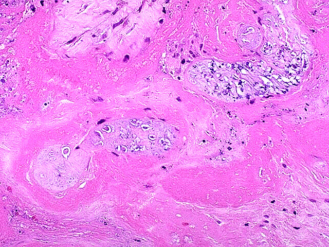
== | ==Chorangioma== | ||
* | ===General=== | ||
*[[Hamartoma]]-like growth in the placenta consisting of [[blood vessel]]s.<ref name=pmid20594143>{{cite journal |author=Amer HZ, Heller DS |title=Chorangioma and related vascular lesions of the placenta--a review |journal=Fetal Pediatr Pathol |volume=29 |issue=4 |pages=199–206 |year=2010 |pmid=20594143 |doi=10.3109/15513815.2010.487009 |url=}}</ref> | |||
===Epidemiology=== | |||
*Often benign. | |||
*May be association with: | |||
**Fetal maternal haemorrhage. | |||
**Hydrops. | |||
**[[IUGR]]. | |||
===Microscopy=== | |||
Features: | |||
*Mass of capillaries. | |||
Image: | |||
*[http://commons.wikimedia.org/wiki/File:Chorangioma_-_intermed_mag.jpg Chorangioma (WC)]. | |||
=See also= | |||
*[[Chorionic villi]]. | *[[Chorionic villi]]. | ||
*[[Endometrium]]. | *[[Endometrium]]. | ||
*[[Pregnancy]]. | *[[Pregnancy]]. | ||
=References= | |||
{{reflist|2}} | {{reflist|2}} | ||
[[Category:Gynecology]] | [[Category:Gynecology]] | ||
Revision as of 18:23, 8 January 2011
The placenta feeds the developing baby, breathes for it and disposes of its waste.
Clinical
Examination of the placenta
- Most placentas are not examined by a pathologist.
Indications for exam by a pathologist:
- Abnormalities in the:
- Fetus:
- Bad fetal outcome.
- Suspected or known congenital abnormalities or chromosomal abnormalities.
- Mother:
- Infection/suspected infection.
- Pre-term labour.
- Maternal disease (e.g. SLE, coagulopathy).
- Complicated pregnancy (preclampsia, pregnancy induced hypertension, gestational diabetes).
- Placenta:
- Unusual gross characteristics.
- Fetus:
Bleeding in late pregnancy
DDx of bleeding in late pregnancy:
- Placental abruption (most common).
- Placenta previa.
- Vasa previa (fetus losing blood).
Clinical screening tests
- PAPP-A - low values seen in aneuploidy.[1]
Normal histology
Amnion
General:
- Next to fetus, surrounds amniotic fluid, avascular.
Characteristics:
- Characterized by a single layer of cells.[2]
- Cuboidal/squamoid shape.
- Eosinophilic cytoplasm.
- Central nucleus.
- Squamous metaplasia may be seen at cord insertion.
- Basement membrane.
- 'Compact layer'.[2]
- 'Fibroblastic layer'.[2]
Chorion
General:
- Surrounds amnion.
Characteristics:
- Layers:[3]
- 'Reticular layer' - cellular (inner aspect).
- 'Pseudo-basemement membrane'.
- 'Outer trophoblastic layer'.
- Has blood vessels.
- Opposed to "trophoblastic X cells" on side opposite of amnion.[2]
- Beneath of the "trophoblastic X cells" is decidua (mnemonic NEW = nucleus central, eosinophilic, well-defined cell border), which is maternal tissue.
Common terms
- Chorionic plate - fetal aspect of placenta.
- Basal plate - maternal aspect of placenta.
- Has extravillous trophoblast.
- Place to look for maternal vessels.
Grossing
This is often very quick. The gross is quite important, as some things cannot be diagnosed microscopically.
General
- Dimensions:
- Disc.
- Length of cord, diameter of cord.
- Mass (weight) -- should be done 'trimmed' (cord cut-off, membrane cut-off).
- Umbilical cord
- Attachment.
- Location: central, eccentric, marginal.
- Marginal attachment assoc. with hypertension[4]
- Membranous or velamentous (veil-like) insertion.
- Vessels separate/branch prior to reaching placental disc.
- Furcate insertion - vessel run on fetal surface (more exposed to trauma).
- Location: central, eccentric, marginal.
- Knots (false vs. true).
- False knots are nothing to worry about -- look like a knot but aren't really one.
- Twisting/coiling.
- Number of vessels.
- Normal: 2 arteries, 1 vein.
- Attachment.
- Membranes - shiny, thin, translucent
- Attachment: marginal (normal), circummarginate (inside edge), circumvallated (folding on self).
- Placental disc.
- Fetal surface - normal is shinny (dull in chorioamnionitis).
- Maternal surface - are the cotyledons intact?
Sections
- Cord two sections.
- Cord at insertion.
- Membranes (rolled).
- Placenta - full thickness (maternal and fetal surface).
Placental membranes
Appearance:[5]
- Normal - shiny.
- Choriomnionitis - opaque/dull.
- Meconium - green.
- Amnion nodosum.
Placental mass
Placental mass by gestational age:[8]
| Gest. Age/Percentile | 25% | 50% | 75% |
| 32 weeks | 275 g | 318 g | 377 g |
| 36 weeks | 369 g | 440 g | 508 g |
| 40 weeks | 440 g | 501 g | 572 g |
Linear regression - placental mass-gestational age
Based on the table in the AFIP book[9] I generated the following regression lines:
| 50% | 10% | 90% | |
| slope (g/week) | 21.58088235 | 19.70588235 | 25.40196078 |
| y-intercept (g) | -357.4558824 | -397.2352941 | -366.7254902 |
| Pearson (r) | 0.988670724 | 0.988268672 | 0.982206408 |
placental mass = slope x gestational age + intercept
What to remember...
Extrapolated from the linear regression (see above):
- 50% at term = 500 grams.
- 50% at 26 weeks = 200 grams.
- The change in mass/week is approximately linear and equal to 300 grams / 14 weeks ~ 20 grams/week.
- The spread in mass between 10% and 90%, crudely estimated, is 200 grams (for GA=26-40).
Sign-out
What should be commented on...
- Placenta:
- Maturity of villi (2nd or 3rd trimester).
- Infarction?
- Subchorionic less important than maternal aspect.
- Peripheral aspect of placental disc less important than central region of disc.
- Blood vessels.
- Maternal.
- Fetal.
- Membranes.
- Membranitis?
- Chorioamnionitis?
- Cord:
- 3 vessel?
- Vasculitis/inflammation?
Mnemonic: chorio, cord, vessels, villi (maturity, infarction).
Twin placentas
These are often submitted... even if they are normal.
General
No membrane between fetuses.
- Split at approx. 7th day.
Diamnionic-monochorionic (DiMo)
- No interposed chorion.[10]
- Always monozygotic.
- Highest risk of TTTS (twin-to-twin transfusion syndrome).
Diamnionic-dichorionic (DiDi)
- Most dizygotic (70%), may be monozygotic (30%).
- If monozygotic -- split before 3 days.
Diseases of the placental attachment
Placenta acreta/percreta/increta
Placenta attaches to the uterus deeper than it should.
Placental abruption
General
Classic clinical manifestations:[11]
- Vaginal bleeding (~70%).
- Abdominal pain (~50%).
- Fetal heart rate abnormalities (~70%).
Pathologic findings
Features:
- Gross pathology: depression on maternal side, large blood clot.
- Central haemorrhage is the most worrisome.
Note:
- There are no good microscopic findings for placental abruption.
Infection
General[12]
- Infection usually ascending, i.e. from vagina up through cervix.
- Assoc. with intercourse.
- Hematogenous rare - manifest as villitis.
- Think TORCH infections (toxoplasmosis, others (syphilis, TB, listeriosis), rubella, cytomegalovirus, herpes simplex virus).
- Funisitis usually follows chorioamnionitis.
- Inflammatory cells in umbilical cord are fetal (trivia).
Types (by site)[12]
- Fetal membranes: chorioamnionitis, membranitis.[13]
- Umbilical cord: funisitis.
- Placenta: placentitis, villitis.
Grading infection (chorioamnionitis, membranitis, funisitis)
Membranitis:[13]
- PMNs - decidua only.
- PMNs - in subamniotic tissue.
- 1 or 2 + necrosis in decidua or chorion/subamniotic tissue.
Chorioamnionitis:[13]
- placental chorionic plate only.
- 1 + subamniotic tissue.
- 1 or 2 + necrosis or abscess.
Sternberg separates vasculitis and funisitis without really explaining the terms[13] -- I presume: vasculitis = inflammation of vessels in the umbilical cord. funisitis = inflammation of the cord (vessels and Wharton jelly).
Umbilical cord vasculitis:[13]
- +0.5 for each vessel.
- +0.5 for each vessel with severe involvement.
Umbilical funisitis:[13]
- focal inflammation.
- diffuse inflammation.
- necrosis - in vessels or Wharton jelly.
Note: There is no such thing as chorionitis.[14]
Infarction
True infarcts
General
- Associated with retroplacental hematoma.
Gross
Features:[15]
- Early - red.
- Late - white/grey.
Images:
Microscopic
Features:
- Loss of intervillous space.[15]
- Villi appear to be crowded.[16]
- Prominent syncytial knots.
- Thickened trophoblastic basement membrance (below cytotrophoblasts).
- +/-Acute atherosis (vaguely like atherosclerosis).
- Fibrioid necrosis.
- Vessel wall lipid deposition.
Images:
- Recent infarct (pathweb.uchc.edu).
- Placental infarct (umpmc.edu).[17]
- Placental infarct - necrotic villi (mda-sy.com).
Significant infarcts
- > 3cm --or-- central location --or-- in 1st or 2nd trimester.
- Small foci are accepted in term placentae - typically at periphery.
Perivillous fibrin deposition
- Massive perivillous fibrin deposition is assoc. with anti-phospholipid antibody (APLA) syndrome.[18]
- APLA is assoc. with recurrent miscarriage - can be treated with heparin + ASA.[18]
- Thought to be an immunologic problem - resulting in platelet activation and fibrin deposition.[18]
Gross
- Pale (white).
- Firm.
- White fibrous sepatae.
Microscopy
- Acellular eosinophilic material around formed villi.
- Obliteration of intervillous space.
Other
Passage of meconium
General
- Associated with fetal distress.
Gross
- Green/green discolourization.
Microscopy
Features:[19]
- Macrophages with brown fine granular pigment.
- Columnar morphology (normally cuboidal).
- "Drop-out" of individual cell -- the loss of individual cells.
Level of staining and time:[20]
- <1 h - no staining of membranes.
- 1-3 h - amnion is stained.
- >3 h - chorion is stained.
DDx:
- Hemosiderin-laden macrophages.
Images:
Special stains
- Hemosiderin +ve in hemosiderin-laden macrophages.
- PAS +ve in meconium-laden macrophages.[21]
Useful to differentiate hemosiderin-laden macrophages and meconium laden macrophages:
- Hemosiderin stain -- +ve for old blood.
- Prussian-blue stain = hemosiderin stain.[22]
- PAS-D -- +ve in chorioamnionitis???
Note:
- Meconium contains bile.[23]
Maternal disease
Hypertensive changes
Features:[24]
- Enlarged endothelial cells - fetal capillaries.
- Atherosis of the spiral arteries - placental bed (maternal).
Associated changes:[24]
- Placental infarcts.
- Increased syncytial knots.
- Hypovascularity of the villi.
- Cytotrophoblastic proliferation.
- Thickening of the trophoblastic basement membrane.
Hypertrophic decidual vasculopathy
Features:[25]
- Mild or moderate:
- Perivascular inflammatory cells.
- +/-Vascular thrombosis.
- Smooth muscle hypertrophy.
- Endothelial hyperplasia.
- Above two lead to narrowing of the decidual spiral arteries[26] -- key feature.
- Severe:[25]
- Atherosis of maternal blood vessels.
- Foamy macrophages within vascular wall.
- Fibrinoid necrosis of vessel wall (amorphous eosinophilic material vessel wall).
- Atherosis of maternal blood vessels.
General:
- Seen in intrauterine growth restriction (IUGR).
Images:
HELLP syndrome
General
- Diagnosed clinically.
- Pathologically not the same as severe preclampsia.[27]
Definition:
- H = hemolysis.
- EL = elevated liver enzymes.
- LP = low platelets.
Microscopic
Features:[28]
- Thrombotic microangiopathic vasculopathy.
- In essence: severe hypertrophic decidual vasculopathy. (???)
Tumours
Chorangioma
General
- Hamartoma-like growth in the placenta consisting of blood vessels.[29]
Epidemiology
- Often benign.
- May be association with:
- Fetal maternal haemorrhage.
- Hydrops.
- IUGR.
Microscopy
Features:
- Mass of capillaries.
Image:
See also
References
- ↑ URL: http://www.ncbi.nlm.nih.gov/sites/entrez?Db=gene&Cmd=ShowDetailView&TermToSearch=5069. Accessed on: 7 July 2010.
- ↑ 2.0 2.1 2.2 2.3 Sternberg, Stephen S. (1997). Histology for Pathologists (2nd ed.). Lippincott Williams & Wilkins. pp. 974. ISBN 978-0397517183.
- ↑ Sternberg, Stephen S. (1997). Histology for Pathologists (2nd ed.). Lippincott Williams & Wilkins. pp. 977. ISBN 978-0397517183.
- ↑ J Anat. Soc. India 49(2) 149-152 (2000). Available at: http://www.indmedica.com/anatomy/aindex1.cfm?anid=41. Accessed on: January 21, 2009.
- ↑ Lester, Susan Carole (2005). Manual of Surgical Pathology (2nd ed.). Saunders. pp. 461. ISBN 978-0443066450.
- ↑ URL: http://medical-dictionary.thefreedictionary.com/amnion+nodosum. Accessed on: 18 November 2010.
- ↑ http://library.med.utah.edu/WebPath/PLACHTML/PLAC042.html
- ↑ AFIP Placental pathol. ISBN: 1-881041-89-1. P.312
- ↑ AFIP Placental pathol. ISBN: 1-881041-89-1. P.312
- ↑ Sternberg, Stephen S. (1997). Histology for Pathologists (2nd ed.). Lippincott Williams & Wilkins. pp. 979. ISBN 978-0397517183.
- ↑ Tikkanen M, Nuutila M, Hiilesmaa V, Paavonen J, Ylikorkala O (2006). "Clinical presentation and risk factors of placental abruption". Acta Obstet Gynecol Scand 85 (6): 700–5. doi:10.1080/00016340500449915. PMID 16752262.
- ↑ 12.0 12.1 Cotran, Ramzi S.; Kumar, Vinay; Fausto, Nelson; Nelso Fausto; Robbins, Stanley L.; Abbas, Abul K. (2005). Robbins and Cotran pathologic basis of disease (7th ed.). St. Louis, Mo: Elsevier Saunders. pp. 1106. ISBN 0-7216-0187-1.
- ↑ 13.0 13.1 13.2 13.3 13.4 13.5 Mills, Stacey E; Carter, Darryl; Greenson, Joel K; Oberman, Harold A; Reuter, Victor E (2004). Sternberg's Diagnostic Surgical Pathology (4th ed.). Lippincott Williams & Wilkins. pp. 2311. ISBN 978-0781740517.
- ↑ ALS. February 2009.
- ↑ 15.0 15.1 Humphrey, Peter A; Dehner, Louis P; Pfeifer, John D (2008). The Washington Manual of Surgical Pathology (1st ed.). Lippincott Williams & Wilkins. pp. 465. ISBN 978-0781765275.
- ↑ Cotran, Ramzi S.; Kumar, Vinay; Fausto, Nelson; Nelso Fausto; Robbins, Stanley L.; Abbas, Abul K. (2005). Robbins and Cotran pathologic basis of disease (7th ed.). St. Louis, Mo: Elsevier Saunders. pp. 1109. ISBN 0-7216-0187-1.
- ↑ URL: http://path.upmc.edu/cases/case75/micro.html. Accessed on: 6 January 2011.
- ↑ 18.0 18.1 18.2 Sebire NJ, Backos M, Goldin RD, Regan L (May 2002). "Placental massive perivillous fibrin deposition associated with antiphospholipid antibody syndrome". BJOG 109 (5): 570–3. PMID 12066949. http://www3.interscience.wiley.com/resolve/openurl?genre=article&sid=nlm:pubmed&issn=1470-0328&date=2002&volume=109&issue=5&spage=570.
- ↑ ALS. 6 Feb 2009.
- ↑ 3 Apr 2009.
- ↑ Povýsil C, Bennett R, Povýsilová V (January 2001). "CD 68 positivity of the so-called meconium corpuscles in human foetal intestine". Cesk Patol 37 (1): 7–9. PMID 11268705.
- ↑ Sienko A, Altshuler G (September 1999). "Meconium-induced umbilical vascular necrosis in abortuses and fetuses: a histopathologic study for cytokines". Obstet Gynecol 94 (3): 415?0. PMID 10472870.
- ↑ Sienko A, Altshuler G (September 1999). "Meconium-induced umbilical vascular necrosis in abortuses and fetuses: a histopathologic study for cytokines". Obstet Gynecol 94 (3): 415?0. PMID 10472870.
- ↑ 24.0 24.1 Soma H, Yoshida K, Mukaida T, Tabuchi Y (1982). "Morphologic changes in the hypertensive placenta". Contrib Gynecol Obstet 9: 58–75. PMID 6754249.
- ↑ 25.0 25.1 Roberts, DJ.; Post, MD. (Dec 2008). "The placenta in pre-eclampsia and intrauterine growth restriction.". J Clin Pathol 61 (12): 1254-60. doi:10.1136/jcp.2008.055236. PMID 18641412.
- ↑ AFIP - Placental Pathology. P.122. ISBN: 1-881041-89-1. 2004.
- ↑ Vinnars MT, Wijnaendts LC, Westgren M, Bolte AC, Papadogiannakis N, Nasiell J (May 2008). "Severe preeclampsia with and without HELLP differ with regard to placental pathology". Hypertension 51 (5): 1295–9. doi:10.1161/HYPERTENSIONAHA.107.104844. PMID 18362224.
- ↑ Ornstein MH, Rand JH (July 1994). "An association between refractory HELLP syndrome and antiphospholipid antibodies during pregnancy; a report of 2 cases". J. Rheumatol. 21 (7): 1360–4. PMID 7966086.
- ↑ Amer HZ, Heller DS (2010). "Chorangioma and related vascular lesions of the placenta--a review". Fetal Pediatr Pathol 29 (4): 199–206. doi:10.3109/15513815.2010.487009. PMID 20594143.