Difference between revisions of "Nucleolus"
Jump to navigation
Jump to search
m (+pl) |
|||
| (12 intermediate revisions by the same user not shown) | |||
| Line 1: | Line 1: | ||
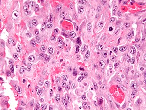
[[Image:Epithelioid_sarcoma_-_cropped_-_very_high_mag.jpg|thumb|right|[[Micrograph]] showing a tumour (epithelioid sarcoma) with prominent nucleoli. [[H&E stain]].]] | |||
The '''nucleolus''' (plural '''nucleoli''') is a thingy in the nucleus that may give the pathologist a clue to what they are looking at. | The '''nucleolus''' (plural '''nucleoli''') is a thingy in the nucleus that may give the pathologist a clue to what they are looking at. | ||
Generally speaking, large nucleoli | Generally speaking, large nucleoli suggest something is happening - they are associated with gene transcription. Large nucleoli are seen in malignancies and reactive conditions. | ||
==Macronucleolus== | ==Macronucleolus== | ||
| Line 8: | Line 9: | ||
Example: | Example: | ||
*Reed-Sternberg cell ([[Hodgkin lymphoma]]) ~ 5-7 micrometers.<ref name=Ref_PCPBoD8_329>{{Ref PCPBoD8|329}}</ref> | *Reed-Sternberg cell ([[Hodgkin lymphoma]]) ~ 5-7 micrometers.<ref name=Ref_PCPBoD8_329>{{Ref PCPBoD8|329}}</ref> | ||
===Image=== | |||
<gallery> | |||
Image:Glioblastoma_with_extreme_nuclear_enlargement_-_very_high_mag.jpg | Extreme nuclear enlargement with huge macronucleoli. (WC) | |||
</gallery> | |||
==Red nucleolus== | ==Red nucleolus== | ||
| Line 15: | Line 21: | ||
*[[Melanoma]]. | *[[Melanoma]]. | ||
*[[Serous carcinoma]]. | *[[Serous carcinoma]]. | ||
*[[Hereditary leiomyomatosis and renal cell carcinoma|Hereditary leiomyomatosis renal cell carcinoma syndrome associated renal cell carcinoma]]. | |||
==Large | ==Large nucleolus== | ||
Large - can be seen with 10x objective. | Large - can be seen with 10x objective. | ||
Examples: | Examples: | ||
*[[Melanoma]]. | |||
*Carcinoma. | |||
**Serous carcinoma. | |||
*Adenocarcinoma. | *Adenocarcinoma. | ||
**High-grade [[renal cell carcinoma]]. | **High-grade [[renal cell carcinoma]]. | ||
| Line 27: | Line 37: | ||
**[[Atypical teratoid/rhabdoid tumour]]. | **[[Atypical teratoid/rhabdoid tumour]]. | ||
**Epithelioid [[angiosarcoma]]. | **Epithelioid [[angiosarcoma]]. | ||
*[[Ganglion cell]]: | |||
**Ganglion (benign). | |||
**[[Gangliocytic paraganglioma]]. | |||
**[[Ganglioneuroma]]. | |||
==Medium-sized nucleolus== | ==Medium-sized nucleolus== | ||
| Line 32: | Line 47: | ||
Examples: | Examples: | ||
*Prostatic | *[[Prostatic adenocarcinoma]]. | ||
*Oncocytoma. | *[[Oncocytoma]]. | ||
*Mammary carcinoma, no special type. | *[[Invasive ductal carcinoma of the breast|Mammary carcinoma, no special type]]. | ||
*[[ | *[[Embryonal carcinoma]]. | ||
*[[Squamous metaplasia of the uterine cervix]]. | |||
==Small== | ==Small== | ||
| Line 49: | Line 65: | ||
Examples: | Examples: | ||
*[[Small cell carcinoma]]. | *[[Small cell carcinoma]]. | ||
*[[Neuroendocrine carcinoma]]. | |||
==See also== | ==See also== | ||
| Line 56: | Line 73: | ||
{{Reflist|1}} | {{Reflist|1}} | ||
[[Category: | [[Category:Histology]] | ||
Latest revision as of 05:47, 21 October 2015

The nucleolus (plural nucleoli) is a thingy in the nucleus that may give the pathologist a clue to what they are looking at.
Generally speaking, large nucleoli suggest something is happening - they are associated with gene transcription. Large nucleoli are seen in malignancies and reactive conditions.
Macronucleolus
Almost the size of RBC ~ 6-7 micrometers.
Example:
- Reed-Sternberg cell (Hodgkin lymphoma) ~ 5-7 micrometers.[1]
Image
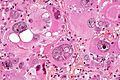
Extreme nuclear enlargement with huge macronucleoli. (WC)
Red nucleolus
Large - can be seen with 10x objective.
Examples:
- Melanoma.
- Serous carcinoma.
- Hereditary leiomyomatosis renal cell carcinoma syndrome associated renal cell carcinoma.
Large nucleolus
Large - can be seen with 10x objective.
Examples:
- Melanoma.
- Carcinoma.
- Serous carcinoma.
- Adenocarcinoma.
- High-grade renal cell carcinoma.
- Sarcoma:
- Ganglion cell:
- Ganglion (benign).
- Gangliocytic paraganglioma.
- Ganglioneuroma.
Medium-sized nucleolus
Medium - can be seen well with 20x objective.
Examples:
- Prostatic adenocarcinoma.
- Oncocytoma.
- Mammary carcinoma, no special type.
- Embryonal carcinoma.
- Squamous metaplasia of the uterine cervix.
Small
Small - hard to see at 20x objective, seen with 40x objective.
Examples:
Indistinct nucleolus
Not present - cannot see with 40x objective.
Examples:
See also
References
- ↑ Mitchell, Richard; Kumar, Vinay; Fausto, Nelson; Abbas, Abul K.; Aster, Jon (2011). Pocket Companion to Robbins & Cotran Pathologic Basis of Disease (8th ed.). Elsevier Saunders. pp. 329. ISBN 978-1416054542.