Ganglioneuroma
| Ganglioneuroma | |
|---|---|
| Diagnosis in short | |
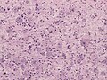
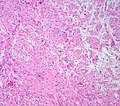
 Adrenal Ganglioneuroma. H&E stain. | |
|
| |
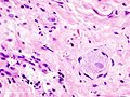
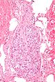
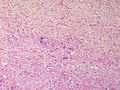
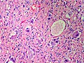
| LM | ganglion cells (large cells with large nucleus and prominent nucleolus), disordered fibrinous-like material, eosinophilic granular bodies |
| LM DDx | ganglioneuroblastoma |
| IHC | synaptophysin +ve, S-100 +ve |
| Gross | solid, firm, white |
| Site | usually adrenal or retroperitoneal, paraspinal |
|
| |
| Prevalence | uncommon |
| Prognosis | good |
Ganglioneuroma is a benign neuroblasic tumour. It is also known as ganglioma.[1]
It should not to be confused with ganglioglioma.
General
- May be retroperitoneal.
- Occasionally found in the GI tract - may form colonic polyp.
- Multiple ganglioneuromas may be due to multiple endocrine neoplasia IIb.
Classification:
- In a grouping known as neuroblastic tumours which includes:[2]
- Ganglioneuroma (benign).
- Ganglioneuroblastoma (intermediate).
- Neuroblastoma (aggressive).
Gross
- Solid.
- White.
- Firm.
- Well-circumscribed.
- May be nodular.
Images
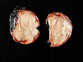
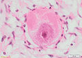
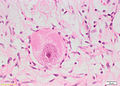
Adrenal ganglioneuroma. (WC)

www:
Microscopic
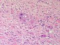
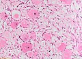
Features:
- Ganglion cells - key feature.
- Large cells with large nucleus.
- Prominent nucleolus.
- Large cells with large nucleus.
- Disordered fibrinous-like material.
- Eosinophilic granular bodies.[3]
See: adrenal ganglioneuroma, colonic ganglioneuroma.
Images
Ganglioneuroma. (WC)
A normal ganglion. (WC)
Adrenal Ganglioneuroma - Medium power (SKB)
Adrenal Ganglioneuroma - Medium power (SKB)
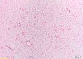
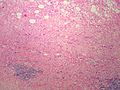
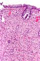
Adrenal Ganglioneuroma - Low power (SKB)
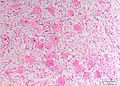
Adrenal Ganglioneuroma - Medium power (SKB)
Adrenal Ganglioneuroma - Medium power (SKB)
Adrenal Ganglioneuroma - Medium power (SKB)
Adrenal Ganglioneuroma - High power (SKB)
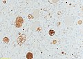
Adrenal Ganglioneuroma - synaptophysin (SKB)
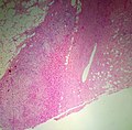
Adrenal Ganglioneuroma - Low power (SKB)
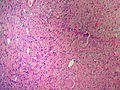
Adrenal Ganglioneuroma - Medium power (SKB)
Adrenal Ganglioneuroma - Medium power (SKB)
Adrenal Ganglioneuroma - Medium power (SKB)
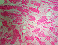
Adrenal Ganglioneuroma - Medium power (SKB)
Abdominal Ganglioneuroma, HE stain. (WC/jensflorian)
Retroperitoneal Ganglioneuroma. (Ed Uthman)
Histopathological image of ganglioneuroma in the posterior wall of pharynx. (WC/KGH)
Ganglioneuroma in colonic biopsy (WC/Nephron)
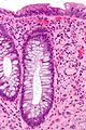
Ganglioneuroma in colonic biopsy, high mag (WC/Nephron)
.jpg)


.jpg)


www:
IHC
Features:[4]
- Spindle cells: S-100 +ve.
- Ganglion cells: NSE, synaptophysin, NF.
Sign out
Paraspinal Lesion, Right, Core Biopsy:
- Ganglioneuroma.

Comment:
The lesion stains as follows:
POSITIVE: S-100 & vimentin (stroma, ganglion cells), synaptophysin (ganglion cells only).
NEGATIVE: AE1/AE3, CD34.
PROLIFERATION (Ki-67): <1% of cells.
See also
References
- ↑ URL: http://medical-dictionary.thefreedictionary.com/ganglioma. Accessed on: 8 November 2010.
- ↑ Shimada H, Ambros IM, Dehner LP, Hata J, Joshi VV, Roald B (July 1999). "Terminology and morphologic criteria of neuroblastic tumors: recommendations by the International Neuroblastoma Pathology Committee". Cancer 86 (2): 349–63. PMID 10421272.
- ↑ R. Kiehl. 8 November 2010.
- ↑ Iacobuzio-Donahue, Christine A.; Montgomery, Elizabeth A. (2005). Gastrointestinal and Liver Pathology: A Volume in the Foundations in Diagnostic Pathology Series (1st ed.). Churchill Livingstone. pp. 217. ISBN 978-0443066573.