Libre Pathology:Image annotations added to Libre Pathology
Jump to navigation
Jump to search
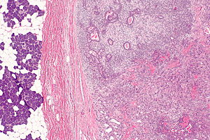

Carcinoma ex pleomorphic adenoma. H&E stain.
Libre Pathology now displays the image annotations of the WikiCommons.
Images with annotations can be identified by text, appearing below the images, that reads "This file has annotations. Move the mouse pointer over the image to see them."
We are excited about the possibilities of this tool for explaining pathology better.
Details and development
The image annotator is based on javascript and was developed by Lupo on the WikiCommons. Javascript in the web browser has to enabled for the annotations to appear. In most cases, this is the default.